. 2022 Feb 20;55(1):7.
doi: 10.1186/s40659-022-00373-7.

The OxyR and SoxR transcriptional regulators are involved in a broad oxidative stress response in Paraburkholderia xenovorans LB400

Affiliations

The OxyR and SoxR transcriptional regulators are involved in a broad oxidative stress response in Paraburkholderia xenovorans LB400

Valentina Méndez et al. Biol Res. .

Abstract

Background: Aerobic metabolism generates reactive oxygen species that may cause critical harm to the cell. The aim of this study is the characterization of the stress responses in the model aromatic-degrading bacterium Paraburkholderia xenovorans LB400 to the oxidizing agents paraquat and H2O2.

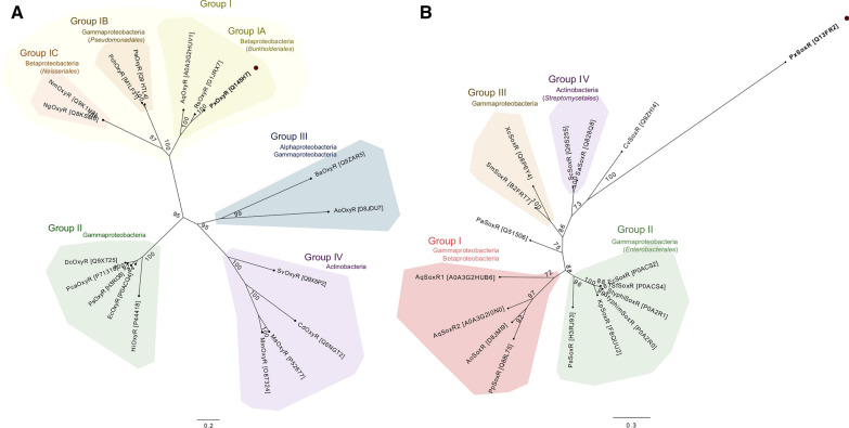
Methods: Antioxidant genes were identified by bioinformatic methods in the genome of P. xenovorans LB400, and the phylogeny of its OxyR and SoxR transcriptional regulators were studied. Functionality of the transcriptional regulators from strain LB400 was assessed by complementation with LB400 SoxR of null mutant P. aeruginosa ΔsoxR, and the construction of P. xenovorans pIZoxyR that overexpresses OxyR. The effects of oxidizing agents on P. xenovorans were studied measuring bacterial susceptibility, survival and ROS formation after exposure to paraquat and H2O2. The effects of these oxidants on gene expression (qRT-PCR) and the proteome (LC-MS/MS) were quantified.

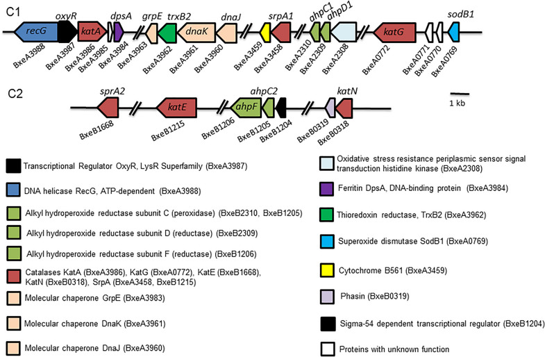
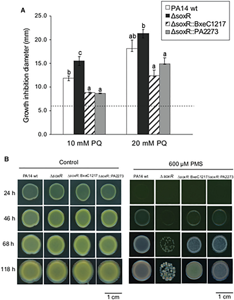
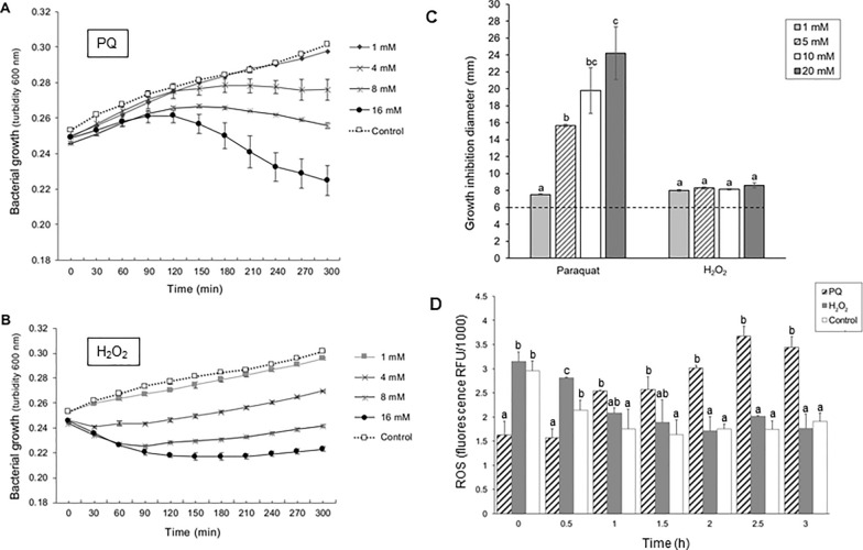
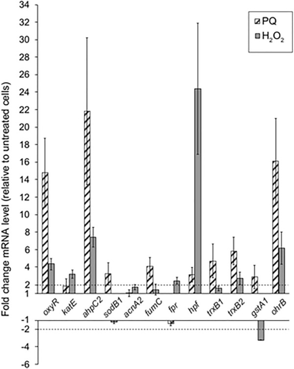
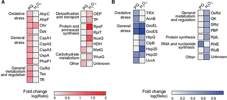
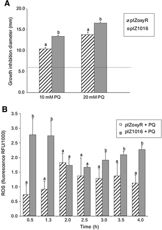
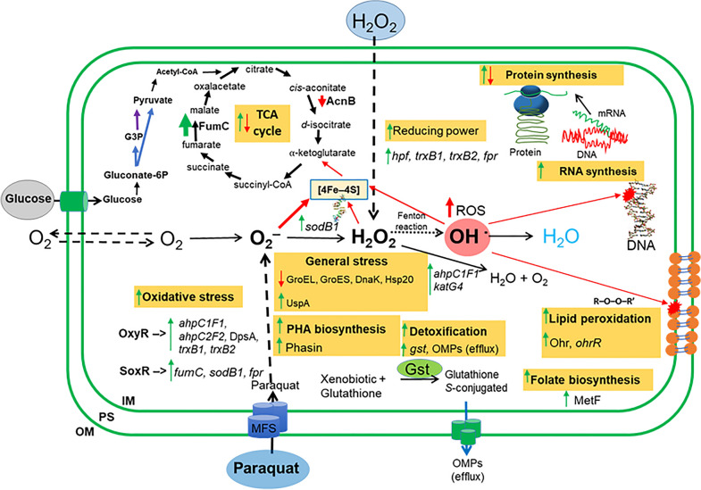
Results: P. xenovorans LB400 possesses a wide repertoire of genes for the antioxidant defense including the oxyR, ahpC, ahpF, kat, trxB, dpsA and gorA genes, whose orthologous genes are regulated by the transcriptional regulator OxyR in E. coli. The LB400 genome also harbors the soxR, fumC, acnA, sodB, fpr and fldX genes, whose orthologous genes are regulated by the transcriptional regulator SoxR in E. coli. The functionality of the LB400 soxR gene was confirmed by complementation of null mutant P. aeruginosa ΔsoxR. Growth, susceptibility, and ROS formation assays revealed that LB400 cells were more susceptible to paraquat than H2O2. Transcriptional analyses indicated the upregulation of the oxyR, ahpC1, katE and ohrB genes in LB400 cells after exposure to H2O2, whereas the oxyR, fumC, ahpC1, sodB1 and ohrB genes were induced in presence of paraquat. Proteome analysis revealed that paraquat induced the oxidative stress response proteins AhpCF and DpsA, the universal stress protein UspA and the RNA chaperone CspA. Both oxidizing agents induced the Ohr protein, which is involved in organic peroxide resistance. Notably, the overexpression of the LB400 oxyR gene in P. xenovorans significantly decreased the ROS formation and the susceptibility to paraquat, suggesting a broad OxyR-regulated antioxidant response.

Conclusions: This study showed that P. xenovorans LB400 possess a broad range oxidative stress response, which explain the high resistance of this strain to the oxidizing compounds paraquat and H2O2.

Keywords: Hydrogen peroxide; Oxidative stress; OxyR; Paraburkholderia xenovorans; Paraquat; SoxR; Superoxide.


Conflict of interest statement

The authors declare that they have no competing interests.

Figures

Fig. 1
Fig. 1
Genomic context of genes involved in oxidative stress response in P. xenovorans LB400. C1 and C2 indicate the major and the minor chromosome, respectively
Fig. 2
Fig. 2
Evolutionary relationships of OxyR and SoxR of P. xenovorans LB400 and other bacteria. Mid-rooted Bayesian Inference trees calculated by MrBayes of the OxyR and SoxR regulators with experimental evidence. P. xenovorans LB400 OxyR (PxOxyR) and SoxR (PxSoxR) are shown in bold letters and marked with a dark circle. Node values represent the bootstrapping value (%) of the analysis. Each group determined was identified at the class rank level. If the amino acid sequences within each group correspond to the same family rank, the taxon name is included in parentheses. A Phylogenetic tree of the H2O2-sensing transcriptional regulator OxyR homologs. Four major groups were identified, predominantly clustered by taxonomic relatedness. PxOxyR is encompassed within the Group IA, represented by Burkholderiales. B Phylogenetic tree of the redox-sensitive transcriptional regulator SoxR homologs. Four major groups are identified, including three singleton sequences, not observing a clear taxonomic distribution within the groups. PxSoxR is a singleton closer to the SoxR regulator of Chromobacterium violaceum DSM 30191 than to other groups
Fig. 3
Fig. 3
The soxR gene of P. xenovorans LB400 complements faulty oxidative response in P. aeruginosa ∆soxR. A Susceptibility assays of P. aeruginosa strains PA14 wt, and mutant strains (∆soxR, ∆soxR::BxeC1217 and ∆soxR::PA2273) exposed to paraquat (PQ). Paper disks soaked with paraquat (10 or 20 mM) were placed on P. aeruginosa bacterial lawns. Growth inhibition zones around the disks were recorded after 24 h at 37 °C. The dotted line at 6 mm corresponds to the disk diameter and indicates no detectable inhibition. Inhibition zone values were calculated as the mean ± SD of three independent experiments. Letters under the error bars indicate significant differences between strains in each treatment. B Growth of P. aeruginosa wt and mutants strains exposed to phenazine methosulfate (PMS) (600 µM). Control cells were grown in absence of PMS. Images are representative of three independent experiments
Fig. 4
Fig. 4
Effects of paraquat and H2O2 on P. xenovorans LB400 growth and ROS formation. Strain LB400 grown until exponential growth phase on glucose was exposed to paraquat (PQ) (A) or H2O2 (B). Growth was monitored by measuring turbidity at 600 nm. C Paper disks soaked with paraquat or H2O2 solutions (1–20 mM) were placed on P. xenovorans LB400 bacterial lawns. Growth inhibition zones around the disks were recorded after 24 h incubation at 30 °C. The dotted line at 6 mm corresponds to the disk diameter and indicates no inhibition. Letters under the error bars indicate significant differences between each concentration of each oxidizing agent. D Exponential glucose-grown cells were exposed to paraquat or H2O2. After incubation with HPF, fluorescence was monitored at 515 nm (emission) and 490 nm (excitation). Cells incubated in absence of oxidizing agents were used as control. Letters under the error bars indicate significant differences between treatments in each time. All values were calculated as the mean ± SD of three independent experiments
Fig. 5
Fig. 5
Expression of oxidative stress genes in P. xenovorans LB400 upon exposure to paraquat or H2O2. Expression levels of oxidative stress genes were measured by RT-qPCR after strain LB400 exposure to paraquat (PQ) or H2O2 (1 mM) for 1 h. The gyrB gene was used as the reference gene. Fold-change levels were calculated as the mean ± SD of three independent experiments. The dotted lines indicate the level of expression from which a significant increase (2) or decrease (-2) of expression is observed
Fig. 6
Fig. 6
Differentially expressed proteins of P. xenovorans LB400 after exposure to paraquat or H2O2. Heatmap showing upregulated (A) or downregulated (B) protein levels of P. xenovorans LB400 after exposure to paraquat (PQ) or H2O2 (1 mM) for 1 h compared to untreated cells. Changes in protein levels were established when changes of ≥ twofold were observed on treated cells versus control cells. Red represents higher expression, whereas blue represents lower expression. Three biological replicates were performed. AhpC, alkyl hydroperoxide reductase subunit C2; organic hydroperoxide resistance protein OhrB; Dps, ferritin DPS family DNA-binding protein DpsA. OxRd oxidoreductase, Tas Tas oxidoreductase, TR transcriptional regulator of LysR family, OEP outer membrane efflux protein related to copper resistance, TP transport protein, HDH homoserine dehydrogenase, GK glycerate kinase, SRd sulphite reductase (NADPH) beta-subunit, PBP penicillin binding-protein. Additional file 1: Tables S3 and S4 provide more details of the proteins
Fig. 7
Fig. 7
Susceptibility and ROS formation in P. xenovorans pIZoxyR exposed to paraquat. A Susceptibility assays of P. xenovorans strains exposed to paraquat (PQ). Paper disks soaked with paraquat (1–20 mM) were placed on P. xenovorans strain bacterial lawns. Zones of growth inhibition around the disks were recorded after 24 h incubation at 30 °C. The diameter of 6 mm corresponds to the disk diameter and indicates no inhibition. Inhibition zone values were calculated as the mean ± SD of three independent experiments. Letters under the error bars indicate significant differences between strains in each treatment. B ROS quantification assay. P. xenovorans pIZoxyR was grown on glucose (5 mM) until exponential growth phase and exposed to paraquat (20 mM). ROS formation of non-treated cells was also monitored. Fluorescence was monitored after incubation with HPF at 490 nm (excitation) and 515 nm (emission). Fluorescence values were calculated as the mean ± SD of three independent experiments. Letters under the error bars indicate significant differences between strains in each time
Fig. 8
Fig. 8
Molecular oxidative stress response of P. xenovorans LB400 during exposure to paraquat and H2O2. Green arrows indicate upregulation of genes and/or proteins, while red arrows indicate downregulation. Dashed arrows indicate the transport of O2, H2O2 and paraquat through the cell membranes. Dotted arrow indicates Fenton reaction, a process during which metal ions (mainly Fe2+) react with H2O2 forming hydroxyl radical. Glucose is converted into gluconate 6-phosphate (gluconate-6P) by glucokinase and the Pentose Phosphate (PP) pathway. Blue arrow indicates the transformation of the gluconate-6P to glyceraldehyde-3-phosphate (G3P) and pyruvate via the Entner–Doudoroff pathway. Purple arrow indicates the conversion of G3P into pyruvate via the lower Embden–Meyerhof–Parnas (EMP) pathway. OM outer membrane, PS periplasmic space, IM inner membrane, FumC/fumC fumarate hydratase C, AcnB aconitate hydratase, hpf high potential Fe-S protein, trxB1 thioredoxin 1, trxB2 thioredoxin 2, fpr flavodoxin/ferredoxin NADP oxidoreductase, sodB1 superoxide dismutase, DnaK GroEL and GroES, molecular chaperones, Hsp20 heat shock protein, UspA universal stress protein, ahpC1D1 and ahpC2F alkyl hydroperoxide reductases, katE catalase, DpsA ferritin protein of the DPS family, Gst/gstA1 glutathione S-transferase, Ohr organic hydroperoxide resistance protein, OhrR organic hydroperoxide resistance transcriptional regulator, MetF 5,10-methylenetetrahydrofolate reductase

Similar articles

Cited by

References

    1. Álvarez-Santullano N, Villegas P, Mardones MS, Durán RE, Donoso R, González A, et al. Genome-wide metabolic reconstruction of the synthesis of polyhydroxyalkanoates from sugars and fatty acids by Burkholderia sensu lato species. Microorganisms. 2021;9:1290. - PMC - PubMed
    1. Agulló L, Cámara B, Martínez P, Latorre-Reyes V, Seeger M. Response to (chloro)biphenyls of the PCB-degrader Burkholderia xenovorans LB400 involves stress proteins induced also by heat shock and oxidative stress. FEMS Microbiol Lett. 2007;267:167–175. - PubMed
    1. Agulló L, Romero-Silva MJ, Domenech M, Seeger M. p-Cymene promotes its catabolism through the p-cymene and the p-cumate pathways, activates a stress response and reduces the biofilm formation in Burkholderia xenovorans LB400. PLoS ONE. 2017;12:e0169544. - PMC - PubMed
    1. Altschul SF, Gish W, Miller W, Myers EW, Lipman DJ. Basic local alignment search tool. J Mol Biol. 1990;215:403–410. - PubMed
    1. Apweiler R, Bairoch A, Wu CH. Protein sequence databases. Curr Opin Che Biol. 2004;8:76–80. - PubMed

MeSH terms

Supplementary concepts

LinkOut - more resources