. 2022 May;23(5):707-719.
doi: 10.1111/mpp.13192. Epub 2022 Feb 20.

The nonstructural protein NSs encoded by tomato zonate spot virus suppresses RNA silencing by interacting with NbSGS3

Affiliations

The nonstructural protein NSs encoded by tomato zonate spot virus suppresses RNA silencing by interacting with NbSGS3

Jianbin Chen et al. Mol Plant Pathol. 2022 May.

Abstract

Viral suppressors of RNA silencing (VSRs) are encoded by diverse viruses to counteract the RNA silencing-mediated defence mounted by the virus-infected host cells. In this study, we identified the NSs protein encoded by tomato zonate spot virus (TZSV) as a potent VSR, and used a potato virus X (PVX)-based heterologous expression system to demonstrate TZSV NSs as a viral pathogenicity factor that intensified PVX symptoms in Nicotiana benthamiana. We then used a yeast two-hybrid screen to identify the suppressor of gene silencing 3 protein of N. benthamiana (NbSGS3), a known component of the plant RNA silencing pathway, as an interaction partner of TZSV NSs. We verified this interaction in plant cells with bimolecular fluorescence complementation, subcellular colocalization, and co-immunoprecipitation. We further revealed that the NSs-NbSGS3 interaction correlated with the VSR activity of TZSV NSs. TZSV NSs reduced the concentration of NbSGS3 protein in plant cells, probably through the ubiquitination and autophagy pathways. Interestingly, TZSV infection, but not NSs overexpression, significantly up-regulated the NbSGS3 transcript levels. Our data indicate that TZSV NSs suppresses RNA silencing of the host plant and enhances TZSV pathogenicity through its interaction with NbSGS3. This study reveals a novel molecular mechanism of NSs-mediated suppression of plant host antiviral defence.

Keywords: Suppressor of gene silencing 3; RNA silencing; VSRs; nonstructural protein NSs; tomato zonate spot virus (TZSV).


Conflict of interest statement

The authors declare that there is no conflict of interest.

Figures
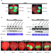
FIGURE 1
FIGURE 1
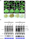
TZSV NSs blocks RNA silencing triggered by single‐stranded green fluorescent protein (GFP) and double‐stranded GFP. (a) Suppression of GFP silencing in Nicotiana benthamiana 16c plants. N. benthamiana leaf patches were co‐infiltrated with Agrobacterium tumefaciens cultures containing GFP (35S‐GFP) and empty vector (EV), NSs, NSm, N, NSs (TSWV) or p19 representative leaves were photographed at 6 days postinoculation (dpi) under UV light. (b) Northern blots of GFP mRNA accumulation and western blots (WB) of GFP accumulation in agroinfiltrated leaf patches from the samples indicated in (a). Ethidium bromide staining of rRNA served as loading control for northern blots and Coomassie Brilliant blue (CBB) staining of the large subunit of RuBisCO was used as loading controls for western blots. (c) Suppression of dsGFP silencing in N. benthamiana leaf patches were co‐infiltrated with A. tumefaciens cultures containing GFP and dsGFP and EV, NSs, NSs (TSWV) or p19 representative leaf patches were photographed under UV light at 6 dpi. (d) Northern blot of GFP mRNA accumulation and western blot of GFP accumulation in agroinfiltrated leaf patches were performed as indicated in (c). Ethidium bromide staining of rRNA and Coomassie Brilliant blue staining of the large subunit of RuBisCO were used as loading controls for northern blots and western blots, respectively
FIGURE 2
FIGURE 2
The NSs of tomato zonate spot virus (TZSV) induces systemic symptoms in Nicotiana benthamiana plants. (a) Symptoms of N. benthamiana plants at 6 days postinoculation (dpi) and 12 dpi infected with PVX, PVX‐NSs, PVX‐NSm, and PVX‐N. The infected leaves were photographed at 6 and 12 dpi (upper panels) or photographed after 3,3′‐diaminobenzidine staining (lower panels). (b) Northern blots and western blots (WB) analysis of PVX coat protein (CP) mRNA and protein accumulation in plants infected with PVX, PVX‐NSs, PVX‐NSm, and PVX‐N, at 6 and 12 dpi. Ethidium bromide staining of rRNA and Coomassie Brilliant blue (CBB) staining of RuBisCO were used as loading controls for northern blots and western blots, respectively. The migration of genomic RNA (gRNA), subgenomic RNA1 (sgRNA1), CP subgenomic RNA (CP sgRNA) is indicated
FIGURE 3
FIGURE 3
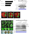
The NSs C‐terminal domain is responsible for gene silencing suppression, severe symptoms and necrotic responses. (a) Schematic representation of NSs mutant. (b) Plants were inoculated with green fluorescent protein (GFP) and P19, pBin‐NSs, pBin‐NSs(1–444aa) or pBin‐NSs(1–423aa), and photographed under UV light at 6 days postinoculation (dpi). (c) Northern blot analysis of GFP mRNA and western blot (WB) analysis of GFP. Ethidium bromide staining of rRNA and Coomassie Brilliant blue staining (CBB) of RuBisCO were used as loading controls for northern and western blots, respectively. (d) Nicotiana benthamiana plants infected with PVX, PVX‐NSs, PVX‐NSs(1–444aa) or PVX‐NSs(1–423aa) were photographed at 6 and 12 dpi. The lower panels show 3,3′‐diaminobenzidine‐stained leaves of infected plants to show H2O2 induction at 12 dpi. (e) Northern blots and western blots analysis of PVX coat protein (CP) mRNA and PVX CP in systemically infected leaves of the N. benthamiana plants. The positions of genomic RNA (gRNA), subgenomic RNA1 and RNA2 (sgRNA1/2), and CP subgenomic RNA (CP sgRNA) are indicated
FIGURE 4
FIGURE 4
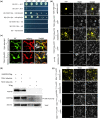
TSWV NSs or TZSV NSs interact with NbSGS3. (a) Interactions between TZSV NSs and NbSGS3 and between TSWV NSs and NbSGS3 identified by yeast two‐hybrid assays. Y2Hgold cells were diluted 10−1 to 10−5 and plated onto QDO (SD−Trp−Leu−His−Ade) medium. Controls: AD‐T + BD‐53 (positive control) or AD‐NbSGS3 + BD or AD + BD‐NSs or AD + BD‐NSs (TSWV) (negative control). (b) Bimolecular fluorescence complementation (BiFC) assays in Nicotiana benthamiana leaves at 72 h postinoculation (hpi). Yellow fluorescence (green) was observed as a consequence of the complementation of the YN and YC tagged with NbSGS3 and NSs or NbSGS3 and NSs (TSWV). Bars, 25 μm. (c) Co‐localization of NbSGS3‐mCherry with NSs‐GFP and NbSGS3‐mCherry with NSs (TSWV)‐GFP in N. benthamiana leaf cells. Infiltrated leaves were treated at 72 hpi after confocal images. Bars, 25 μm. (d) NbSGS3‐3FLAG was co‐expressed with TZSV NSs‐3HA or TSWV NSs‐3HA in N. benthamiana. Total protein was extracted and immunoprecipitated with anti‐FLAG magnetic beads. Western blots were carried out using monoclonal anti‐FLAG or anti‐HA antibodies to explore FLAG‐tagged proteins or co‐immunoprecipitated HA‐tagged protein, respectively. (e) BiFC of NbSGS3‐cYFP with NSs‐nYFP encoded by TZSV or TSWV were transiently co‐expressed in N. benthamiana leaves. Fluorescence was detected by confocal microscopy at 48 hpi. Bar, 25 μm
FIGURE 5
FIGURE 5
The viral suppressors of RNA silencing (VSR) activity of TZSV NSs is attenuated by NbSGS3 overexpression. (a) Suppression of green fluorescent protein (GFP) silencing in Nicotiana benthamiana plants. N. benthamiana leaf patches were co‐infiltrated with Agrobacterium tumefaciens cultures containing NbSGS3 and GFP (35S‐GFP) and empty vector (EV), NSs, NSs (TSWV). Representative leaves were photographed at 6 days postinoculation (dpi) under UV light. (b) Western blot analysis of GFP accumulation in plants at 6 dpi. Coomassie Brilliant blue (CBB) staining of RuBisCO was used as loading control
FIGURE 6
FIGURE 6
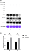
NSs induces the degradation of NbSGS3 protein. (a) NbSGS3‐3FLAG, green fluorescent protein (GFP), and TZSV NSs‐HA was transiently co‐expressed in Nicotiana benthamiana leaves by Agrobacterium infiltration. Infiltrated leaf areas were harvested for protein extraction at 56 h postinoculation (hpi). At 40 hpi, the infiltrated leaf areas were treated with MG132 or E64d or dimethyl sulphoxide (DMSO) for an additional 16 h. Total protein was probed with anti‐FLAG or anti‐HA or anti‐GFP antibodies. The bottom panel shows equal protein loading by Commassie Brilliant blue (CBB) staining. The band intensities were determined by ImageJ. (b) The expression of NbSGS3 in transiently expressed plants with NSs (TZSV) or GFP was analysed by reverse transcription quantitative PCR. Error bars represent the mean ± SEM of three independent experiments. (Student's t test, *p <0 .05)
FIGURE 7
FIGURE 7
NbSGS3 is required for antiviral RNAi defence against TZSV. (a) The expression of NbSGS3 in virus‐induced gene silencing (VIGS) plants was analysed by reverse transcription quantitative PCR (RT‐qPCR). Error bars represent the mean ± SEM of three independent experiments. Student's t test was performed to assess differences between TRV‐NbSGS3 and TRV infected plants, significant differences (*p < 0.05, **p < 0.01). (b) and (c) Detection of TZSV accumulation in systemically infected leaves of TRV‐NbSGS3 and TRV preinfected Nicotiana benthamiana plant by RT‐qPCR analysis (b) and western blot (c). TZSV N‐specific primers and TZSV N‐specific antibody were used to detect accumulation of TZSV RNA and N protein, respectively. Coomassie Brilliant blue (CBB) staining of RuBisCO was used as loading control. (d) The expression of NbSGS3 in virus‐infected plants was analysed by RT‐qPCR. Error bars represent the mean ± SEM of three independent experiments. Student's t test was performed to assess differences between TRV‐NbSGS3‐ and TRV‐infected plants. Data are mean ± SEM, asterisks indicate a significant difference (Student's t test, *p < 0.05, **p < 0.01)

Similar articles

Cited by

References

    1. Bucher, E. , Sijen, T. , De Haan, P. , Goldbach, R. & Prins, M. (2003) Negative‐strand tospoviruses and tenuiviruses carry a gene for a suppressor of gene silencing at analogous genomic positions. Journal of Virology, 77, 1329–1336. - PMC - PubMed
    1. Cheng, X. , Wang, A. & Simon, A.E. (2017) The potyvirus silencing suppressor protein VPg mediates degradation of SGS3 via ubiquitination and autophagy pathways. Journal of Virology, 91, e01478‐16. - PMC - PubMed
    1. Csorba, T. , Kontra, L. & Burgyan, J. (2015) viral silencing suppressors: tools forged to fine‐tune host–pathogen coexistence. Virology, 479–480, 85–103. - PubMed
    1. Dalmay, T. , Hamilton, A. , Rudd, S. , Angell, S. & Baulcombe, D.C. (2000) An RNA‐dependent RNA polymerase gene in Arabidopsis is required for posttranscriptional gene silencing mediated by a transgene but not by a virus. Cell, 101, 543–553. - PubMed
    1. Ding, S.W. (2010) RNA‐based antiviral immunity. Nature Reviews Immunology, 10, 632–644. - PubMed

Publication types

LinkOut - more resources