. 2022 Feb 18;5(6):e202101103.
doi: 10.26508/lsa.202101103. Print 2022 Jun.

Human immunodeficiency virus type 1 impairs sumoylation

Affiliations

Human immunodeficiency virus type 1 impairs sumoylation

Bilgül Mete et al. Life Sci Alliance. .

Abstract

During infection, the human immunodeficiency virus type 1 (HIV-1) manipulates host cell mechanisms to its advantage, thereby controlling its replication or latency, and evading immune responses. Sumoylation is an essential post-translational modification that controls vital cellular activities including proliferation, stemness, or anti-viral immunity. SUMO peptides oppose pathogen replication and mediate interferon-dependent anti-viral activities. In turn, several viruses and bacteria attack sumoylation to disarm host immune responses. Here, we show that HIV-1 impairs cellular sumoylation and targets the host SUMO E1-activating enzyme. HIV-1 expression in cultured HEK293 cells or in CD4+ Jurkat T lymphocytes diminishes sumoylation by both SUMO paralogs, SUMO1 and SUMO2/3. HIV-1 causes a sharp and specific decline in UBA2 protein levels, a subunit of the heterodimeric SUMO E1 enzyme, which likely serves to reduce the efficiency of global protein sumoylation. Furthermore, HIV-1-infected individuals display a significant reduction in total leukocyte sumoylation that is uncoupled from HIV-induced cytopenia. Because sumoylation is vital for immune function, T-cell expansion and activity, loss of sumoylation during HIV disease may contribute to immune system deterioration in patients.


Conflict of interest statement

The authors declare that they have no conflict of interest.

Figures
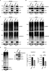
Figure S1.
Figure S1.. Viral protein expression in HEK293 or Jurkat cells, and HIV-1–induced down-regulation of cellular sumoylation.
(A, B) Assessment of EGFP, HIV-1 Rev, and HIV-1 integrase expression by Western blot in HEK293 cells (A), or in CD4+ Jurkat T lymphocytes (B), transfected or electroporated with the lentiviral vector pfNL43-dE-EGFP encoding HIV-1. The EGFP cassette was inserted within the HIV-1 env gene. (C) HEK293 cells or CD4+ Jurkat T lymphocytes were transfected or electroporated with a vector encoding EGFP only. Global SUMO1 and SUMO2/3 conjugates were analyzed by Western blot. (D) HeLa cells were transfected with the lentiviral vector pfNL43-dE-EGFP encoding HIV-1, and lysed 72 h post-transfection to assess global sumoylation levels, as well as UBA2, EGFP, HIV-1 Rev, and HIV-1 integrase expression by Western blot. Densitometric quantifications of SUMO and UBA2 signals were normalized to GAPDH expression (which serves as a loading control). Data are presented as mean ± SEM (n = 3), P-values were calculated using t test assuming unequal variances. Control: cells transfected with an empty vector backbone devoid of HIV-1 genes, HIV: cells expressing the HIV-1 genome.
Figure 1.
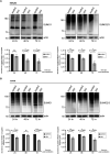
Figure 1.. HIV-1 diminishes the abundance of cellular SUMO conjugates.
(A, B) A lentiviral vector (pfNL43-dE-EGFP) encoding HIV-1 (containing all nine viral genes: gag, pol, tat, rev, vif, vpr, vpu, nef, and env) was transfected into HEK293 cells (A) or electroporated to CD4+ Jurkat T lymphocytes (B), allowing reverse transcription and integration, followed by viral protein expression in cells. Neither the envelope protein nor any viral particles are produced because of the insertion of an EGFP cassette within the env gene. Expression levels of EGFP and two viral proteins, Rev and integrase, are shown in Fig S1A and B. This system allows in vitro simulation of HIV-1 infection in a safe, reproducible and quantifiable manner. Cells were lysed at indicated times post-transfection, and global sumoylation levels were assessed by Western blot, using human anti-SUMO1 and anti-SUMO2/3 antibodies. Representative blots are shown. Densitometric quantifications of SUMO signals were normalized to actin expression (which serves as a loading control). Data are presented as mean ± SEM (n = 4 for HEK293 cells, n = 7 for Jurkat cells), asterisks denote statistical significance (P-values were calculated using t test assuming unequal variances, ns, not significant). Control: cells transfected with an empty vector backbone devoid of HIV-1 genes, HIV, cells expressing the HIV-1 genome.
Figure 2.
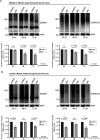
Figure 2.. HIV-1 induces proteasome-independent loss of cellular SUMO conjugates.
(A, B) HEK293 cells (A) or CD4+ Jurkat T lymphocytes (B) were transfected or electroporated with the lentiviral vector pfNL43-dE-EGFP encoding the HIV-1 genome as described above, then treated for 24 h with 3 μM MG132, a proteasome inhibitor drug, before lysis (at indicated times post-transfection/electroporation). Global SUMO1 and SUMO2/3 conjugates were analyzed by Western blot. Densitometric quantifications of SUMO signals were normalized to actin expression (which serves as a loading control). Data are presented as mean ± SEM (n = 5 for HEK293 cells, n = 4 for Jurkat cells), asterisks denote statistical significance (P-values were calculated using t test assuming unequal variances, ns: not significant). Control: cells transfected with an empty vector backbone devoid of HIV-1 genes, HIV: cells expressing the HIV-1 genome. Ubiquitin blots are shown in Fig S2.
Figure S2.
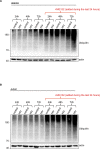
Figure S2.. Ubiquitin conjugates in MG132-treated cells.
(A, B) Western blot analysis of global ubiquitin conjugates of HEK293 cells (A) and CD4+ Jurkat T lymphocytes (B) shown in Fig 2. Cells were transfected or electroporated with the lentiviral vector pfNL43-dE-EGFP encoding HIV-1, and treated for 24 h with 3 μM MG132 before lysis (at indicated times post-transfection/electroporation). The ubiquitylation profiles of untreated cells are also shown for comparison.
Figure 3.
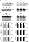
Figure 3.. HIV-1 targets UBA2, a subunit of the SUMO E1–activating enzyme.
(A) HEK293 cells or CD4+ Jurkat T lymphocytes were transfected or electroporated with the lentiviral vector pfNL43-dE-EGFP encoding HIV-1 as described above, followed by lysis at indicated times post-transfection to assess the level of free (unconjugated) SUMO1 and SUMO2/3 proteins by Western blot. (B) Western blots show UBA2, SAE1 and UBC9 protein levels in HEK293 and Jurkat cells. Cells were lysed and proteins were analyzed at indicated times after transfection or electroporation with HIV-1. HIV-1 specifically down-regulates the host UBA2 protein, a subunit of the heterodimeric SUMO E1 enzyme. Densitometric quantifications of UBA2, SAE1, and UBC9 signals were normalized to tubulin expression, which serves as a loading control. Data are presented as mean ± SEM (n ≥ 3 for HEK293 and Jurkat cells), asterisks denote statistical significance (P-values were calculated using t test assuming unequal variances, ns, not significant). Control, cells transfected with an empty vector backbone devoid of HIV-1 genes; HIV, cells expressing the HIV-1 genome.
Figure S3.
Figure S3.. Further analysis of HIV-1–induced UBA2 loss.
(A) Real-time PCR analyses of UBA2 mRNA levels in HEK293 cells or in CD4+ Jurkat T lymphocytes transfected or electroporated with the lentiviral vector pfNL43-dE-EGFP encoding the HIV-1 genome. Data are presented as mean ± SEM (n = 4 for HEK293 cells, n = 3 for Jurkat cells). P-values were calculated using t test assuming unequal variances. Control, cells transfected with an empty vector backbone devoid of HIV-1 genes; HIV, cells expressing the HIV-1 genome. Analyses were performed at indicated time points post-transfection/electroporation. (B) Western blot analysis of UBA2 protein in HIV-1–expressing HEK293 or Jurkat cells treated with the proteasome inhibitor drug MG132, as in Fig 2. Densitometric quantifications of UBA2 protein were normalized to actin expression, which serves as a loading control. Data are presented as mean ± SEM (n = 3), asterisks denote statistical significance (P-values were calculated using t test assuming unequal variances, ns, not significant). Control, cells transfected with an empty vector backbone devoid of HIV-1 genes; HIV, cells expressing the HIV-1 genome.
Figure 4.
Figure 4.. Global leukocyte sumoylation is impaired during HIV disease.
(A) Western blots show SUMO conjugate levels in total leukocytes of a representative ART-naive HIV-1–infected individual (HIV(+)), in comparison with an uninfected individual (HIV(−)). Western blots were performed as described in Fig 1 and in the Materials and Methods section; sumoylated proteins were detected using human anti-SUMO1 and anti-SUMO2/3 antibodies. Actin: loading control (important note: equal amounts [20 μg] of protein were loaded in each well to allow fair comparison of SUMO profiles between HIV(−) and HIV(+) samples). Graphs (right panel) show densitometric quantifications of SUMO signals normalized to actin expression. Data are presented as mean ± SEM (n = 15 individuals for SUMO1, n = 19 individuals for SUMO2/3), P-values are indicated (using t test assuming unequal variances). Asterisks denote statistical significance. (B) The contribution of CD4+ cells to total leukocyte sumoylation was assessed by Western blot after ex vivo depletion of the former from the peripheral blood (leukocytes) of HIV-negative individuals. Total leukocytes (total: before depletion) and the CD4+ cell-deprived fraction (CD4(−)) are shown for comparison for a representative individual. Graphs show densitometric quantifications of SUMO signals normalized to actin expression. Data are presented as mean ± SEM (n = 4 individuals), P-values were calculated using t test assuming unequal variances, ns, not significant. Sumoylation in the CD4+ fraction is shown in Fig S4B. (C) Western blot shows leukocyte sumoylation profiles of a representative HIV-1–infected individual before (labeled as naive) and after anti-retroviral therapy (labeled as ART), with respect to that of an uninfected control individual (HIV(−)). Patient received ART for 3 mo after which the HIV-1 viral load became undetectable, yet the CD4+ count did not considerably rise. Graph shows densitometric quantifications of SUMO signals normalized to actin expression. Data are presented as mean ± SEM (n = 3 individuals), P-values are indicated (using t test assuming unequal variances). Asterisks denote statistical significance. (D) Western blot analysis of UBA2 in total leukocytes of a representative ART-naive HIV-1–infected individual (HIV(+)), in comparison with an uninfected individual (HIV(−)). 3 HIV(+) and 3 HIV(−) individuals were analyzed; graph shows densitometric quantifications of UBA2 signals normalized to actin expression (data are presented as mean ± SEM, n = 3 individuals, asterisks denote statistical significance; P-value is indicated, using t test assuming unequal variances).
Figure S4.
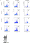
Figure S4.. Further analysis of sumoylation in leukocytes.
(A) CD4+ T-cell counts of the four HIV(−) individuals described in Fig 4B. Flow cytometry analyses indicate that CD4+ cells constitute about 8–15% of total leukocytes (PMBCs and granulocytes) of these individuals. Left, middle and right columns show CD4 positivity in total leukocytes, CD4+ cell-depleted (CD4(−)), and CD4+ cell populations, respectively. (B) Sumoylation in CD4+ cells, in comparison with that of total leukocytes. CD4+ cells were removed ex vivo from the peripheral blood (leukocytes) of an HIV(−) individual analyzed in Fig 4B. Total leukocytes (total: before depletion) and the CD4+ fraction (CD4(+)) are shown for comparison.

Similar articles

Cited by

References

    1. Cohen MS, Shaw GM, McMichael AJ, Haynes BF (2011) Acute HIV-1 infection. N Engl J Med 364: 1943–1954. 10.1056/NEJMra1011874 - DOI - PMC - PubMed
    1. Pope M, Haase AT (2003) Transmission, acute HIV-1 infection and the quest for strategies to prevent infection. Nat Med 9: 847–852. 10.1038/nm0703-847 - DOI - PubMed
    1. Fenwick C, Joo V, Jacquier P, Noto A, Banga R, Perreau M, Pantaleo G (2019) T-cell exhaustion in HIV infection. Immunol Rev 292: 149–163. 10.1111/imr.12823 - DOI - PMC - PubMed
    1. Burgos J, Ribera E, Falcó V (2018) Antiretroviral therapy in advanced HIV disease: Which is the best regimen? AIDS Rev 20: 3–13. 10.24875/aidsrev.m17000010 - DOI - PubMed
    1. Goff SP (2007) Host factors exploited by retroviruses. Nat Rev Microbiol 5: 253–263. 10.1038/nrmicro1541 - DOI - PubMed

Publication types

Substances