. 2022 Feb 17;10(1):15.
doi: 10.1038/s41413-022-00189-x.

Estrogen-mediated downregulation of HIF-1α signaling in B lymphocytes influences postmenopausal bone loss

Affiliations

Estrogen-mediated downregulation of HIF-1α signaling in B lymphocytes influences postmenopausal bone loss

Xianyi Meng et al. Bone Res. .

Abstract

In the bone marrow, B cells and bone-resorbing osteoclasts colocalize and form a specific microenvironment. How B cells functionally influence osteoclasts and bone architecture is poorly understood. Using genetically modified mice and high-throughput analyses, we demonstrate that prolonged HIF-1α signaling in B cells leads to enhanced RANKL production and osteoclast formation. In addition, deletion of HIF-1α in B cells prevents estrogen deficiency-induced bone loss in mice. Mechanistically, estrogen controls HIF-1α protein stabilization through HSP70-mediated degradation in bone marrow B cells. The stabilization of HIF-1α protein in HSP70-deficient bone marrow B cells promotes RANKL production and osteoclastogenesis. Induction of HSP70 expression by geranylgeranylacetone (GGA) administration alleviates ovariectomy-induced osteoporosis. Moreover, RANKL gene expression has a positive correlation with HIF1A expression in human B cells. In conclusion, HIF-1α signaling in B cells is crucial for the control of osteoclastogenesis, and the HSP70/HIF-1α axis may serve as a new therapeutic target for osteoporosis.


Conflict of interest statement

The authors declare no competing interests.

Figures
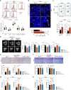
Fig. 1
Fig. 1
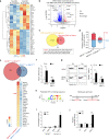
Sustained activation of HIF-1α signaling in B cells enhances osteoclastogenesis and osteoporosis. a Quantification and histograms of HIF-1α expression in CD4 T cells, CD8 T cells, B cells, monocytes and neutrophils from the bone marrow of sham-operated (Sham) and ovariectomized (OVX) mice (n = 8). b Representative immunofluorescence microscopy images of tibial sections from Sham and OVX mice (HIF-1α, green; B220, purple), along with the average number of HIF-1α+B220+ B cells per high-power field (HPF). Asterisks indicate HIF-1α+B220+ B cells in the bone niche. GP growth plate. Scale bars, 50 μm. c Representative plots and quantification of HIF-1α expression in Pro-B and Pre-B populations from Sham and OVX mice (n = 6). d Representative μCT images of tibial trabecular bone and structural parameters (BV/TV, Tb.N, Tb.Th, Tb.Sp) in Mb1cre/+, Vhlf/fMb1cre/+ and Vhlf/fHif1af/fMb1cre/+ mice (n = 5). Scale bars, 500 μm. Representative TRAP staining (e) and TB staining (f) in tibias from mice shown in (d). Bone resorption parameters (N.Oc/T.Ar, Oc.S/BS) and bone formation parameters (N.Ob/T.Ar, Ob.S/BS) in metaphyseal regions of the tibia were assessed by histomorphometric analyses. Scale bars, 100 μm. Trap, CathK (g), Col1a1 and Runx2 (h) expression in bone from mice shown in (d). Values for control group were set as 1. *P < 0.05, **P < 0.01, ***P < 0.001
Fig. 2
Fig. 2
HIF-1α signaling activation in B cells enhances RANKL production. a Differentially expressed genes in total B cells from bone marrow of sham control (Sham) or ovariectomized (OVX) mice (n = 3). b Volcano plot of RNA-seq analysis of total B cells from bone marrow of Sham and OVX mice showing upregulated and downregulated genes (n = 3). c Experimental outline that led to the discovery of RANKL (Tnfsf11) as a HIF-1α target gene during ovariectomy-induced bone loss. d Number of differentially expressed genes in WT and Hif1a-deficient bone marrow B cells after normoxic or hypoxic culture for 12 h. WT B cells under hypoxia, BWT (H); WT B cells under normoxia, BWT (N); Hif1a-deficient B cells under hypoxia, BKO (H); Hif1a-deficient B cells under normoxia, BKO (N). e Venn diagram depicting overlap of differentially expressed genes between hypoxic WT B cells versus normoxic WT B cells (red circle) and hypoxic Hif1a-deficient B cells versus hypoxic WT B cells (blue circle). Heatmap showing the commonly differentially expressed genes. Color scale indicates Log2 fold change. f Rankl mRNA expression in Hif1a-deficient B cells or control B cells after normoxic (Nx) or hypoxic (Hx) culture for 12 h (n = 6). g RANKL expression in B cells isolated from the bone marrow of Hif1af/f or Hif1af/fMb1cre/+ mice after normoxic (Nx) or hypoxic (Hx) culture for 24 h (n = 6). h Transcription factor HIF-1α binding sequence in the JASPAR database (left) and schematic analysis of hypoxia response elements (HREs) on the Rankl promoter (right). i ChIP assays showing the binding of HIF-1α to the Rankl promoter in total bone marrow B cells after normoxic (Nx) or hypoxic (Hx) culture for 12 h (n = 3). j Luciferase reporter assay in CH12F3 cells transfected with empty vector (EV) or HRE constructs (I and II) after normoxic (Nx) or hypoxic (Hx) culture for 24 h (n = 3). **P < 0.01, ***P < 0.001
Fig. 3
Fig. 3
Loss of HIF-1α in B cells partially inhibits ovariectomy-induced bone loss through RANKL-mediated osteoclastogenesis. a Representative μCT images of tibial trabecular bone and structural parameters (BV/TV, Tb.N, Tb.Th, Tb.Sp) in sham-operated (Sham) and ovariectomized (OVX) Hif1af/fMb1cre/+ mice and Hif1af/f littermate control mice (n = 5, 6). Scale bars, 500 μm. Representative TRAP staining (b) and TB staining (c) in tibia from mice shown in (a). Bone resorption parameters (N.Oc/T.Ar, Oc.S/BS) and bone formation parameters (N.Ob/T.Ar, Ob.S/BS) in metaphyseal regions of the tibia were assessed by histomorphometric analyses. Scale bars, 100 μm. Trap, CathK (d), Col1a1 and Runx2 (e) mRNA expression in bone from mice shown in (a). f RANKL and OPG levels and the RANKL/OPG ratio in bone marrow fluid from mice shown in (a). g Representative plots and frequencies of RANKL+ cells in B cell subpopulations from mice as in (a). *P < 0.05, **P < 0.01, ***P < 0.001
Fig. 4
Fig. 4
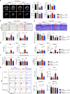
Estrogen controls HIF-1α stabilization via the HSP70-mediated degradation pathway. a Volcano plot of RNA-seq analysis of estrogen (E2) (1 μmol·L1)-treated versus vehicle (Veh)-treated WT bone marrow B cells under hypoxic conditions showing genes upregulated and downregulated, respectively, with a fold change (FC) higher than 2. b Heatmap of differentially expressed genes found within gene sets displayed in (a). c Hspa1a, Hsp90aa1, Hsp90ab1 and Hif1a mRNA expression in B cells treated with the indicated concentration of estrogen (E2) under hypoxia for 12 h (n = 3). Values for the control group were set as 1. d Hif1a, Vhl, Phd1, Phd2, Phd3, Hspa1a, Hsp90aa1, and Hsp90ab1 mRNA expression in B cells from the bone marrow of sham control (Sham) and ovariectomized (OVX) mice. e Levels of VHL, PHD1, PHD2, PHD3, HSP70, and HSP90 protein in isolated B cells from the bone marrow of OVX and Sham control mice (n = 3). f HIF-1α, HSP70, HSP90, hydroxylated HIF-1α (P564 and P402 sites), PHD1, PHD2, PHD3, VHL and β-actin expression in isolated bone marrow B cells with the indicated concentration of estrogen (E2) treatment under hypoxic culture for 24 h. g HIF-1α, HSP70 and β-actin expression in bone marrow B cells transfected with control or HSP70 siRNA lentivirus under hypoxic culture for 24 h. MG132 (100 μmol·L1) was added to HIF-1α immunoprecipitation samples. *P < 0.05, **P < 0.01, ***P < 0.001
Fig. 5
Fig. 5
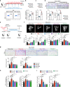
The HSP70/HIF-1α axis controls RANKL production in B cells and modulates osteoclastogenesis. a Scheme of adoptive transfer of siControl or siHSP70 lentivirus-transfected bone marrow B cells (5 × 106 cells per week) into female recipient Mb1cre/+/iDTR mice treated daily i.p. injection of DT (200 ng·d−1) for 28 days. b HIF-1α expression in B cells from the bone marrow of Mb1cre/+/iDTR recipient mice. c Frequencies of RANKL+ B cells from the bone marrow of Mb1cre/+/iDTR recipient mice. d Representative TRAP staining and histomorphometry of N.Oc/T.Ar, Oc.S/BS were analyzed in tibia from mice shown in (b). Scale bars, 200 μm. e Experimental scheme of pharmacological induction of HSP70 by GGA in osteoporotic mice. f Representative μCT images of tibial trabecular bone and structural parameters (BV/TV, Tb.N, Tb.Th, Tb.Sp) in mice post sham surgery with vehicle (Veh) or GGA treatment, as well as OVX surgery with vehicle (Veh), GGA or estrogen (E2) treatment (n = 6, 7). Scale bars, 500 μm. g Representative TRAP staining and bone resorption parameters (N.Oc/T.Ar, Oc.S/BS) were analyzed in tibia from mice shown in (f) Scale bars, 100 μm. h HIF-1α expression in Pro-B and Pre-B populations from mice shown in (f). i Frequencies of RANKL+ Pro-B and Pre-B populations from mice shown in (f). *P < 0.05, **P < 0.01, ***P < 0.001
Fig. 6
Fig. 6
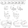
RANKL gene expression is associated with HIF1A gene expression in human B cells. a Progressive development of cord blood-derived human CD34+ cells under B cell differentiation conditions for 49 days. RANKL and HIF1A gene expression was examined at the indicated time points (n = 3). RANKL (b) and HIF-1α (c) expression in human CD34+ cell-derived Pro-B, Pre-B, and immature B populations. d Normalized expression values of RANKL and HIF1A in human CLP, Pro-B, Pre-B, and immature B populations from the public gene expression database GSE14714. Bounds of boxes and whiskers represent the min-to-max normalized value of gene expression in each population. Medians are indicated in each box as centerline. e Correlation between HIF1A and RANKL expression values in human CLP and bone marrow B cells from the public gene expression database GSE14714 (R2 and P values are indicated). **P < 0.01 by Pearson’s test (e)
Fig. 7
Fig. 7
HIF-1α signaling in B lymphocytes regulates bone homeostasis through RANKL-mediated osteoclastogenesis.

Similar articles

Cited by

References

    1. Zaidi M. Skeletal remodeling in health and disease. Nat. Med. 2007;13:791–801. doi: 10.1038/nm1593. - DOI - PubMed
    1. Mellis DJ, Itzstein C, Helfrich MH, Crockett JC. The skeleton: a multi-functional complex organ: the role of key signalling pathways in osteoclast differentiation and in bone resorption. J. Endocrinol. 2011;211:131–143. doi: 10.1530/JOE-11-0212. - DOI - PubMed
    1. Nakashima T, Takayanagi H. New regulation mechanisms of osteoclast differentiation. Ann. N. Y. Acad. Sci. 2011;1240:E13–E18. doi: 10.1111/j.1749-6632.2011.06373.x. - DOI - PubMed
    1. Ross FP. M-CSF, c-Fms, and signaling in osteoclasts and their precursors. Ann. N. Y. Acad. Sci. 2006;1068:110–116. doi: 10.1196/annals.1346.014. - DOI - PubMed
    1. Devlin RD, Reddy SV, Savino R, Ciliberto G, Roodman GD. IL-6 mediates the effects of IL-1 or TNF, but not PTHrP or 1,25(OH)2D3, on osteoclast-like cell formation in normal human bone marrow cultures. J. Bone Miner. Res. 1998;13:393–399. doi: 10.1359/jbmr.1998.13.3.393. - DOI - PubMed