. 2022 Feb 17;17(2):e0263262.
doi: 10.1371/journal.pone.0263262. eCollection 2022.

Considerations and practical implications of performing a phenotypic CRISPR/Cas survival screen

Affiliations

Considerations and practical implications of performing a phenotypic CRISPR/Cas survival screen

Ator Ashoti et al. PLoS One. .

Abstract

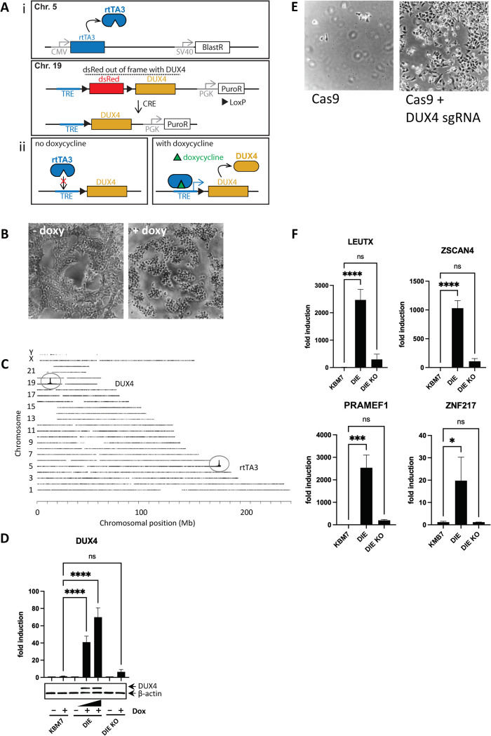
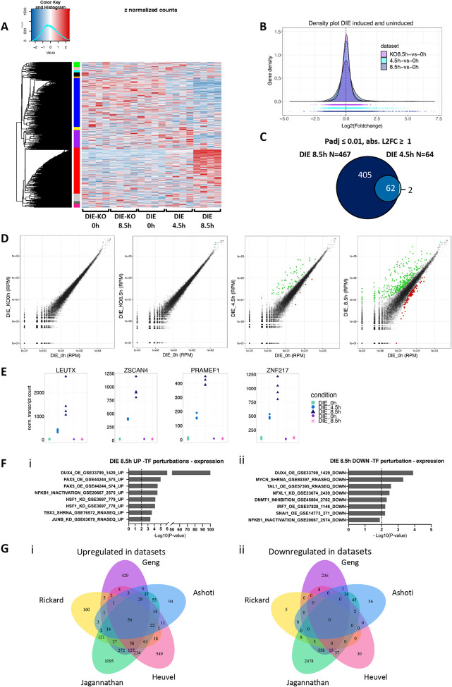
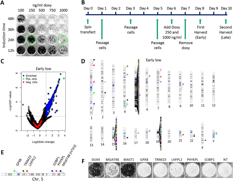
Genome-wide screens that have viability as a readout have been instrumental to identify essential genes. The development of gene knockout screens with the use of CRISPR-Cas has provided a more sensitive method to identify these genes. Here, we performed an exhaustive genome-wide CRISPR/Cas9 phenotypic rescue screen to identify modulators of cytotoxicity induced by the pioneer transcription factor, DUX4. Misexpression of DUX4 due to a failure in epigenetic repressive mechanisms underlies facioscapulohumeral muscular dystrophy (FHSD), a complex muscle disorder that thus far remains untreatable. As the name implies, FSHD generally starts in the muscles of the face and shoulder girdle. Our CRISPR/Cas9 screen revealed no key effectors other than DUX4 itself that could modulate DUX4 cytotoxicity, suggesting that treatment efforts in FSHD should be directed towards direct modulation of DUX4 itself. Our screen did however reveal some rare and unexpected genomic events, that had an important impact on the interpretation of our data. Our findings may provide important considerations for planning future CRISPR/Cas9 phenotypic survival screens.


Conflict of interest statement

The authors have declared that no competing interests exist.

Figures

Fig 1
Fig 1. Creation and validation of the DIE cell line.
(A) Constructs expressed in the DIE cell line to induce expression of DUX4. i) Top: Schematic representation of the rtTA3 construct (integrated on chromosome 5), constitutively expressing rtTA3 (under control of the CMV promoter) with selection marker blasticidin (BlastR, SV40 promoter). Bottom: LSL (LoxP-dsRed-stop-LoxP)-DUX4 cassette under control of TRE, tetracycline-responsive element, inducible by tetracycline-derivative doxycycline, together with selection marker puromycin (PuroR, PGK promoter), integrated on chromosome 19. dsRed but not DUX4 can be induced, as DUX4 is out of frame with dsRed. Action of CRE recombinase removed dsRed between the LoxP sites, enabling doxycycline-inducible DUX4 expression. ii) In the absence of doxycycline, rtTA3 does not bind to the TRE; addition of doxycycline results in rtTA3-TRE binding and subsequent transcription of DUX4. (B) Phase-contrast images of DIE cells without doxycycline and 24h after doxycycline exposure (1000ng/ml). (C) Schematic representation of transgene integration sites within human genome, by TLA analysis. The inducible DUX4 cassette maps to the p-arm of chromosome 19, and the rtTA3 transgene maps to the end of the q-arm of chromosome 5. (D) Analysis of DUX4 expression by qRT-PCR and western blot in KBM7, DIE and DIE-KO cells (DIE cells with knockout for DUX4) with or without doxycycline addition (500ng/ml and 1000ng/ml in DIE cells, 1000ng/ml in KBM7 and DIE KO cells), as detected by qRT-PCR (top panel) and western blot analysis (bottom panel). β -actin was used as a loading. Fold induction was calculated by 2^-(ddCT) of untreated or doxycycline-treated cells normalized by HPRT expression. Statistical significance was determined by ANOVA analysis. (E) Phase-contrast images of DIE cells which were transduced with either Cas9 protein, or Cas9 protein with DUX4 sgRNA prior to treatment with doxycycline (1000ng/ml) for at least 24h. Dead cells were removed by a DPBS wash to expose the surviving population. (F) Induction of mRNA expression of known downstream targets of DUX4 upon doxycycline treatment (1000ng/ml) in KBM7, DIE and DIE-KO cells, as measured by qRT-PCR. Fold induction was calculated by 2^-(ddCT) of uninduced or doxycycline-induced cells normalized by HPRT expression. Statistical significance was determined by ANOVA analysis.
Fig 2
Fig 2. RNA-sequencing data reveals differentially expressed genes upon DUX4 expression.
(A) Heatmap showing differentially expressed genes between DIE cells and DIE-KO (knockout for DUX4) cells, untreated or treated with doxycycline (1000ng/ml) for 4.5h or 8.5h, with gene clusters (color-coded) on the y-axis, and samples on the x-axis. (B) Gene density plot demonstrating the effects of DUX4 expression on the transcriptome of the DIE cell line. Doxycycline addition (1000ng/ml) for 4.5h or 8.5h results in an increase of differentially expressed genes compared to uninduced DIE cells, as indicated by the bell shape widening and shortening. This effect was not seen doxycycline-treated DIE-KO cells. (C) Venn diagram showing the overlap and the number of differentially expressed genes after 4.5h and 8.5h of doxycycline addition (1000ng/ml) (adjusted P-value ≤ 0.01, and absolute Log2FC ≥ 1). (D) Scatter plots of gene expression (RPM: Reads per million) of doxycycline-treated (1000ng/ml) DIE cells versus untreated DIE cells. The left two panels represent uninduced DIE cells (DIE_0h) on the x-axis versus doxycycline-treated or untreated DIE-KO samples (KO_0h and KO_8.5h) on the y-axis and show that addition of doxycycline has no effect on gene expression in DIE-KO cells. The right two panels compare the doxycycline-treated DIE cells with untreated DIE samples (4.5h and 8.5h). Green and red points represent the differentially expressed genes with an adjusted P-value ≤ 0.01, and absolute Log2FC ≥ 1. Green points represent upregulated genes, and the red points represent downregulated genes. (E) Count plots showing UMI and between sample normalized transcript counts of 4 known DUX4 targets genes: LEUTX, ZSCAN4, PRAMEF1 and ZNF217, in uninduced and doxycycline-treated (1000ng/ml) DIE cells and doxycycline-treated (1000ng/ml) DIE-KO cells. Every sample has 4 technical replicates, represented by 4 symbols. (F) Transcription factor perturbations analysis identifying transcription factors that are linked to the i) upregulation and ii) downregulation of the differentially expressed genes found in this study. Activation: OE or ACTIVATION, inhibition: KO, KD, SIRNA, SHRNA, INACTIVATION, or INHIBITION. (G) Quintuple Venn diagram comparing genes that are following DUX4 expression i) upregulated and ii) downregulated in this study (Ashoti) to those found in previous transcriptomic studies (Geng with P-value ≤ 0.01, FDR ≤ 0.05, abs L2FC ≥ 1; Rickard with Padj value of < 0.005 and abs L2FC > 2; Jagannathan with P-value ≤ 0.01, FDR ≤ 0.05, abs L2FC ≥ 1, Heuvel with P-value ≤ 0.005, FDR ≤ 0.05, abs L2FC ≥ 1).
Fig 3
Fig 3. CRISPR Screen setup and discovery of a CRISPR/Cas9 screening artefact.
(A) Viability staining of DIE cells treated with doxycycline (Doxy) concentrations (100, 250, 500 or 1000ng/ml) and for different exposure times (2, 4, 6, or 8h) to determine the optimal concentration and exposure time to induce sufficient cell death rates in DIE cells. Green circles indicate which conditions (low Doxy: 250ng/ml high Doxy: 1000ng/ml); were used for the genome-wide CRIPSR/Cas9 screen. (B) The CRISPR/Cas9 screen timeline from the time of library transfection (Day 0) to the final harvest of surviving DIE cells (Day 10). 6 days after transfection of the library, Doxycycline (Doxy) was added for 24h to induce DUX4 expression. Low Doxy: 250ng/ml; high Doxy: 1000ng/ml; early harvest: 24h after Doxy removal; late harvest: 48h after Doxy removal. (C) Volcano plot showing the enrichment of sets of guides of the low doxycycline 250ng/ml) -early harvest (24h after doxycycline removal) screen (early-low). Blue points represent guide sets that are significantly enriched (P-value ≤ 0.01), LFC ≥ 1), green points are the positive controls (DUX4, MAST1, MGAT4B), red points represent the non-target/negative control guides. (D) Chromosomal ideogram indicating the location of enriched hits in the human genome, of the low doxycycline-early harvest screen (see panel B). (E) Schematic representation of the location of a small number of false positive hits on chromosome 5 and chromosome 19. (F) Viability staining demonstrating surviving DIE-Cas9 cells (DIE cells constitutively expressing Cas9) after 250ng/ml doxycycline exposure, containing knockouts of the same genes mentioned in (E), but also DUX4, MGAT4B and MAST1. Media did not contain any selection markers (blasticidin or puromycin) to select for the presence of the rtTA3 or the DUX4 transgene. NT: Non-target controls.
Fig 4
Fig 4. Filtered CRISPR screen data and validation of potential hits.
(A) Adjusted volcano plot of screen data with low doxycycline (250ng/ml)/early harvest (24h after doxycycline removal, see Fig 3B) showing the enrichment of sets of guides targeting genes not located on chromosome 5q or chromosome 19p. Blue points represent guide sets that are significantly enriched (P-value ≤ 0.01), Log2(fold change) ≥ 1), the green point is the positive control (DUX4), red points represent the non-target control guides. (B) Venn diagram showing the overlap of filtered hits between the four screens (EL: Early harvest-low Doxy, LL: Late harvest-Low doxy, EH: Early harvest-high Doxy, LH: Late harvest-High doxy), see also Fig 3B. (C) Viability staining showing surviving DIE cells containing single knockouts of potentials hits, identified in the CRISPR screen. Knockouts of individual genes were generated by transfection of sgRNA; 6 days later, cells were left untreated or treated with 3 different concentrations of doxycycline (100, 250 and 1000ng/ml) for and incubated for an additional 48–96 hours prior to visualizing surviving cells. Data are representative of at least three independent experiments. (D) Viability staining showing the surviving DIE-ieGFP-Cas9 cells (DIE cells expressing Cas9 constitutively and contain doxycycline-inducible eGFP) with single knockouts of mediator complex subunits. Knockouts of individual genes were generated by transfection of sgRNA; 6 days later cells were treated for doxycycline (250ng/ml) for 24h and incubated for an additional 48–96 hours prior to visualizing surviving cells. Data are representative of at least three independent experiments. (E) FACS data showing GFP-positive cells in surviving populations of DIE-ieGFP-Cas9 (expressing constitutive Cas9 and doxycycline-inducible GFP). cells containing single knockouts as indicated. Knockouts of individual genes were generated by transfection of sgRNA; 6 days later, cells were treated with doxycycline (250ng/ml) for 24h prior to FACS analysis. DIE-ieGFP-Cas9 cells comprised of 42% of eGFP-positive cells after DUX4 knockout. rtTA, MED25, MED24 and MED16 knockouts displayed a lower percentage of eGFP-expressing cells, comprising between 1.2–4% of eGFP-expressing cells. Data are representative of at least three independent experiments.

Similar articles

Cited by

References

    1. Barrangou R, Fremaux R, Deveau H, Richards M, Boyaval P, Moineau S, et al.. CRISPR provides acquired resistance against viruses in prokaryotes. Sciencee. 2007;315: 1709–1712. doi: 10.1126/science.1138140 - DOI - PubMed
    1. Brouns SJ, Jore MM, Lundgren M, Westra ER, Slijkhuis R, Snijders A, et al.. Small CRISPR RNAs guide antiviral defense in prokaryotes. Science. 2008;321: 960–964. doi: 10.1126/science.1159689 - DOI - PMC - PubMed
    1. Christian M, Cermak T, Doyle EL, Schmidt C, Zhang F, Hummel A, et al.. Targeting DNA double-strand breaks with TAL effector nucleases. Genetics. 2010;186: 756–761. doi: 10.1534/genetics.110.120717 - DOI - PMC - PubMed
    1. Li T, Huang S, Jiang WZ, Wright D, Spalding MH, Weeks DP, et al.. TAL nucleases (TALNs): Hybrid proteins composed of TAL effectors and FokI DNA-cleavage domain. Nucleic Acids Research. 2011;39: 359–372. doi: 10.1093/nar/gkq704 - DOI - PMC - PubMed
    1. Mussolino C, Morbitzer R, Lütge F, Dannemann N, Lahaye T, Cathomen T. A novel TALE nuclease scaffold enables high genome editing activity in combination with low toxicity. Nucleic Acids Research. 2011;39: 9283–9293. doi: 10.1093/nar/gkr597 - DOI - PMC - PubMed

Publication types

MeSH terms

Grants and funding

AA was supported by The FSHD Foundation (https://www.fshd.nl) and the SingelSwim Utrecht Foundation (https://www.singelswimutrecht.nl) Funders did not play a role in study design, data collection or publication of the manuscript.