. 2022 Feb 9;17(2):e0263707.
doi: 10.1371/journal.pone.0263707. eCollection 2022.

Optimizing environmental safety and cell-killing potential of oncolytic Newcastle Disease virus with modifications of the V, F and HN genes

Affiliations

Optimizing environmental safety and cell-killing potential of oncolytic Newcastle Disease virus with modifications of the V, F and HN genes

J Fréderique de Graaf et al. PLoS One. .

Abstract

Newcastle Disease Virus (NDV) is an avian RNA virus, which was shown to be effective and safe for use in oncolytic viral therapy for several tumour malignancies. The presence of a multi basic cleavage site (MBCS) in the fusion protein improved its oncolytic efficacy in vitro and in vivo. However, NDV with a MBCS can be virulent in poultry. We aimed to develop an NDV with a MBCS but with reduced virulence for poultry while remaining effective in killing human tumour cells. To this end, the open reading frame of the V protein, an avian specific type I interferon antagonist, was disrupted by introducing multiple mutations. NDV with a mutated V gene was attenuated in avian cells and chicken and duck eggs. Although this virus still killed tumour cells, the efficacy was reduced compared to the virulent NDV. Introduction of various mutations in the fusion (F) and hemagglutinin-neuraminidase (HN) genes slightly improved this efficacy. Taken together, these data demonstrated that NDV with a MBCS but with abrogation of the V protein ORF and mutations in the F and HN genes can be safe for evaluation in oncolytic viral therapy.


Conflict of interest statement

The authors have declared that no competing interests exist.

Figures
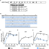
Fig 1
Fig 1. Characterization of NDV F3aa-STOPV viruses in vitro.
(A) Schematic representation of the sequence of the stutter site of NDV F3aa and the NDV F3aa-STOPV mutants. (B) Amino acid sequence of V protein ORFs of NDV F3aa and F3aa-STOPV. (C-E) Cells were inoculated in triplo in (C) DF-1 at an MOI of 0.005 and at an MOI of 0.05 in (D) QT6 and (E) A549 cells. Supernatant samples were collected at the indicated time points and titrated in Vero cells (N = 2). Means and standard deviations of triplicates of representative experiments are plotted. The area under the curve (AUC) was used for statistical analysis. * = p <0.05, ** = p<0.01, *** = p<0.001 (one-way ANOVA + paired t-test).
Fig 2
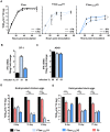
Fig 2. Characterization of NDV F3aa-STOPV viruses in ovo.
(A) DF-1 cells were mock-treated (solid line) or pretreated with 0.4 μg/ml chicken IFN-β for 24 hrs (dotted line) and inoculated in triplo at an MOI of 0.005. Samples were taken at the indicated time points and titrated. The experiment was conducted two times. Means and standard deviations of triplicates of a representative experiment are plotted. The AUC was used for statistical analysis. * = p <0.05, ** = p<0.01, *** = p<0.001 (one-way ANOVA + paired t-test). (B) DF-1 or (C) A549 cells were inoculated with mock or at an MOI of 0.05 and harvested after 24h. The percentage of infected cells was determined by flow cytometry. Results are represented as fold change of IFN mRNA transcription in virus-infected cells versus mock. (D) Chicken and (E) duck eggs of different ages were inoculated and the allantoic fluid was harvested after 48 hours. The amount of infectious virus particles was determined by titration, with a maximal cut off of 8.3E8 TCID50/ml. * = p <0.05, ** = p<0.01, *** = p<0.001 (one-way ANOVA + Dunn’s multiple comparison test). ND: not detected.
Fig 3
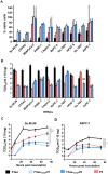
Fig 3. Cell death of human cancer cell lines upon inoculation with NDV F3aa-STOPV viruses.
(A) Indicated HPACs were inoculated at an MOI of 10 in triplo with the indicated viruses. Cell viability was determined by an LDH cytotoxicity assay 120 hours after inoculation. Results are represented as percentage viable cells compared to mock, which were considered as 100% viable. Experiments were conducted two times. Means and standard deviations of triplicates of representative experiments are plotted. (B) Indicated HPACs were inoculated in duplo at an MOI of 0.1. After 48 hours, supernatant samples were collected and titrated in Vero cells. The experiment was conducted two times. Means and standard deviations of triplicates of a representative experiment are plotted. (C) Su.86.86 and (D) AsPC-1 cells were inoculated at an MOI of 0.05 in triplo. Supernatant samples were collected at the indicated time points and titrated in Vero cells. The experiment was conducted two times. Means and standard deviations of triplicates of a representative experiment are plotted. The AUC was used for statistical analysis. * = p <0.05, ** = p<0.01, *** = p<0.001 (one-way ANOVA + paired t-test). ND: not detected.
Fig 4
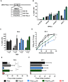
Fig 4. Characterization of F3aa-S mutant viruses.
(A) Schematic representation of the amino acid sequence of the cleavage site in the F protein of recombinant viruses. (B) Indicated HPACs were inoculated at an MOI of 10 or (C) Vero cells at indicated MOIs (Vero cells) in triplo. The percentage viable cells was determined by an LDH cytotoxicity assay 120 hours after inoculation. Results are represented as percentage viable cells compared to mock, which were considered 100% viable. (D) Vero cells were inoculated at an MOI of 0.05 in triplo. Supernatant samples were collected at the indicated time points and titrated. The AUC was used for statistical analysis. * = p <0.05, ** = p<0.01, *** = p<0.001 (one-way ANOVA + paired t-test). (E) Vero and (F) DF-1 cells were inoculated and fixed 16 hours later and stained. The focus index was determined by counting the number of nuclei per foci for N = 30 foci. Experiments were conducted twice. Means and standard deviations of triplicates of a representative experiment are plotted. *** = p<0.001, **** = p<0.0001 (one-way ANOVA + unpaired t test). NDV F0, taken along as control.
Fig 5
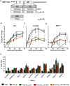
Fig 5. Characterization of NDV F3aa-(S)-STOPV mutants.
(A) Schematic representation of the nucleotide sequence of the TIS and amino acid substitution of the ICD of the F and HN protein. (B) Vero cells, (C) Su.86.86 and (D) AsPC-1 were inoculated at an MOI of 0.05 in triplo. Supernatant samples were collected at the indicated time points and titrated. Means and standard deviations of triplicates of a representative experiment are plotted. The AUC was used for statistical analysis. * = p <0.05, ** = p<0.01, *** = p<0.001 (one-way ANOVA + paired t-test as compared to NDV F3aa-S- STOPV2). (E) The indicated HPACs were inoculated at an MOI of 10 in triplo. The percentage viable cells was determined by an LDH cytotoxicity assay. Results are represented as percentage viable cells compared to mock, which were considered 100% viable. The experiment was conducted two times. Means and standard deviations of triplicates of a representative experiment are plotted.

Similar articles

Cited by

References

    1. Freeman a, Zakay-Rones Z, GomoriI J, Linetsky E, Rasooly L, Greenbaum E, et al.. Phase I/II Trial of Intravenous NDV-HUJ Oncolytic Virus in Recurrent Glioblastoma Multiforme. Mol Ther. 2006;13: 221–228. doi: 10.1016/j.ymthe.2005.08.016 - DOI - PubMed
    1. Pecora AL, Rizvi N, Cohen GI, Meropol NJ, Sterman D, Marshall JL, et al.. Phase I Trial of Intravenous Administration of PV701, an Oncolytic Virus, in Patients With Advanced Solid Cancers. J Clin Oncol. 2002;20: 2251–2266. doi: 10.1200/JCO.2002.08.042 - DOI - PubMed
    1. Hotte SJ, Lorence RM, Hirte HW, Polawski SR, Bamat MK, O’Neil JD, et al.. An Optimized Clinical Regimen for the Oncolytic Virus PV701. Clin Cancer Res. 2007;13: 977–985. doi: 10.1158/1078-0432.CCR-06-1817 - DOI - PubMed
    1. Laurie SA, Bell JC, Atkins HL, Roach J, Bamat MK, O’Neil JD, et al.. A Phase 1 Clinical Study of Intravenous Administration of PV701, an Oncolytic Virus, Using Two-Step Desensitization. Clin Cancer Res. 2006;12: 2555–2562. doi: 10.1158/1078-0432.CCR-05-2038 - DOI - PubMed
    1. Lorence RM, Rood PA, Kelley KW. Newcastle disease virus as an antineoplastic agent: induction of tumor necrosis factor-alpha and augmentation of its cytotoxicity. J Natl Cancer Inst. 1988;80: 1305–12. doi: 10.1093/jnci/80.16.1305 - DOI - PubMed

Publication types

MeSH terms

Grants and funding

JFG and DG obtained funding from the Dutch Foundation “Overleven with alvleesklierkanker”; https://www.supportcasper.nl/nl/over-support-casper/stichting/ RF and SN obtained funding from NWO-TTW grant #15414 (NWO-domein Toegepaste en Technische Wetenschappen; https://www.nwo.nl/toegepaste-en-technische-wetenschappen-ttw). The funders had no role in study design, data collection and analysis, decision to publish, or preparation of the manuscript.