. 2022 Feb 4;18(2):e1010294.
doi: 10.1371/journal.ppat.1010294. eCollection 2022 Feb.

Cellular ESCRT components are recruited to regulate the endocytic trafficking and RNA replication compartment assembly during classical swine fever virus infection

Affiliations

Cellular ESCRT components are recruited to regulate the endocytic trafficking and RNA replication compartment assembly during classical swine fever virus infection

Chun-Chun Liu et al. PLoS Pathog. .

Abstract

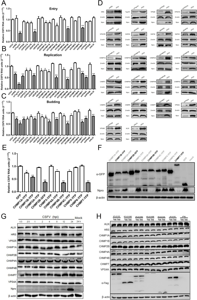
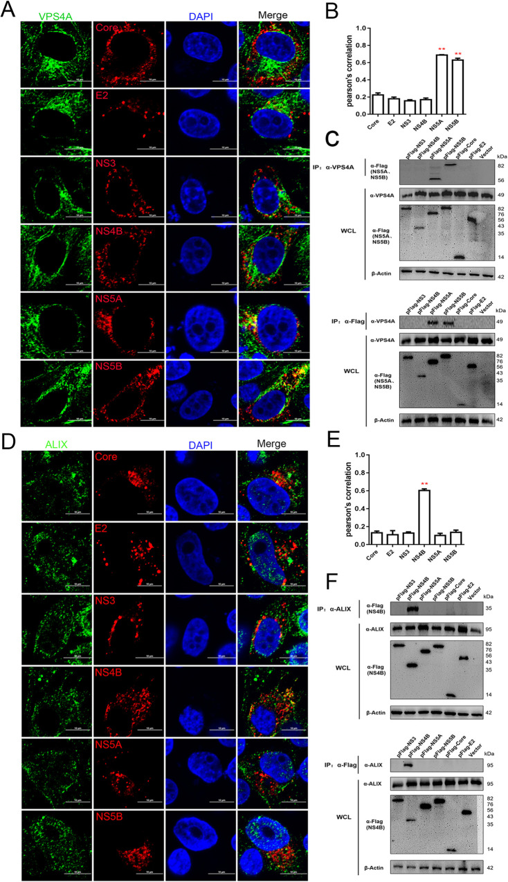
As the important molecular machinery for membrane protein sorting in eukaryotic cells, the endosomal sorting and transport complexes (ESCRT-0/I/II/III and VPS4) usually participate in various replication stages of enveloped viruses, such as endocytosis and budding. The main subunit of ESCRT-I, Tsg101, has been previously revealed to play a role in the entry and replication of classical swine fever virus (CSFV). However, the effect of the whole ESCRT machinery during CSFV infection has not yet been well defined. Here, we systematically determine the effects of subunits of ESCRT on entry, replication, and budding of CSFV by genetic analysis. We show that EAP20 (VPS25) (ESCRT-II), CHMP4B and CHMP7 (ESCRT-III) regulate CSFV entry and assist vesicles in transporting CSFV from Clathrin, early endosomes, late endosomes to lysosomes. Importantly, we first demonstrate that HRS (ESCRT-0), VPS28 (ESCRT-I), VPS25 (ESCRT-II) and adaptor protein ALIX play important roles in the formation of virus replication complexes (VRC) together with CHMP2B/4B/7 (ESCRT-III), and VPS4A. Further analyses reveal these subunits interact with CSFV nonstructural proteins (NS) and locate in the endoplasmic reticulum, but not Golgi, suggesting the role of ESCRT in regulating VRC assembly. In addition, we demonstrate that VPS4A is close to lipid droplets (LDs), indicating the importance of lipid metabolism in the formation of VRC and nucleic acid production. Altogether, we draw a new picture of cellular ESCRT machinery in CSFV entry and VRC formation, which could provide alternative strategies for preventing and controlling the diseases caused by CSFV or other Pestivirus.


Conflict of interest statement

The authors have declared that no competing interests exist.

Figures

Fig 1
Fig 1. ESCRT is involved in CSFV infection.
(A) PK-15 cells were transfected with siESCRTs or siCtrl and then inoculated with CSFV (MOI = 10), and the cells were harvested for RT-qPCR at 1 hpi. These data are presented as the mean + SD of data from three independent experiments. *, P< 0.05; **, P <0.01. (B) PK-15 cells were transfected with siESCRTs or siCtrl and then inoculated with CSFV (MOI = 0.1), then harvested the whole cell cultures at 24 hpi for RT-qPCR. These data are presented as the mean + SD of data from three independent experiments. *, P< 0.05; **, P <0.01. (C) PK-15 cells were transfected with siESCRTs or siCtrl and then inoculated with CSFV (MOI = 0.1), at 24 hpi, the cell supernatant were harvested and used for infected new PK-15 cells, and harvested the new cells at 24 hpi for RT-qPCR. These data are presented as the mean + SD of data from three independent experiments. *, P< 0.05; **, P <0.01. (D) PK-15 cells were treated as described in panel B. At 24 hpi, the cells were harvested and subjected to Western blotting by using the indicated antibodies as follows: rabbit anti-ESCRTs antibody, rabbit anti-Npro antibody, or mouse anti-β-actin antibody. These data are representative of three independent experiments. (E and F) PK-15 cells were transfected YFP-tagged ESCRT-III or vector plasmid and then inoculated with CSFV (MOI = 0.1). At 24 hpi, the cell culture was harvested, respectively, for RT-qPCR and Western blotting described above. These data are presented as the mean + SD of data from three independent experiments. *, P< 0.05; **, P <0.01. (G) PK-15 cells were infected with CSFV (MOI = 1) for indicated time points, then cells were harvested and subjected to Western blotting by using the indicated antibodies against ALIX, HRS, VPS25, CHMP1A, CHMP2B, CHMP4A, CHMP4B, CHMP7, VPS4A, Npro and β-actin. These data are representative of three independent experiments. (H) PK-15 cells were transfected doses of indicated plasmids (pFlag-NS3, -NS4B, -NS5A, -NS5B, -Core, -E2) or vector for 48 hpt, then harvested and subjected to Western blotting by using rabbit anti-ESCRTs antibody or mouse anti-Flag antibody, along with β-actin as a loading control. These data are representative of three independent experiments.
Fig 2
Fig 2. ESCRTs machinery participates in CSFV endocytosis.
(A-D) PK-15 cells were infected with CSFV (MOI = 10) at 4°C for 1 hpi, then shifted to 37°C for indicated time points, respectively. After fixed and subjected to immunofluorescent by using mouse anti-CSFV E2 monoclonal antibody (WH303) and rabbit anti-VPS25 (A); -CHMP4B (B); -CHMP7 (C); -VPS4A (D) antibody. The nuclei were stained with DAPI. The white arrow indicates the co-localized protein. Bars = 10 μm. These data are representative of three independent experiments. (E-G) PK-15 cells were infected with CSFV (MOI = 10) or not for 6 hpi, then harvested for immunoprecipitation by using mouse anti-VPS25 antibody (E) or rabbit anti-CHMP4B (F); -CHMP7 antibody (G), and the whole-cell lysates were subjected to Western blotting by using the antibodies against Clathrin, Rab5, Rab9, LAMP-1, Tsg101, VPS25, CHMP4B, CHMP7, VPS4A and ALIX. These data are representative of three independent experiments.
Fig 3
Fig 3. Visualization of the location of the CSFV replication complex.
(A and B) PK-15 cells were infected with CSFV (MOI = 10) or not for 24 hpi, then harvested and subjected to electron microscopy. (C) This picture is the field of view enlarged by the white box in panel B, the black arrow indicated the virus replication complex (VRC). Additionally, LDs means Lipid droplets, M means mitochondrion. These data are representative of three independent experiments.
Fig 4
Fig 4. ESCRT participates in the formation of the CSFV replication complex.
(A) PK-15 cells were inoculated with CSFV (MOI = 1) for 24 hpi, then fixed and subjected to immunofluorescent by using mouse anti-dsRNA antibody (red) and rabbit anti-ESCRTs antibody (green). The nuclei were stained with DAPI. Bars = 10 μm. These data are representative of three independent experiments. (B) The colocalization analysis was expressed as Pearson’s correlation coefficient, respectively. Results are represented as the mean + SD of data from three independent experiments. (C) PK-15 cells were infected with CSFV at different MOI for 24 hpi and extracted viral replication complex related proteins. The extraction was subjected to Western blotting by using rabbit anti-ESCRTs antibody, rabbit anti-GM130 antibody, or rabbit anti-Calnexin antibody, along with mouse anti-β-actin antibody as control. These data are representative of three independent experiments. (D) Western blotting results were analyzed of grayscale analysis through image J software, respectively. The white column indicates that the MOI is 1, the black column indicates that the MOI is 0.1 and the red column represents mock. These data are presented as the mean + SD of data from three independent experiments. *, P< 0.05; **, P <0.01. (E) PK-15 cells were treated as described in panel C, then harvested and subjected to RT-qPCR. These data are presented as the mean + SD of data from three independent experiments. ***, P <0.001.
Fig 5
Fig 5. Interaction between major subunits of the ESCRT-0/I/II system and CSFV proteins.
(A, D and G) PK-15 cells were transfected with indicated plasmids (pFlag-Core, -E2, -NS3, -NS4B, -NS5A, -NS5B) for 48 hpt, then fixed and subjected to immunofluorescent by using rabbit anti-HRS (A); -VPS28 (D); -VPS25 (G) antibody (green) and mouse anti-Flag antibody (red). The nuclei were stained with DAPI. Bars = 10 μm. These data are representative of three independent experiments. (B, E and H) The colocalization analysis was expressed as Pearson’s correlation coefficient, respectively. Results are represented as the mean + SD of data from three independent experiments. **, P < 0.01. (C, F and I) HEK-293T cells were transfected with indicated plasmids (pFlag-NS3, -NS4B, -NS5A, -NS5B, -Core, -E2) or vector for 48 hpt, then harvested for immunoprecipitation by using mouse anti-Flag antibody or mouse anti-HRS (C); -VPS28 (F); -VPS25 (I) antibody, and whole-cell lysates were subjected to Western blotting by using rabbit anti-HRS/VPS28/VPS25 antibody or mouse anti-Flag antibody, along with β-actin as a loading control. These data are representative of three independent experiments.
Fig 6
Fig 6. Interaction between major subunits of the ESCRT-III system and CSFV proteins.
(A, D and G) PK-15 cells were transfected with indicated plasmids (pFlag-Core, -E2, -NS3, -NS4B, -NS5A, -NS5B) for 48 hpt, then fixed and subjected to immunofluorescent by using rabbit anti-CHMP2B (A); -CHMP4B (D); -CHMP7 (G) antibody (green) and mouse anti-Flag antibody (red). The nuclei were stained with DAPI. Bars = 10 μm. These data are representative of three independent experiments. (B, E and H) The colocalization analysis was expressed as Pearson’s correlation coefficient, respectively. Results are represented as the mean + SD of data from three independent experiments. **, P < 0.01. (C, F and I) HEK-293T cells were transfected with indicated plasmids (pFlag-NS3, -NS4B, -NS5A, -NS5B, -Core, -E2) or vector for 48 hpt, then harvested for immunoprecipitation by using mouse anti-Flag antibody or rabbit anti-CHMP2B (C); -CHMP4B (F); -CHMP7 (I) antibody, and whole-cell lysates were subjected to Western blotting by using rabbit anti-CHMP2B/CHMP4B/CHMP7 antibody or mouse anti-Flag antibody, along with β-actin as a loading control. These data are representative of three independent experiments.
Fig 7
Fig 7. Interaction between endogenous VPS4A/ALIX proteins and CSFV proteins.
(A and D) PK-15 cells were transfected with indicated plasmids (pFlag-Core, -E2, -NS3, -NS4B, -NS5A, -NS5B) for 48 hpt, then fixed and subjected to immunofluorescent by using rabbit anti-VPS4A (A); -ALIX (D) antibody (green) and mouse anti-Flag antibody (red). The nuclei were stained with DAPI. Bars = 10 μm. These data are representative of three independent experiments. (B and E) The colocalization analysis was expressed as Pearson’s correlation coefficient, respectively. Results are represented as the mean + SD of data from three independent experiments. **, P < 0.01. (C and F) HEK-293T cells were transfected with indicated plasmids (pFlag-NS3, -NS4B, -NS5A, -NS5B, -Core, -E2) or vector for 48 hpt, then harvested for immunoprecipitation by using mouse anti-Flag antibody or mouse anti-VPS4A antibody (C); rabbit anti-ALIX antibody (F), and whole-cell lysates were subjected to Western blotting by using rabbit anti-VPS4A/ALIX antibody or mouse anti-Flag antibody, along with β-actin as a loading control. These data are representative of three independent experiments.
Fig 8
Fig 8. ESCRT subunits are involved in the replication complex of CSFV.
(A) PK-15 cells were transfected with indicated plasmids (pFlag-NS3, -NS4B, -NS5A, -NS5B) for 48 hpt, then fixed and subjected to immunofluorescent by using mouse anti-HRS/VPS28/VPS25/CHMP7/VPS4A/ALIX antibody (green), goat anti-Flag antibody (red) and rabbit anti-Calnexin antibody (purple); or rabbit anti-CHMP2B/CHMP4B antibody (green), goat anti-Flag antibody (red) and mouse anti-Calnexin antibody (purple). The nuclei were stained with DAPI. Bars = 10 μm. These data are representative of three independent experiments. (B) The colocalization coefficient of ESCRTs, NS (nonstructural proteins) and ER was expressed as Pearson’s correlation coefficient. The white column indicates the co-localization of the ESCRT subunits and ER, the black column indicates the co-localization of the ESCRT subunits and nonstructural proteins, and the red column indicates the co-localization of the ER and nonstructural proteins. Results are represented as the mean + SD of data from three independent experiments.
Fig 9
Fig 9. VPS4A protein is closely related to lipid droplets (LDs).
(A) PK-15 cells were infected with CSFV (MOI = 1) for 24 hpi, then fixed and subjected to immunofluorescent by using rabbit anti-ESCRTs antibody (red) and dyeing with BODIPY (green). The nuclei were stained with DAPI. Bars = 10 μm. These data are representative of three independent experiments. (B) The colocalization analysis of ESCRT subunits and LDs was expressed as Pearson’s correlation coefficient. Results are represented as the mean + SD of data from three independent experiments. **, P < 0.01. (C) PK-15 cells were infected with CSFV (MOI = 1) for 24 hpi, then fixed and subjected to immunofluorescent by using rabbit anti-VPS4A/CHMP7 antibody (red), mouse anti-dsRNA (purple), and dyeing with BODIPY (green). The nuclei were stained with DAPI. Bars = 10 μm. These data are representative of three independent experiments. (D) The colocalization analysis of VPS4A or CHMP7, dsRNA, and LDs were expressed as Pearson’s correlation coefficient. The white column indicates the co-localization of the ESCRT subunits and LDs, the black column indicates the co-localization of the ESCRT subunits and dsRNA, and the red column indicates the co-localization of the LDs and dsRNA. Results are represented as the mean + SD of data from three independent experiments.
Fig 10
Fig 10. Assembly between major subunit proteins of ESCRT after CSFV infection.
(A, C, E and G) PK-15 cells were infected with CSFV (MOI = 1) for 24 hpi and harvested for immunoprecipitation by using rabbit anti-CHMP2B (A); -CHMP4B (C); -CHMP7 (E) antibody, or mouse anti-VPS4A antibody (G), and the whole-cell lysates were subjected to Western blotting by using the antibodies against HRS, Tsg101, VPS25, CHMP1A, CHMP1B, CHMP2B, CHMP4B, CHMP7, VPS4A and ALIX. These data are representative of three independent experiments. (B, D, F and H) PK-15 cells were infected with CSFV (MOI = 1) for 24 hpi, then fixed and subjected to immunofluorescent by using rabbit anti-CHMP2B (B); -CHMP4B (D) antibody (green) and mouse anti-HRS/VPS4A/Tsg101 antibody (red); or mouse anti-CHMP7 antibody (F) (red) and rabbit anti-Tsg101/VPS4A antibody (green); or mouse anti-VPS4A antibody (H) (red) and rabbit anti-HRS/CHMP2B/CHMP4B/CHMP7 antibody (green). The nuclei were stained with DAPI. Bars = 10 μm. These data are representative of three independent experiments.
Fig 11
Fig 11. Schematic model depicting CSFV life cycle and the role of ESCRT proteins in PK-15 cells.
(1) Clathrin interacted with Tsg101, VPS25, CHMP4B and CHMP7 proteins upon CSFV entry, then transported to the early endosomes. (2) In the early endosomes, CHMP4B and CHMP7 proteins interacted with Rab5 protein and assist the transport of CSFV to the late endosomes. (3) In the late endosomes, Tsg101, CHMP4B, and CHMP7 proteins interacted with Rab9 protein and transport CSFV to the lysosomes. (4) Later, in the lysosomes, the Tsg101 and CHMP4B proteins interacted of with LAMP-1 leads to uncoating and released nucleic acid, then transported to the endoplasmic reticulum area. (5) Finally, ESCRT proteins interacted with nonstructural proteins of CSFV and formed a virus replication complex (VRC) in the ER lumens for viral genome replication. Finally, LDs were closed to the VRC and connected by VPS4A protein.

Similar articles

Cited by

References

    1. Chambers TJ, Hahn CS, Galler R, Rice CM. Flavivirus genome organization, expression, and replication. Annual review of microbiology. 1990;44:649–88. doi: 10.1146/annurev.mi.44.100190.003245 . - DOI - PubMed
    1. White JM, Whittaker GR. Fusion of Enveloped Viruses in Endosomes. Traffic. 2016;17(6):593–614. doi: 10.1111/tra.12389 ; PubMed Central PMCID: PMC4866878. - DOI - PMC - PubMed
    1. Kalia M, Jameel S. Virus entry paradigms. Amino acids. 2011;41(5):1147–57. doi: 10.1007/s00726-009-0363-3 ; PubMed Central PMCID: PMC7088018. - DOI - PMC - PubMed
    1. Haywood AM. Membrane uncoating of intact enveloped viruses. Journal of virology. 2010;84(21):10946–55. doi: 10.1128/JVI.00229-10 ; PubMed Central PMCID: PMC2953184. - DOI - PMC - PubMed
    1. McNamara RP, Dittmer DP. Extracellular vesicles in virus infection and pathogenesis. Current opinion in virology. 2020;44:129–38. Epub 2020/08/28. doi: 10.1016/j.coviro.2020.07.014 ; PubMed Central PMCID: PMC7755726. - DOI - PMC - PubMed

Publication types

MeSH terms

Grants and funding

This work was supported by grant from the National Natural Science Foundation of China (32172840 and 31872471, to B.Z.). The funders had no role in study design, data collection and analysis, decision to publish, or preparation of the manuscript.