. 2022 Jan 18:8:780834.
doi: 10.3389/fmed.2021.780834. eCollection 2021.

Kinin B1 Receptor Mediates Renal Injury and Remodeling in Hypertension

Affiliations

Kinin B1 Receptor Mediates Renal Injury and Remodeling in Hypertension

Debargha Basuli et al. Front Med (Lausanne). .

Abstract

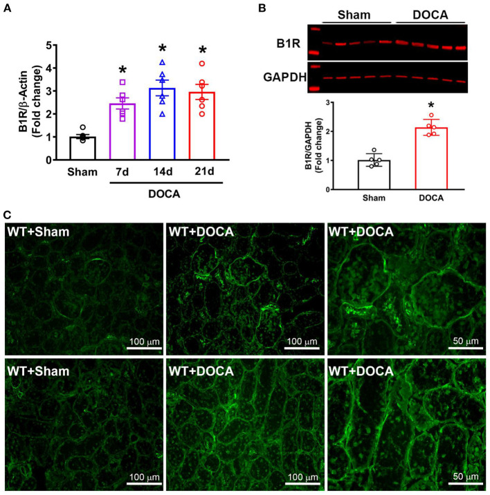
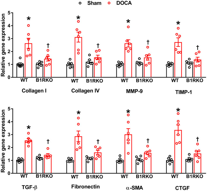
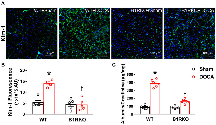
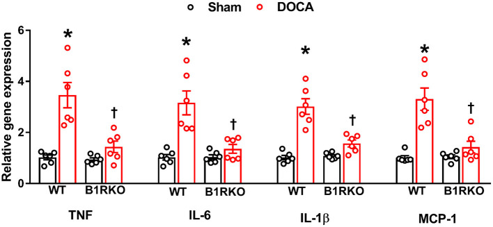
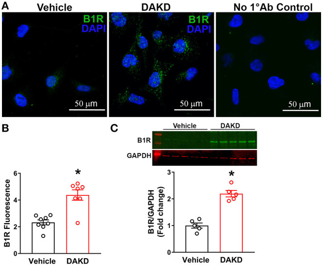
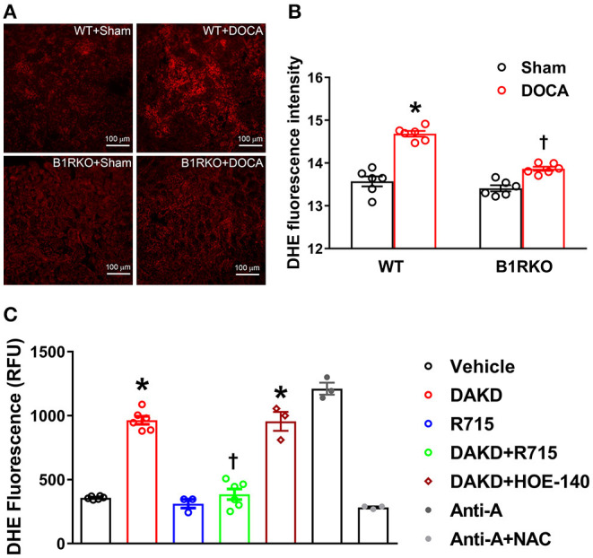
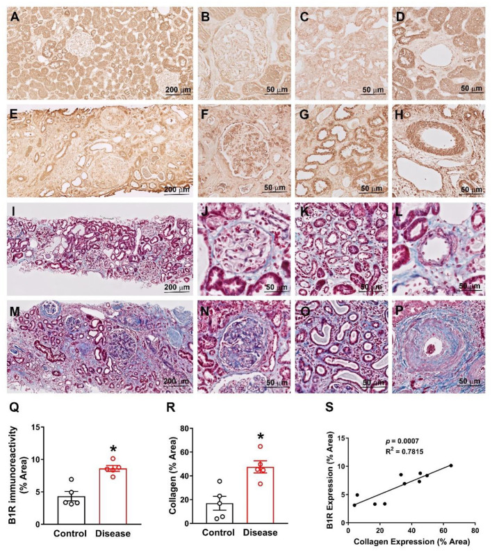
Despite many readily available therapies, hypertensive kidney disease remains the second most prevalent cause of end-stage renal disease after diabetes, and continues to burden patient populations and escalate morbidity and mortality rates. Kinin B1 receptor (B1R) activation has been shown to have a role in the development of hypertension, one of the major etiologies for chronic kidney disease. However, the role of B1R in hypertension induced renal injury and remodeling remains unexplored. Using a DOCA-salt-induced hypertensive mouse model, we investigated whether B1R deficiency reduces hypertensive renal injury and fibrosis. To further recognize the translational role of B1R, we examined the expression of B1R and its correlation with collagen deposition in renal biopsies from control and hypertensive kidney disease patients. Our data indicates that renal B1R expression was upregulated in the kidneys of DOCA-salt hypertensive mice. Genetic ablation of B1R protected the mice from DOCA-salt-induced renal injury and fibrosis by preventing inflammation and oxidative stress in the kidney. Cultured human proximal tubular epithelial cells expressed B1R and stimulation of B1R with an agonist resulted in increased oxidative stress. In human kidney biopsy samples, we found that the B1R immunoreactivity was not only significantly increased in hypertensive patients compared to normotensive patients, but also there is a positive correlation between B1R expression and renal fibrosis levels. Taken together, our results identify a critical role of B1R in the development of inflammation and fibrosis of the kidney in hypertension.

Keywords: hypertension; inflammation; kinin B1 receptor; oxidative stress; renal fibrosis.


Conflict of interest statement

The authors declare that the research was conducted in the absence of any commercial or financial relationships that could be construed as a potential conflict of interest.

Figures

Figure 1
Figure 1
Elevated B1R expression in the kidneys of DOCA-salt hypertensive mice. (A) Gene expression measured by real time RT-PCR showing significantly increased mRNA in the kidneys at 7, 14, and 21 days of DOCA-salt treated hypertensive mice compared with sham mice (n = 6, One-way ANOVA, *P < 0.01 vs. Sham). (B) Representative western blot image and quantification data showing significantly increased B1R protein expression in the kidney cortex of hypertensive mice (n = 5, Unpaired, 2-tailed t-test, *P < 0.01 vs. Sham). (C) Immunofluorescence staining showing increased B1R expression in the glomeruli (top row) and tubules (bottom row) of DOCA-salt treated wild-type (WT) mice compared to sham treated mice (B1R, green).
Figure 2
Figure 2
B1R gene deletion prevents gene expression of renal fibrosis markers in DOCA-salt hypertension. Gene expression measured by real time RT-PCR showing significantly increased mRNA of renal fibrosis markers in the kidney cortex of DOCA-salt treated hypertensive mice compared with sham mice. B1R knockout mice did not show this DOCA-salt-induced increase in renal fibrosis markers. Data was normalized to β-actin and presented as mean ± SEM (n = 6, Two-way ANOVA, *P < 0.01 vs. Sham, P < 0.05 vs. WT + DOCA).
Figure 3
Figure 3
B1R gene deletion abrogates DOCA-salt induced renal myofibroblast transition. (A) Representative immunofluorescence images showing expression of alpha-smooth muscle actin (α-SMA), fibronectin, and transforming growth factor-beta (TGF-β) in the kidney sections. Quantification data presented as mean fluorescence staining showing increased expression of α-SMA (B), fibronectin (C), and TGF-β (D) of DOCA-salt treated hypertensive mice compared with sham mice. These increases in fibrosis markers expression were blunted in B1R deficient mice with 3 weeks of DOCA-salt treatment (n = 6, Two-way ANOVA, *P < 0.01 vs. Sham, P < 0.05 vs. WT + DOCA).
Figure 4
Figure 4
B1R gene deletion prevents kidney injury in DOCA-salt hypertension. (A) Representative immunofluorescence images showing expression of kidney injury molecule-1 (Kim-1) in the kidney sections. (B) Quantification data presented as mean fluorescence staining showing increased expression of Kim-1 in the kidneys of DOCA-salt treated hypertensive mice compared with sham treated wild-type (WT) mice. This increase in Kim-1 expression was blunted in B1R deficient mice (B1RKO) mice with DOCA-salt treatment (n = 5, Two-way ANOVA, *P < 0.01 vs. Sham, P < 0.05 vs. WT + DOCA). (C) At the end of 3 weeks of DOCA-salt treatment, urinary albumin to creatinine levels ratio was significantly increased in WT mice, which was prevented in B1RKO mice (n = 6, Two-way ANOVA, *P < 0.01 vs. Sham, P < 0.05 vs. WT + DOCA).
Figure 5
Figure 5
B1R gene deletion prevents inflammation in the kidney during DOCA-salt-induced hypertension. DOCA-salt treatment for 3 weeks significantly increased mRNA expression of pro-inflammatory cytokines TNF, IL-6, IL-1β and chemokine MCP-1 in the kidney cortex of wild-type (WT) mice compared with sham mice. B1R knockout (B1RKO) mice did not show this DOCA-salt-induced increase in inflammatory markers. Gene expression measured by real time RT-PCR. Data was normalized to β-actin and presented as mean ± SEM (n = 6, Two-way ANOVA, *P < 0.01 vs. Sham, P < 0.05 vs. WT + DOCA).
Figure 6
Figure 6
B1R expression in human kidney proximal tubular epithelial (HK-2) cells. (A) Representative photomicrographs showing immunofluorescence of B1R in HK-2 cells treated vehicle or B1R specific agonist DAKD for 24 h. Control HK-2 cells without primary antibody (No 1°Ab control) incubation and with only secondary antibody incubation shows no staining. (B) The quantification of B1R immunoreactivity shows that the DAKD treatment of HK-2 cells for 24 h significantly upregulated the B1R expression (n =7–8, Unpaired, 2-tailed t-test, *P < 0.01 vs. Vehicle). (C) Representative western blot image and quantification data showing significantly increased B1R protein expression in HK-2 cells following stimulation with DAKD for 24 h (n = 5, Unpaired, 2-tailed t-test, *P < 0.01 vs. Vehicle).
Figure 7
Figure 7
B1R blockade reduces reactive oxygen species (ROS) production in the kidney and HK-2 cells. (A) Representative photomicrographs showing dihydroethidium (DHE) staining in the kidney sections. (B) DHE staining quantified as relative fluorescence intensity showing increased superoxide generation in the kidneys of DOCA-salt treated hypertensive mice compared with sham treated wild-type (WT) mice. This increase in DHE staining was blunted in B1R deficient mice (B1RKO) mice with DOCA-salt treatment (n = 6, Two-way ANOVA, *P < 0.01 vs. Sham, P < 0.05 vs. WT + DOCA). (C) ROS production measured by microplate DHE assay showing B1R specific agonist DAKD stimulation of HK-2 cells induced a significant increase in total ROS production indicating increased oxidative stress, and pre-treatment with a specific B1R antagonist (R715, 10 μM) prevented this DAKD-induced ROS production. Pretreatment with specific B2R antagonist (HOE-140, 10 μM) for 1 h was not able to prevent the ROS production suggesting the B1R specific ROS production (n = 6 independent culture wells/group). Treatment with antimycin A (Anti-A), an inhibitor of complex III of the mitochondrial electron transport chain, is used as a positive control for ROS generation and treatment with N-Acetyl-L-Cysteine (NAC) is used as an antioxidant control. Statistical significance: One-way ANOVA followed by Tukey's multiple comparisons test. *p < 0.05 compared to vehicle, p < 0.05 compared to Ang II (n = 3–6, One-way ANOVA, *P < 0.01 vs. Vehicle, P < 0.05 vs. DAKD).
Figure 8
Figure 8
B1R expression is increased and correlates with collagen expression in human kidney sections from hypertensive chronic kidney disease. Representative immunohistochemistry photomicrographs showing B1R specific immunoreactivity in kidney sections of healthy controls (A), specifically in glomerulus (B), tubules (C) and arteries (D). The B1R specific immunoreactivity is increased in kidney sections from renal disease (E), specifically in glomerulus (F), tubules (G) and arteries (H). Representative photomicrographs showing Masson Trichrome staining for collagen deposition in health control kidney section (I) with in glomerulus (J), tubules (K) and arteries (L). The collagen deposition was increased in kidney sections from the kidney disease subjects (M), specifically in glomerulus (N), tubules (O) and arteries (P). Quantification data showing increased B1R (Q) and collagen (R) expression in kidney sections from renal disease subjects (n = 5, Unpaired, 2-tailed t-test, *P < 0.01 vs. Control). B1R immunoreactivity is positively correlated with collagen deposition (S). Pearson correlation and least-squares linear regression were used to determine correlation coefficients between variables.

Similar articles

References

    1. Meyrier A. Nephrosclerosis: update on a centenarian. Nephrol Dial Transplant. (2015) 30:1833–41. 10.1093/ndt/gfu366 - DOI - PubMed
    1. Klag MJ, Whelton PK, Randall BL, Neaton JD, Brancati FL, Stamler J. End-stage renal disease in african-american and white men. 16-year mrfit findings. JAMA. (1997) 277:1293–8. 10.1001/jama.1997.03540400043029 - DOI - PubMed
    1. Walker WG, Neaton JD, Cutler JA, Neuwirth R, Cohen JD. Renal function change in hypertensive members of the multiple risk factor intervention trial. Racial and treatment effects. The mrfit research group. JAMA. (1992) 268:3085–91. 10.1001/jama.1992.03490210067037 - DOI - PubMed
    1. Liu T, Liu M, Shang P, Jin X, Liu W, Zhang Y, et al. . Investigation into the underlying molecular mechanisms of hypertensive nephrosclerosis using bioinformatics analyses. Mol Med Rep. (2018) 17:4440–8. 10.3892/mmr.2018.8405 - DOI - PMC - PubMed
    1. Wenzel UO, Bode M, Kohl J, Ehmke H. A pathogenic role of complement in arterial hypertension and hypertensive end organ damage. Am J Physiol Heart Circ Physiol. (2017) 312:H349–54. 10.1152/ajpheart.00759.2016 - DOI - PubMed

LinkOut - more resources