. 2022 Feb 2;18(2):e1010233.
doi: 10.1371/journal.ppat.1010233. eCollection 2022 Feb.

cGAS exacerbates Schistosoma japonicum infection in a STING-type I IFN-dependent and independent manner

Affiliations

cGAS exacerbates Schistosoma japonicum infection in a STING-type I IFN-dependent and independent manner

Le Liang et al. PLoS Pathog. .

Abstract

Schistosomiasis, which is caused by infection with Schistosoma spp., is characterized by granuloma and fibrosis in response to egg deposition. Pattern recognition receptors are important to sense invading Schistosoma, triggering an innate immune response, and subsequently shaping adaptive immunity. Cyclic GMP-AMP synthase (cGAS) was identified as a major cytosolic DNA sensor, which catalyzes the formation of cyclic GMP-AMP (cGAMP), a critical second messenger for the activation of the adaptor protein stimulator of interferon genes (STING). The engagement of STING by cGAMP leads to the activation of TANK-binding kinase 1 (TBK1), interferon regulatory factor 3 (IRF3), and the subsequent type I interferon (IFN) response. cGAS is suggested to regulate infectious diseases, autoimmune diseases, and cancer. However, the function of cGAS in helminth infection is unclear. In this study, we found that Cgas deficiency enhanced the survival of mice infected with S. japonicum markedly, without affecting the egg load in the liver. Consistently, Cgas deletion alleviated liver pathological impairment, reduced egg granuloma formation, and decreased fibrosis severity. In contrast, Sting deletion reduced the formation of egg granulomas markedly, but not liver fibrosis. Notably, Cgas or Sting deficiency reduced the production of IFNβ drastically in mice infected with S. japonicum. Intriguingly, intravenous administration of recombinant IFNβ exacerbated liver damage and promoted egg granuloma formation, without affecting liver fibrosis. Clodronate liposome-mediated depletion of macrophages indicated that macrophages are the major type of cells contributing to the induction of the type I IFN response during schistosome infection. Moreover, cGAS is important for type I IFN production and phosphorylation of TBK1 and IRF3 in response to stimulation with S. japonicum egg- or adult worm-derived DNA in macrophages. Our results clarified the immunomodulatory effect of cGAS in the regulation of liver granuloma formation during S. japonicum infection, involving sensing schistosome-derived DNA and producing type I IFN. Additionally, we showed that cGAS regulates liver fibrosis in a STING-type I-IFN-independent manner.


Conflict of interest statement

The authors have declared that no competing interests exist.

Figures
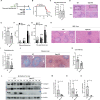
Fig 1
Fig 1. cGAS exacerbates Schistosoma japonicum infection in mice.
(A) Scheme for monitoring of the survival of mice infected with S. japonicum. (B) The survival curve of mice infected with S. japonicum. The Kaplan–Meier method was used for the statistical analysis. (C) The egg loads in the livers of wild-type and Cgas knockout mice infected with S. japonicum. Data are expressed as the mean ± SD of the indicated number of mice from one of three independent experiments. (D-E) Representative imaging showing liver damage in the wild-type and Cgas knockout mice infected with S. japonicum. The quantification of the liver damage is shown in E. Data are expressed as the mean ± SD of the indicated number of mice from one of three independent experiments. (F-G) Measurement of the level of ALT (F) and AST in the sera of wild-type and Cgas knockout mice left uninfected or infected with S. japonicum for the indicated times. Data are expressed as mean ± SD of indicated number of mice from one of three independent experiments. (H-I) Representative imaging showing egg-induced granuloma in the livers of wild-type and Cgas knockout mice infected with S. japonicum. The quantification of the area of the granuloma is shown in (I). Data are expressed as the mean ± SD of the indicated number of granulomas. (J-K) Representative imaging showing Masson staining of the fibrosis in the livers of wild-type and Cgas knockout mice infected with S, japonicum. The quantification of the area of fibrosis is shown in (K). Data are expressed as the mean ± SD of the indicated number of mice from one of three independent experiments. (L-O) Western blotting of the indicated fibrosis-related proteins in the livers of wild-type and Cgas knockout mice infected with S. japonicum. The quantification of gray intensity is shown in (M-O). An unpaired Student’s t-test was used for the statistical analysis in (C, E, I, K and M-O). Two-way ANOVA with Bonferroni’s post hoc test was used for the statistical analysis in (F and G). ns, not significant; *, p < 0.05; **, p < 0.01; ***, p < 0.001. Scale bar, 200 μm.
Fig 2
Fig 2. STING exacerbates Schistosoma japonicum infection in mice.
(A) The scheme for monitoring of the survival of wild-type and Sting knockout mice infected with S. japonicum; (B) The survival curve of wild-type and Sting knockout mice infected with S. japonicum. (C) The egg loads in the livers of wild-type and Sting knockout mice infected with S. japonicum. Data are expressed as the mean ± SD of the indicated number of mice from one of three independent experiments. (D-E) Representative imaging showing liver damage in the wild-type and Sting knockout mice infected with S. japonicum. Scale bar, 1 000 μm. The quantification of the liver damage is shown in E. Data are expressed as the mean ± SD of the indicated number of mice from one of three independent experiments. (F-G) Measurement of the level of ALT (F) and AST in the sera of wild-type and Sting knockout mice left uninfected or infected with S. japonicum for the indicated times. Data are expressed as mean ± SD of indicated number of mice from one of three independent experiments. (H-I) Representative images showing egg-induced granulomas in the livers of wild-type and Sting knockout mice infected with S. japonicum. Scale bar, 200 μm. The quantification of the area of the granuloma is shown in (I). Data are expressed as the mean ± SD of the indicated number of granulomas from one of three independent experiments. (J-K) Representative images showing Masson staining of the fibrosis in the livers of wild-type and Sting knockout mice infected with S. japonicum. Scale bar, 200 μm. The quantification of the area of fibrosis is shown in (K). Data are expressed as the mean ± SD of the indicated number of mice from one of three independent experiments. (L-N) Western blotting detection of the indicated fibrosis-related proteins in the livers of wild-type and Sting knockout mice infected with Schistosoma japonicum. The quantification of gray intensity is shown in (M and N). An unpaired Student’s t-test was used for the statistical analysis in (C, E, I, K, M and N). Two-way ANOVA with Bonferroni’s post hoc test was used for the statistical analysis in (F and G). ns, not significant; *, p < 0.05; **, p < 0.01; ***, p < 0.001.
Fig 3
Fig 3. Schistosoma infection induces a type I IFN response.
(A-B) qRT-PCR measurement of Ifnb1 transcripts in the liver (A) and lung tissues (B) of mice infected with S. japonicum for the indicated times. (C) ELISA detection of IFNβ in serum harvested from mice infected with S. japonicum for the indicated times. (D-E) qRT-PCR measurement of the transcripts of Cgas (D) and Sting (E) in the liver tissues of mice infected with S. japonicum for the indicated times. (F) Measurement of the abundance of cGAMP in the liver tissues of mice infected with S. japonicum for the indicated times using a cGAMP Enzyme Immunoassay Kit. One-way ANOVA with Bonferroni’s post hoc test were used for the statistical analysis. ns, not significant; *, p < 0.05; **, p < 0.01, ***, p < 0.001.
Fig 4
Fig 4. IFNβ exacerbates Schistosoma japonicum infection in mice.
(A) Schematic diagram showing the experimental procedure for the tail vein injection of phosphate-buffered saline (PBS) control and 2 μg of recombinant interferon beta once a week for 7 weeks in mice infected with S. japonicum. The liver tissues were harvested for further experiments. (B) The egg loads in the liver of mice infected with S. japonicum left untreated or treated with interferon beta. Data are expressed as the mean ± SD of the indicated number of mice from one of three independent experiments. (C-D) Representative images showing liver damage in mice infected with S. japonicum left untreated or treated with interferon beta. Scale bar, 1 000 μm. The quantification of the liver damage is shown in D. Data are expressed as the mean ± SD of the indicated number of mice from one of three independent experiments. (E-F) Measurement of the level of ALT (E) and AST (F) in the sera of mice infected with S. japonicum left untreated or treated with interferon beta by tail vein injection. Data are expressed as the mean ± SD of the indicated number of mice from one of three independent experiments. (G-H) Representative imaging showing egg-induced granulomas in the livers of mice infected with S. japonicum left untreated or treated with interferon beta by tail vein injection. Scale bar, 200 μm. The quantification of the area of the granulomas is shown in (H). Data are expressed as the mean ± SD of indicated number of granulomas of mice from one of three independent experiments. (I-J) Representative images showing Masson staining of the fibrosis in the liver of mice infected with S. japonicum left untreated or treated with interferon beta. Scale bar, 200 μm. The quantification of the area of fibrosis is shown in (J). Data are expressed as the mean ± SD of the indicated number of mice from one of three independent experiments. An unpaired Student’s t-test was used for the statistical analysis in (B, D, H and J). Two-way ANOVA with Bonferroni’s post hoc test was used for the statistical analysis in (E and F). ns, not significant; *, p < 0.05; **, p < 0.01.
Fig 5
Fig 5. The cGAS-STING axis is essential for Schistosoma japonicum infection-induced type I IFN response.
(A) qRT-PCR measurement of Ifnb1 transcripts in the liver of wild-type and Cgas KO mice infected with S. japonicum for the indicated times (B) ELISA detection of IFNβ in the serum of wild-type and Cgas KO mice infected with S. japonicum for the indicated times. (C-D) Western blotting detection of the phosphorylation of TBK1 in the liver of wild-type and Cgas KO mice infected with Schistosoma japonicum for 7 weeks. (D) The quantification of the gray intensity of pTBK1 is shown in (D). (E) qRT-PCR measurement of Ifnb1 transcripts in the liver of wild-type and Sting KO mice infected with S. japonicum for the indicated times. (F) ELISA detection of IFNβ in the serum of wild-type and Sting KO mice infected with S. japonicum for the indicated times. (G-H) Western blotting detection of the phosphorylation of TBK1 in the liver of wild-type and Sting KO mice infected with S. japonicum for 7 weeks. The quantification of the gray intensity of pTBK1 is shown in (H). Two-way ANOVA with Bonferroni’s post hoc test were used for the statistical analysis in (A, B, E and F). An unpaired Student’s t-test was used for the statistical analysis in (D and H). ns, not significant; *, p < 0.05, **, p < 0.01, ***, p < 0.001. Scale bar, 200 μm.
Fig 6
Fig 6. Sensing of schistosome-derived DNA by cGAS in macrophages mediates type I IFN response to S. japonicum infection.
(A) Scheme for the clodronate liposome-mediated depletion of macrophages in mice and detection of interferon beta in the liver and sera of mice infected with S. japonicum. Mice were treated with control liposomes or clodronate liposomes twice a week for 1 week before infection and administered continuously for 4 weeks post infection at an interval of twice a week. The liver tissue and sera were collected from mice left uninfected or infected with S. japonicum for 4 weeks for further detection of interferon beta. (B) qRT-PCR measurement of Ifnb1 transcripts in the liver tissues of mice infected with S. japonicum for the indicated times treated with control or clodronate liposomes. (C) ELISA detection of IFNβ in serum harvested from mice infected with S. japonicum for the indicated times treated with control or clodronate liposomes. (D-E) qRT-PCR measurement of transcripts of Ifnb1 and Cxcl10 in wild-type and Cgas knockout peritoneal macrophages transfected with DNA from egg of S. japonicum for the indicated times. (F-G) qRT-PCR measurement of the transcripts of Ifnb1 and Cxcl10 in wild-type and Cgas knockout peritoneal macrophages transfected with DNA from adult schistosomes for the indicated times. (H-J) Western blotting detection of the indicated proteins in the lysates of mouse peritoneal macrophages isolated from wild-type and Cgas KO mice stimulated with S. japonicum egg DNA and ISD for the indicated times (H). The quantification data is shown in (I and J). (K-M) Western blotting detection of the indicated proteins in the lysates of mouse peritoneal macrophages isolated from wild-type and Cgas KO mice stimulated with adult S. japonicum DNA and LPS for the indicated times (K). The quantification data is shown in (L and M). Two-way ANOVA with Bonferroni’s post hoc test were used for the statistical analysis in (D-G). ns, not significant; *, p < 0.05, **, p < 0.01, ***, p < 0.001.

Similar articles

Cited by

References

    1. Colley DG, Bustinduy AL, Secor WE, King CH. Human schistosomiasis. Lancet. 2014;383(9936):2253–64. Epub 2014/04/05. doi: 10.1016/S0140-6736(13)61949-2 ; PubMed Central PMCID: PMC4672382. - DOI - PMC - PubMed
    1. Hotez PJ, Bottazzi ME, Bethony J, Diemert DD. Advancing the development of a human schistosomiasis vaccine. Trends Parasitol. 2019;35(2):104–8. Epub 2018/11/21. doi: 10.1016/j.pt.2018.10.005 . - DOI - PubMed
    1. Wilson RA. Schistosomiasis then and now: what has changed in the last 100 years? Parasitology. 2020;147(5):507–15. Epub 2020/01/23. doi: 10.1017/S0031182020000049 . - DOI - PMC - PubMed
    1. Hambrook JR, Hanington PC. Immune evasion strategies of schistosomes. Front Immunol. 2020;11:624178. Epub 2021/02/23. doi: 10.3389/fimmu.2020.624178 ; PubMed Central PMCID: PMC7889519. - DOI - PMC - PubMed
    1. Chow J, Franz KM, Kagan JC. PRRs are watching you: Localization of innate sensing and signaling regulators. Virology. 2015;479–480:104–9. Epub 2015/03/25. doi: 10.1016/j.virol.2015.02.051 ; PubMed Central PMCID: PMC4424080. - DOI - PMC - PubMed

Publication types

Grants and funding

This work was supported by the National Nature Science Foundation of China (Nos. 81772225 and 81971969 to JC) and the Fifth Round of Three-Year Public Health Action Plan of Shanghai [grant number GWV-10.1-XK13 to JC]. The funders had no role in the study design, data collection, and analysis, decision to publish, or preparation of the manuscript.