. 2022 Apr;59(4):2605-2619.
doi: 10.1007/s12035-022-02760-3. Epub 2022 Feb 2.

The Putative Drosophila TMEM184B Ortholog Tmep Ensures Proper Locomotion by Restraining Ectopic Firing at the Neuromuscular Junction

Affiliations

The Putative Drosophila TMEM184B Ortholog Tmep Ensures Proper Locomotion by Restraining Ectopic Firing at the Neuromuscular Junction

Tiffany S Cho et al. Mol Neurobiol. 2022 Apr.

Abstract

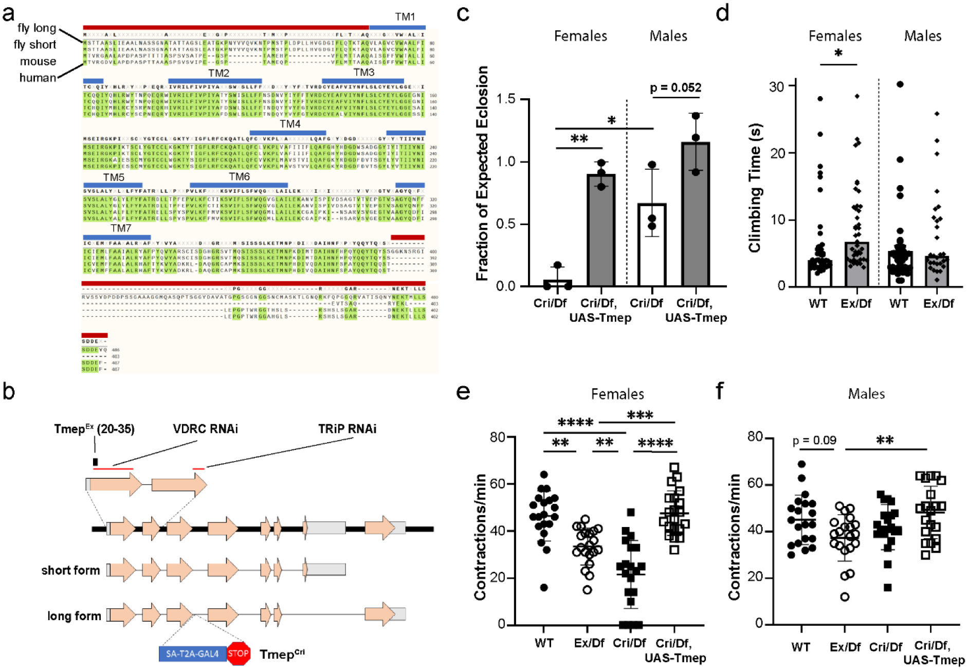
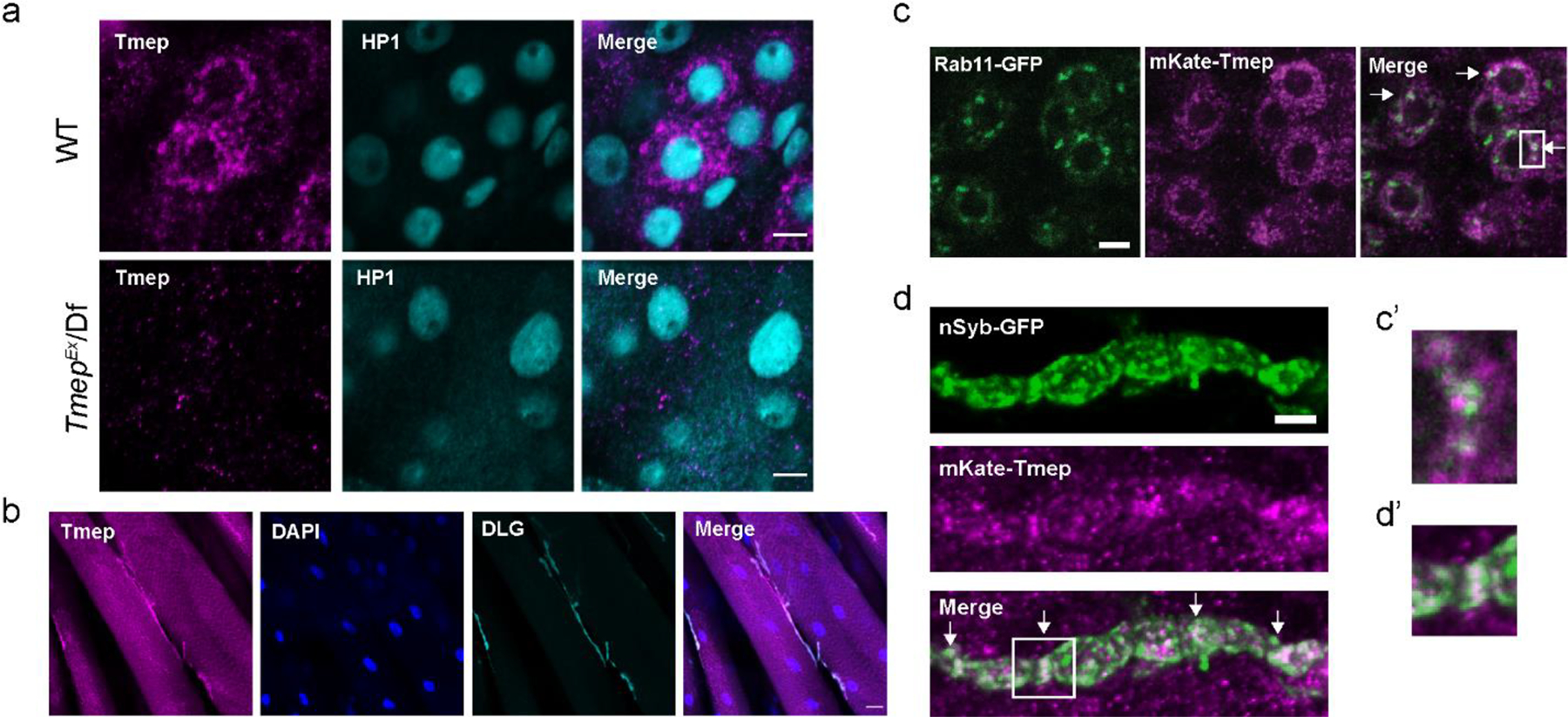
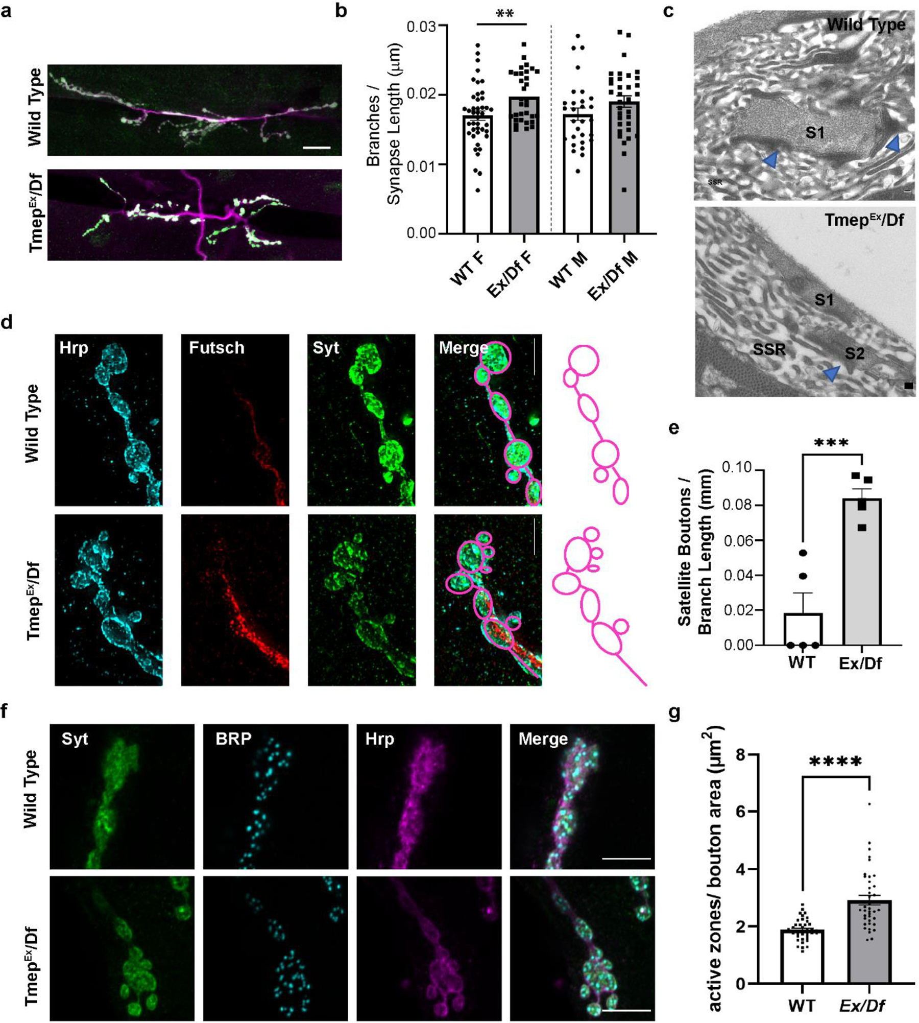
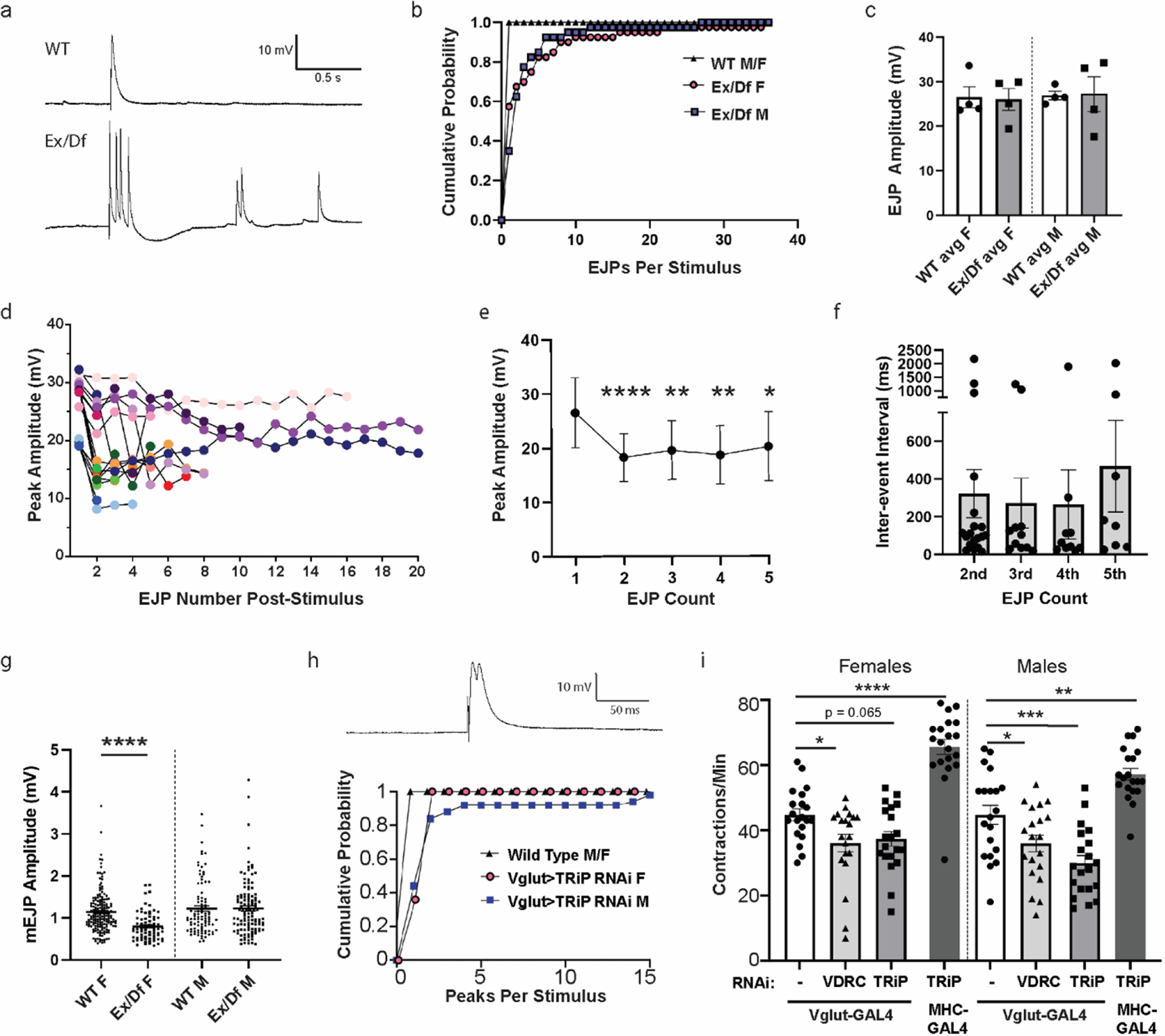
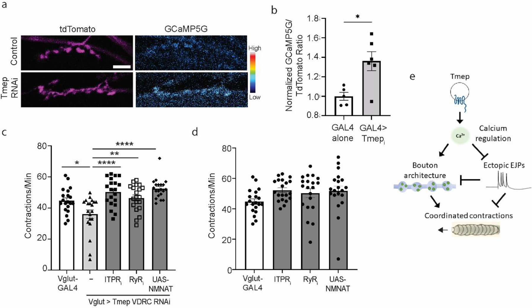
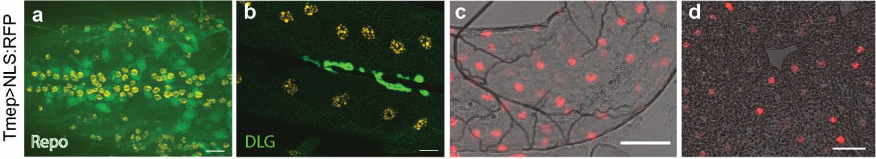
TMEM184B is a putative seven-pass membrane protein that promotes axon degeneration after injury. TMEM184B mutation causes aberrant neuromuscular architecture and sensory and motor behavioral defects in mice. The mechanism through which TMEM184B causes neuromuscular defects is unknown. We employed Drosophila melanogaster to investigate the function of the closely related gene, Tmep (CG12004), at the neuromuscular junction. We show that Tmep is required for full adult viability and efficient larval locomotion. Tmep mutant larvae have a reduced body contraction rate compared to controls, with stronger deficits in females. In recordings from body wall muscles, Tmep mutants show substantial hyperexcitability, with many postsynaptic potentials fired in response to a single stimulation, consistent with a role for Tmep in restraining synaptic excitability. Additional branches and satellite boutons at Tmep mutant neuromuscular junctions are consistent with an activity-dependent synaptic overgrowth. Tmep is expressed in endosomes and synaptic vesicles within motor neurons, suggesting a possible role in synaptic membrane trafficking. Using RNAi knockdown, we show that Tmep is required in motor neurons for proper larval locomotion and excitability, and that its reduction increases levels of presynaptic calcium. Locomotor defects can be rescued by presynaptic knockdown of endoplasmic reticulum calcium channels or by reducing evoked release probability, further suggesting that excess synaptic activity drives behavioral deficiencies. Our work establishes a critical function for Tmep in the regulation of synaptic transmission and locomotor behavior.

Keywords: Calcium; Epilepsy; Excitability; Neuromuscular junction; Synapse; TMEM184B.


Conflict of interest statement

Conflicts of interest/Competing interests

The authors declare no competing financial interests.

Figures

Figure 1.
Figure 1.. CG12004 (Tmep), a Drosophila protein with high homology to mammalian TMEM184b, is needed for full viability and locomotor function.
a, Alignment of Drosophila, mouse, and human TMEM184B proteins. Top to bottom: CG12004 long isoform, CG12004 short isoform, mouse TMEM184b isoform, human TMEM184b isoform. Transmembrane segments (blue lines) and sequences used for antibody generation (red lines) are shown. b, Schematic drawing of the CG12004 genomic region on Chromosome 3. Mutations and regions targeted by knockdown constructs used in this study are indicated. c, Rates of eclosion of TmepCri/Df (Cri/Df) in both sexes and rescue by UAS-Tmep (N-terminal mKate-tagged). P values: Male vs Female TmepCri/Df, p = 0.016; Male TmepCri/Df vs Rescue, p = 0.052; Female TmepCri/Df vs Rescue, p = 0.0024. N = 3 independent crosses with 5 male and 5 female parents in each cross. Statistical evaluation was done using one-way ANOVA with Sidak’s multiple comparisons test. d, climbing assay after disorientation of wild type and TmepEx/Df (Ex/Df) females and males. N = 43, 41, 42, 29 flies (left to right). For females, p = 0.045, unpaired t-test. e-f, larval contraction rate by genotype and sex (e shows females, f shows males). P values for females: wild type vs TmepEx/Df, p = 0.0017; wild type vs TmepCri/Df, p < 0.0001; TmepEx/Df vs TmepCri/Df, p = 0.005; TmepEx/Df vs rescue, p = 0.0005; TmepCri/Df vs Rescue, p < 0.0001; wild type vs rescue, p = 0.98. For males, all comparisons were p > 0.05 except TmepEx/Df vs rescue, where p = 0.008. N = 20 for all genotypes. Statistical significance was calculated by one-way ANOVA with Bonferroni’s multiple comparison correction.
Figure 2.
Figure 2.. Tmep localizes to neuronal endosomes and synaptic vesicles.
a, Antibody to Drosophila Tmep shows neuronal staining in the larval ventral nerve cord. Magenta, Tmep; Teal, HP1 (nuclei). Image is from the area where motor neurons reside. Limited staining is observed in Tmep mutants (TmepEx/Df). Scale bar = 5 μm. b, Tmep protein levels at the neuromuscular junction. Magenta, Tmep; Blue, DAPI; Teal, discs large (DLG) showing the postsynaptic area. Scale bar = 20 μm. c, Tmep shows partial colocalization with Rab11-positive endosomes. Motor neuron expression of mKate-Tmep (magenta) and Rab11-GFP (green) is driven by BG380-GAL4. Image is a single confocal slice of cell bodies in the ventral neve cord; arrows in the merged image show areas of partial colocalization. Scale bar = 5 μm. c’, expanded view of the boxed region in c. d, Tmep shows partial colocalization with synaptic vesicles at the larval NMJ. Image is a single confocal slice. Nsyb-GFP and mKate were expressed as in c. Scale bar = 5 μm. d’, expanded view of the boxed region in d.
Figure 3.
Figure 3.. Tmep mutant larvae show extra branches, boutons, and active zones at the neuromuscular junction.
All images show wandering female third instar larvae. a, Representative images of muscle 6/7 NMJs stained with antibodies to Synaptotagmin (Syt, green) and HRP (magenta) illustrating branching phenotypes. Scale bar = 20 μm. b, Quantification of branches in wild type or Ex/Df larvae. Branch count is normalized to the total synapse length (sum of all branches). c, Representative transmission EM sections of segment A3, muscle 6/7 from 3rd instar larvae. Blue arrows indicate presynaptic dense bodies (T-bars). Synapses (electron-dense areas) are labelled by S (S1, S2). Subsynaptic reticulum area is marked as SSR. Ex/Df synapse shows two boutons’ sections in the same plane, suggesting possible bouton fragmentation at the synaptic terminal. Scale bar = 200 nm. d, Synaptic terminal boutons in wild type and Ex/Df showing an example of satellite boutons at the mutant NMJ. HRP (blue), Futsch (red), and Synaptotagmin (Syt, green) are shown. Scale bar = 5 μm. Drawings illustrate the bouton structure in the images. e, Satellite bouton quantification normalized to the length of the branch upon which the boutons are located. N = 5 female larvae per genotype. p = 0.0009 using unpaired t-test. f, representative images showing active zone density at presynaptic terminal boutons. Syt (green), Bruchpilot (BRP, light blue) and HRP (magenta) are shown. g, quantification of active zone density per square micron of bouton area in female larvae. N = 6 animals, n= 38 total boutons analyzed per genotype. Statistical comparison used unpaired t-test (p < 0.0001).
Figure 4.
Figure 4.. Tmep is required for proper synaptic transmission.
a, Representative examples of evoked junctional potentials (EJPs) evoked by stimulation of wild type (WT) or TmepEx/Df (Ex/Df) female neuromuscular junctions (1 ms, 3.3 mV). b-e, Analysis of EJPs from N=4 larvae/genotype and n=10 stimuli/larvae. b, Cumulative probability of single and multiple EJPs following stimulation in wild type and Ex/Df larvae (over the 5 sec post-stimulation), separated by sex. All recordings were done in 0.4 mM Ca2+ HL3.1. c, EJP amplitudes of the first response recorded following each stimulus. d, Peak amplitudes of EJPs per stimulus. 17 of 40 EJPs measured in 4 Ex/Df females showed multiple peaks and are displayed on the graph. e, Relative sizes of multi-EJP responses to a single stimulus from female Ex/Df larvae. N=17, 17, 13, 12, and 10 stimuli showed at least 5 responses and were included in analysis. Differences from the first peak’s amplitude were analyzed by fitting a mixed model using paired comparisons (responses in each larvae), with Dunnett’s correction for multiple comparisons. P values: 1st-2nd, p <0.0001; 1st-3rd, p = 0.0072; 1st-4th, p = 0.0016; 1st-5th, p = 0.0336. f, Inter-event interval from females shown in e. Only the first five inter-event intervals were analyzed, and each point represents the interval from the previous spike (i.e. 2nd is from spike 1 to spike 2). No significant differences were observed (p > 0.05, one-way ANOVA). g, mEJP amplitudes from wild type and Ex/Df larvae of each sex. N=4 larvae per group. Fifteen seconds of baseline recording were analyzed per animal. T-tests within each sex were used to evaluate statistical significance (females, p <0.0001; males, p = 0.98). h, representative trace (above) and cumulative probability (below) of multi-peak events in Tmep knockdown (TRiP) larvae using Vglut-GAL4. Note that controls (black triangles) do not show any multi-peak responses. N=5 larvae and 10 stimuli/larvae per genotype. i, Crawling analysis of larvae in which Tmep is knocked down in motor neurons (Vglut-GAL4) or muscle (MHC-GAL4) with one of two different RNAi constructs (sequence targets are shown in Figure 1). N=20 animals per genotype and sex. One-way ANOVA with Dunnett’s post-hoc test within sex; p = 0.025 (Vglut F alone vs VDRC F), p = 0.065 (Vglut F alone vs TRiP F), p = <0.0001 (Vglut F alone vs MHC> TRiP F), p = 0.035 (Vglut M alone vs TRiP M), p = 0.0002 (Vglut M alone vs TRiP M), p = 0.0016 (Vglut M alone vs MHC> TRiP).
Figure 5.
Figure 5.. Larval crawling deficiencies are restored by reduction of presynaptic excitability.
a, Representative images of baseline calcium in GAL4 alone (UAS-Dicer2, Vglut-GAL4, UAS-GCaMP5G-T2A-tdTomato) and Tmep RNAi larvae (UAS-Dicer2, Vglut-GAL4, UAS-Tmepi (VDRC), UAS-GcaMP5G-T2A-tdTomato). TdTomato images are pseudocolored with a Magenta look-up table, and GCaMP is pseudocolored with the Cyan Hot lookup table (Fiji), for which the scale is shown to the right. b, Quantification of baseline calcium in the genotypes shown in a. N = 5–6 larvae per genotype. Unpaired t-test, p = 0.012. c, Crawling deficiency of Tmep knockdown larvae can be restored by knockdown of IPTR, RyR, or by over-expression of NMNAT. Female data is shown. One-way ANOVA with Sidak’s multiple comparison correction was used to calculate p values as follows: p = 0.014 (Vglut vs Tmep RNAi), p = 0.0014 (Tmep RNAi vs Tmep/RyR), p < 0.0001 (Tmep RNAi vs Tmepi/ITPRi or NMNAT). d, Individual RNAi expression of IPTR, RyR, or UAS-NMNAT does not change baseline contraction rates (for all, p>0.05). Female data is shown. e, Model for Tmep influence on presynaptic calcium, synaptic activity, synaptic structure, and locomotion.
Online Resource 1.
Online Resource 1.. Tmep is expressed broadly throughout the larvae.
RedStinger:NLS (red and orange) shows nuclei of cells in which endogenous Tmep is expressed. a, Ventral nerve cord. Green is Repo (glial nuclei). b, Neuromuscular junction. Green is discs large (DLG). c, Gut expression. d, Fat body expression. For all figures, scale bars = 20 μm.

Similar articles

Cited by

References

    1. Schneggenburger R, Neher E (2005) Presynaptic calcium and control of vesicle fusion. Curr. Opin. Neurobiol 15:266–274 - PubMed
    1. Kwon SK, Sando R, Lewis TL, et al. (2016) LKB1 Regulates Mitochondria-Dependent Presynaptic Calcium Clearance and Neurotransmitter Release Properties at Excitatory Synapses along Cortical Axons. PLoS Biol 14:. 10.1371/journal.pbio.1002516 - DOI - PMC - PubMed
    1. Brusich DJ, Spring AM, James TD, et al. (2018) Drosophila CaV2 channels harboring human migraine mutations cause synapse hyperexcitability that can be suppressed by inhibition of a Ca2+store release pathway. PLoS Genet 14:. 10.1371/journal.pgen.1007577 - DOI - PMC - PubMed
    1. Stern M, Ganetzky B (1989) Altered synaptic transmission in drosophila hyperkinetic mutants. J Neurogenet 5:215–228. 10.3109/01677068909066209 - DOI - PubMed
    1. Uytterhoeven V, Kuenen S, Kasprowicz J, et al. (2011) Loss of Skywalker reveals synaptic endosomes as sorting stations for synaptic vesicle proteins. Cell 145:117–132. 10.1016/j.cell.2011.02.039 - DOI - PubMed

Substances

LinkOut - more resources