. 2022 Mar 23;96(6):e0007422.
doi: 10.1128/jvi.00074-22. Epub 2022 Feb 2.

p17-Modulated Hsp90/Cdc37 Complex Governs Oncolytic Avian Reovirus Replication by Chaperoning p17, Which Promotes Viral Protein Synthesis and Accumulation of Viral Proteins σC and σA in Viral Factories

Affiliations

p17-Modulated Hsp90/Cdc37 Complex Governs Oncolytic Avian Reovirus Replication by Chaperoning p17, Which Promotes Viral Protein Synthesis and Accumulation of Viral Proteins σC and σA in Viral Factories

Wei-Ru Huang et al. J Virol. .

Erratum in

Abstract

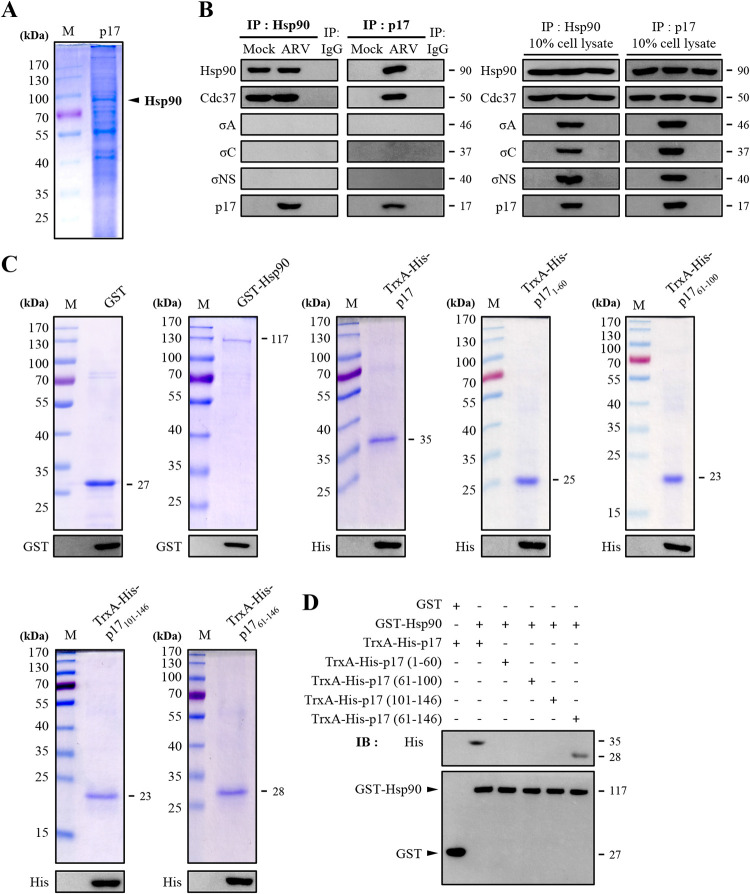
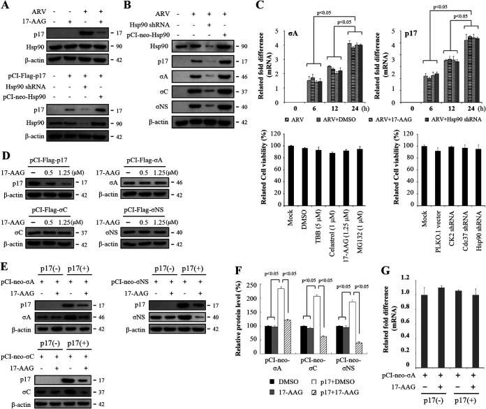
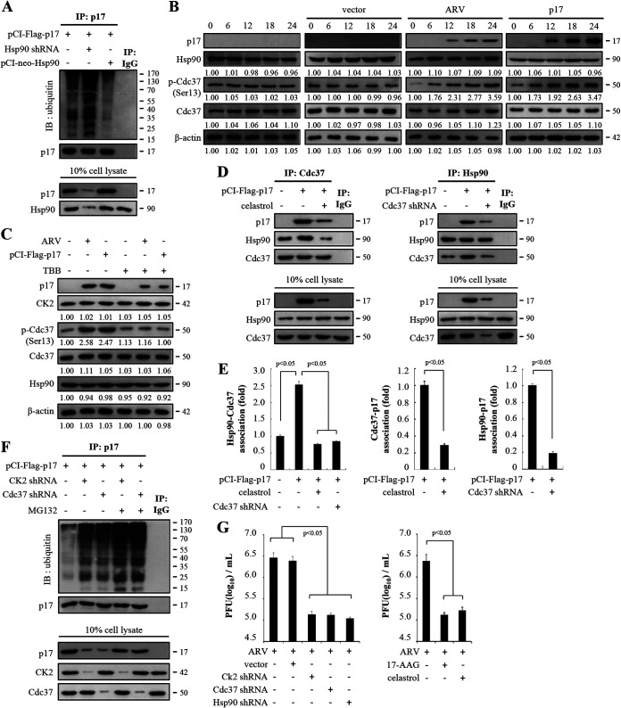
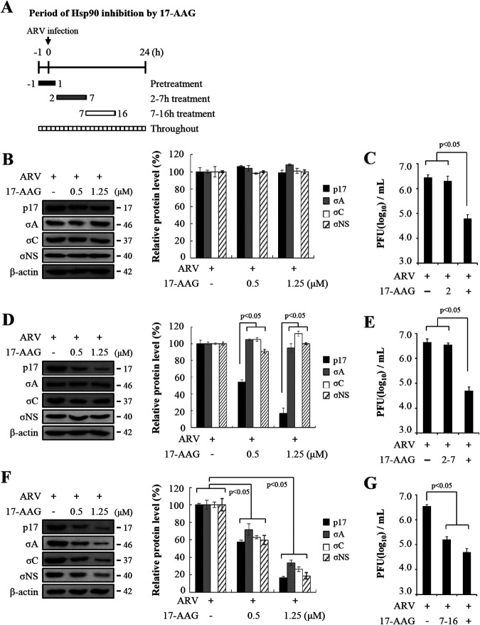
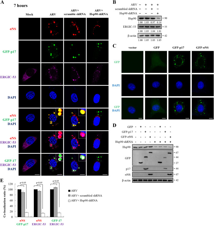
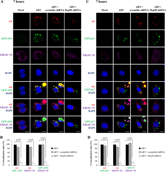
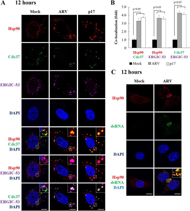
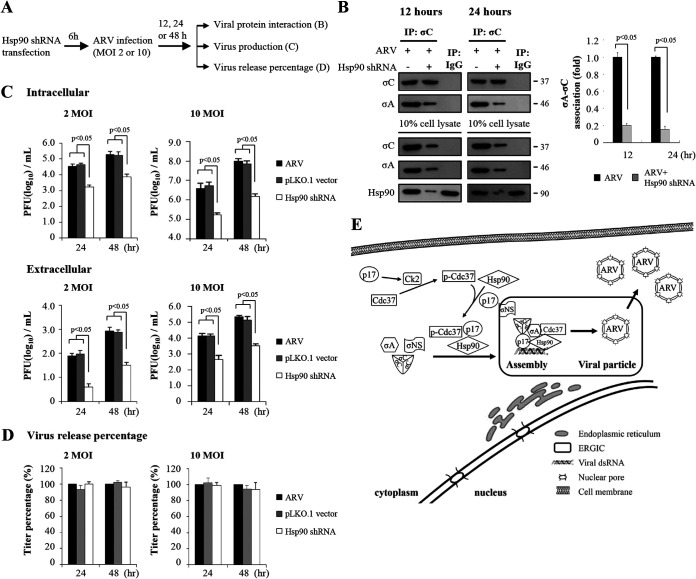
In this work we have determined that heat shock protein 90 (Hsp90) is essential for avian reovirus (ARV) replication by chaperoning the ARV p17 protein. p17 modulates the formation of the Hsp90/Cdc37 complex by phosphorylation of Cdc37, and this chaperone machinery protects p17 from ubiquitin-proteasome degradation. Inhibition of the Hsp90/Cdc37 complex by inhibitors (17-N-allylamino-17-demethoxygeldanamycin 17-AGG, and celastrol) or short hairpin RNAs (shRNAs) significantly reduced expression levels of viral proteins and virus yield, suggesting that the Hsp90/Cdc37 chaperone complex functions in virus replication. The expression levels of p17 were decreased at the examined time points (2 to 7 h and 7 to 16 h) in 17-AAG-treated cells in a dose-dependent manner while the expression levels of viral proteins σA, σC, and σNS were decreased at the examined time point (7 to 16 h). Interestingly, the expression levels of σC, σA, and σNS proteins increased along with coexpression of p17 protein. p17 together with the Hsp90/Cdc37 complex does not increase viral genome replication but enhances viral protein stability, maturation, and virus production. Virus factories of ARV are composed of nonstructural proteins σNS and μNS. We found that the Hsp90/Cdc37 chaperone complex plays an important role in accumulation of the outer-capsid protein σC, inner core protein σA, and nonstructural protein σNS of ARV in viral factories. Depletion of Hsp90 inhibited σA, σC, and p17 proteins colocalized with σNS in viral factories. This study provides novel insights into p17-modulated formation of the Hsp90/Cdc37 chaperone complex governing virus replication via stabilization and maturation of viral proteins and accumulation of viral proteins in viral factories for virus assembly. IMPORTANCE Molecular mechanisms that control stabilization of ARV proteins and the intermolecular interactions among inclusion components remain largely unknown. Here, we show that the ARV p17 is an Hsp90 client protein. The Hsp90/Cdc37 chaperone complex is essential for ARV replication by protecting p17 chaperone from ubiquitin-proteasome degradation. p17 modulates the formation of Hsp90/Cdc37 complex by phosphorylation of Cdc37, and this chaperone machinery protects p17 from ubiquitin-proteasome degradation, suggesting a feedback loop between p17 and the Hsp90/Cdc37 chaperone complex. p17 together with the Hsp90/Cdc37 complex does not increase viral genome replication but enhances viral protein stability and virus production. Depletion of Hsp90 prevented viral proteins σA, σC, and p17 from colocalizing with σNS in viral factories. Our findings elucidate that the Hsp90/Cdc37 complex chaperones p17, which, in turn, promotes the synthesis of viral proteins σA, σC, and σNS and facilitates accumulation of the outer-capsid protein σC and inner core protein σA in viral factories for virus assembly.

Keywords: Hsp90/Cdc37 chaperone machinery; avian reovirus; inner core protein σA; nonstructural protein p17; nonstructural protein σNS; outer-capsid protein σC; viral factory.


Conflict of interest statement

The authors declare no conflict of interest.

Figures

FIG 1
FIG 1
Identification of potential cellular factors that interact with p17. (A) Cellular proteins coimmunoprecipitated by the p17 antibody were analyzed by SDS-PAGE. Vero cells were transfected with pcDNA3.1-p17 plasmid for 24 h, followed by a coimmunoprecipitation assay with p17 antibody. Cellular proteins coimmunoprecipitated by the p17 antibody were analyzed by SDS-PAGE. (B) Coimmunoprecipitation assays in ARV-infected Vero cells were performed with the Hsp90 and p17 antibodies, respectively. Following immunoblotting, Hsp90, Cdc37, σA, σC, σNS, and p17 were detected by the respective antibodies. (C) To define the region within p17 that was involved in Hsp90 binding, a series of truncated versions of His-tagged p17 constructs were established. The purified protein TrxA-His-p17, TrxA-His-p17 mutants, and GST-Hsp90 fusion proteins were analyzed by SDS-PAGE. The expected sizes of purified fusion proteins are indicated. The expressed fusion proteins were confirmed by Western blotting assays with the anti-GST or anti-His antibodies. (D) An in vitro GST pulldown assay was carried out with purified His- or GST-tagged fusion proteins (2 μg). Elution fractions were examined by Western blotting with the indicated antibodies (GST and His). The representative data are from three independent experiments. The predicted size of each protein was labeled at the right of gels and blots in KDa in each figure.
FIG 2
FIG 2
The Hsp90/Cdc37 chaperone complex protects p17 from ubiquitin-proteasome-mediated degradation, and p17 increases the expression levels of σA, σC, and σNS proteins of ARV. (A) To study whether Hsp90 stabilizes p17, inhibition of Hsp90 by 17-AAG (1.25 μM) in ARV-infected cells as well as depletion of Hsp90 with an shRNA or overexpression of Hsp90 in p17-transfected Vero cells was carried out. Vero cells were infected with an MOI of 10 for 24 h. The expression levels of p17 and Hsp90 were examined by Western blot analysis with the indicated antibodies. β-Actin was used as an internal control. (B) To examine whether Hsp90 affects other structural proteins, inhibition of Hsp90 with an shRNA or overexpression of Hsp90 in ARV-infected cells was carried out. The expression levels of viral proteins and Hsp90 were examined by Western blot analysis with the indicated antibodies. β-Actin was used as an internal control. (C) To further study whether Hsp90 transcriptionally regulates the σA and p17 genes of ARV, mRNA levels of the σA-encoding and p17-encoding genes of ARV in Vero cells treated with 17-AAG and Hsp90 shRNA were analyzed. Vero cells were infected at different time points as indicated. The mRNA levels of σA-encoding and p17-encoding genes of ARV in Vero cells treated with 17-AAG (1.25 μM) and Hsp90 shRNA were measured by qRT‐PCR, respectively. Vero cells were transfected with the Hsp90 shRNA plasmid for 6 h, followed by infection with ARV at an MOI of 10. To examine whether the compounds or shRNAs had deleterious effects on cells, cell viability was determined using the MTT assay. Each value represents mean ± SE from three independent experiments. DMSO, dimethyl sulfoxide. (D) 17-AAG was also used to inhibit Hsp90 in the indicated plasmid-transfected Vero cells. The expression levels of viral proteins p17, σA, σC, and σNS were examined by Western blotting with the indicated antibodies. β-Actin was used as an internal control. All experiments were conducted in three replicate experiments. (E) Expression plasmids (pCI-Flag-σA, pCI-Flag-σC, and pCI-Flag-σNS) were expressed in Vero cells with or without the p17 protein. The medium was replaced with fresh medium containing 1.25 μM 17-AAG at 6 h posttransfection. At 24 h posttransfection, the cell lysates were prepared and subjected to immunoblotting with the indicated antibodies. (F) Densitometry analysis results for Western blotting are expressed as percentages representing p17, σA, σC, and σNS protein levels, normalized to β-actin. The results were calculated from the data shown in panel D. Mock cells were considered 100%. All data shown represent the means ± standard deviations (SD) calculated from three independent experiments. (G) To investigate whether p17 affected the mRNA levels of the σA-encoding gene of ARV, the mRNA levels of the σA-encoding gene of ARV were quantified by real-time qRT-PCR in p17-transfected cells in the presence or absence of the indicated drugs. Each value represents the mean ± SE from three independent experiments. Signals were quantified using ImageJ software.
FIG 3
FIG 3
The ARV p17 protein enhances the formation of Hsp90/Cdc37 chaperone machinery. (A) To investigate whether Hsp90 protects p17 protein from ubiquitin-proteasome-mediated degradation, the signals of polyubiquitinated p17 were examined in Hsp90 knockdown and Hsp90 overexpression in p17-transfected Vero cells. Rabbit IgG served as negative control. (B) To investigate whether the p17 protein modulates phosphorylation of Cdc37, levels of Hsp90 and p-Cdc37 in ARV-infected and p17-transfected Vero cells were analyzed. The expression levels of p17, Hsp90, Cdc37, and p-Cdc37 in Vero cells infected with ARV at an MOI of 10 or transfected with the pCI-Flag-p17 plasmid were examined by Western blotting assays. Vero cells were ARV infected and pCI-p17 transfected at the indicated time points (hours). The levels of the indicated proteins in the mock-control group (0 h) were considered 1-fold. The fold changes indicated below each lane were normalized against values for the mock-control group. Protein levels were normalized to those for β‐actin. Signals in all Western blots were quantified with ImageJ software. All experiments were conducted in three independent experiments. (C) To study whether the p17 protein modulates phosphorylation of Cdc37, the effect of CK2 inhibitor TBB on p17-mediated phosphorylation of Cdc37 was assayed. The expression levels of p17, CK2, p-Cdc37, Cdc37, and Hsp90 were examined in p17-transfected and ARV-infected Vero cells. Vero cells were transfected with the pCI-Flag-p17 plasmid for 6 h, followed by treatment with CK2 inhibitor TBB (5 μM) for 24 h. In the ARV-infected group, Vero cells were simultaneously treated with CK2 inhibitor TBB and infected with ARV at an MOI of 10 for 24 h. The levels of indicated proteins in the mock-control group were considered 1-fold. The fold changes indicated below each lane were normalized against values for the mock-control group. Protein levels were normalized to those for β‐actin. Signals in Western blots were quantified with ImageJ software. All experiments were conducted in three independent experiments. (D) To investigate whether p17 modulates the formation of the Hsp90/Cdc37 complex and whether p17 associates with Hsp90 and Cdc37, the celastrol inhibitor and Cdc37 shRNA were used in p17-transfected Vero cells. p17 and cellular proteins coimmunoprecipitated by Hsp90 or Cdc37 antibodies were analyzed. Vero cells were transfected with the pCI-Flag-p17 plasmid for 6 h, followed by treatment with celastrol for 24 h. Furthermore, Vero cells were simultaneously transfected with the pCI-Flag-p17 and Cdc37 shRNA plasmid DNA. The amounts of p17/Hsp90, p17/Cdc37, and Hsp90/Cdc37 association were examined in the indicated treatments. The protein levels were detected by the indicated antibodies. (E) Densitometry analysis results for Western blotting are expressed as percentages representing the amount of protein association in panel D. Mock cells were considered 1-fold. Signals for all blots were quantified using ImageJ software. All experiments were conducted in three independent experiments. (F) To assess whether CK2 and Cdc37 are involved in p17 stabilization, Vero cells were transfected with the respective pCI-Flag-p17 or cotransfected with pCI-Flag-p17 and shRNA plasmid DNA for 6 h, followed by treatment with MG132 (1 μM). The amounts of p17 polyubiquitylation were examined by Western blotting assays. Similar results were obtained in three independent experiments. (G) To examine whether Hsp90/Cdc37 chaperone machinery plays a critical role in virus propagation, cells treated with celastrol (1 μM) or knockdown of CK2, Cdc37, and Hsp90 were analyzed. Vero cells were transfected with the respective Hsp90 shRNA plasmid for 6 h, followed by infection with ARV at an MOI of 10 for 24 h. In drug-treated groups, Vero cells were simultaneously treated with celastrol and infected with ARV at an MOI of 10 for 24 h. Cell lysates were collected 24 h postinfection for determination of virus titers by plaque assays. Each value represents the mean ± SE from three independent experiments.
FIG 4
FIG 4
The Hsp90/Cdc37 chaperone machinery is required for ARV replication. (A) Experimental design for pulse treatment with 17-AAG in ARV-infected cells at different time points in the viral life cycle is shown. To elucidate inhibition of Hsp90 in the presence of different concentrations of 17-AAG, Vero cells were treated with 17-AAG during different time windows before and after ARV infection at an MOI of 10. After treatment with 17-AAG on different time windows, cells were washed to remove the drug. (B to G) All 17-AAG-treated and untreated cell lysates were collected at 24 h postinfection for Western blotting (B, D, and F) and determination of virus titers (C, E, and G). The expression levels of viral proteins p17, σA, σC, and σNS were analyzed by Western blotting assays. Densitometry analysis results for Western blotting are expressed as percentages representing p17, σA, σC, and σNS protein levels, normalized to β-actin. Untreated cells were considered 100%. Signals for all blots were quantified using ImageJ software. All experiments were conducted in three independent experiments. (C, E, and G) The effect of Hsp90 on ARV replication was also analyzed by determining the virus titers of the groups treated for different time points. Vero cells were infected with ARV at an MOI of 10 for 24 h and treated with 17-AAG (1.25 μM) for different time windows. All 17-AAG-treated and untreated cell lysates were collected at 24 h postinfection for determination of virus titers. Each value represents the mean ± SE from three independent experiments.
FIG 5
FIG 5
Distribution of GFP-p17, σNS, and ERGIC-53 in cytoplasmic viral inclusions and Hsp90 promotion of accumulation of p17 in viral inclusions. (A) Confocal immunofluorescence images of ARV-infected Vero cells (MOI of 10, 7 h postinfection) stained with DAPI (blue) and antibodies specific for σNS (red) and ERGIC-53 (purple). For transient expression of GFP-p17, the recombinant plasmid pcDNA3.1-GFP-p17 was used. The subcellular localization of GFP-p17, σNS, and ERGIC-53 was visualized by confocal microscopy. To examine whether Hsp90 enhances accumulation of p17 and σNS in viral inclusions and ERGIC, the subcellular localization of p17, σNS, and ERGIC-53 was visualized in Hsp90-knockdown Vero cells. Vero cells were transfected with the Hsp90 shRNA plasmid for 6 h, followed by infection with ARV at an MOI of 10. Viral inclusions are identifiable by σNS. ERGIC-53 is a marker protein for ERGIC. Enlarged images correspond to the region indicated by the white box in the merged image. The representative images are from three independent experiments. Scale bars, 20 μm. (B) The expression levels of Hsp90 and ERGIC-53 were examined by Western blotting. To study whether knockdown of Hsp90 influences the expression levels of ERGIC-53, Vero cells were transfected with the Hsp90 shRNA plasmid for 6 h, followed by infection with ARV at an MOI of 10. The levels of the indicated proteins in the mock-control group were considered 1-fold. The fold changes indicated below each lane were normalized against values for the mock-control group. Protein levels were normalized to those for β‐actin. Signals in all Western blots were quantified with ImageJ software. Experiments were conducted in three independent experiments. (C) Immunofluorescence images of GFP, GFP-p17, and GFP-σNS in Vero cells were observed at 7 h posttransfection. Vero cells were stained with DAPI (blue). (D) To examine whether the GFP-p17 and GFP-σNS fusion proteins affect stabilization of p17 and σNS, the expression levels of GFP, GFP-p17, GFP-σNS, and Hsp90 in Hsp90-depleted cells were assayed by Western blotting. (E) Quantification ratios of the colocalization of GFP-p17/σNS, σNS/ERGIC-53, and GFP-p17/ERGIC-53 in ARV-infected, scrambled shRNA-transfected, and Hsp90 shRNA-transfected Vero cells (A). ARV-infected cells were considered 100%. At least 10 fields of 1 to 2 cells per field were taken for each sample per experiment while the exposure settings were unchanged during acquisition of various samples. The colocalization was quantified with ImageJ software. Each experiment was performed at least three times. Statistical analysis was performed using Student’s t test.
FIG 6
FIG 6
Distribution of inner core protein σA, GFP-p17, and GFP-σNS in cytoplasmic viral inclusions and Hsp90 promotion of accumulation of inner core protein σA in viral inclusions. (A and C) Confocal immunofluorescence images of ARV-infected Vero cells (MOI of 10, 7 h postinfection) stained with DAPI (blue) and antibodies specific for inner core protein σA (red). For transient expression of GFP-p17 and GFP-σNS, the recombinant plasmids pcDNA3.1-GFP-p17 and pcDNA3.1-GFP-σNS were used. The subcellular localization of σA with GFP-σNS (A) or GFP-p17 (C) was visualized by confocal microscopy. To examine whether Hsp90 enhances accumulation of σA, p17, and σNS proteins in viral inclusions and ERGIC, the subcellular localization of these proteins was visualized in Hsp90-knockdown Vero cells. Viral inclusions are identifiable by σNS. Enlarged images correspond to the region indicated by the white box in the merged image. The representative images are from three independent experiments. Scale bars, 20 μm. (B and D) Quantification ratios of the colocalization of GFP-σNS/σA, σA/ERGIC-53, and GFP-σNS/ERGIC-53 (B) as well as the colocalization of GFP-p17/σA, σA/ERGIC-53, and GFP-p17/ERGIC-53 (D) in ARV-infected, scrambled shRNA-transfected, and Hsp90 shRNA-transfected Vero cells. Quantification of the colocalization was done with ImageJ software. ARV-infected cells were considered 100%. At least 10 fields of 1 to 2 cells per field were taken for each sample per experiment. All experiments were conducted in three independent experiments. Statistical analysis was performed using Student’s t test.
FIG 7
FIG 7
Distribution of outer-capsid protein σC, GFP-p17, and GFP-σNS in cytoplasmic viral inclusions and Hsp90 promotion of accumulation of outer-capsid protein σC in viral inclusions. Confocal immunofluorescence images of ARV-infected Vero cells (MOI of 10, 7 h postinfection) stained with DAPI (blue) and antibodies specific for outer-capsid protein σC (red). For transient expression of GFP-p17 and GFP-σNS, the recombinant plasmids pcDNA3.1-GFP-p17 and pcDNA3.1-GFP-σNS were used. (A and C) The subcellular localization of σC with GFP-σNS (A) or GFP-p17 (C) was visualized by confocal microscopy. To examine whether Hsp90 enhances accumulation of viral proteins σC, p17, and σNS in viral inclusions, the subcellular localization of outer-capsid protein σC, GFP-σNS, and GFP-p17 was visualized in Hsp90-knockdown Vero cells. Viral inclusions are identifiable by σNS. Enlarged images correspond to the region indicated by the white box in the merged image. The representative images are from three independent experiments. Scale bars, 20 μm. (B and D) Quantification ratios of the colocalization of GFP-σNS/σC, σC/ERGIC-53, and GFP-σNS/ERGIC-53 (B) as well as the colocalization of GFP-p17/σC, σC/ERGIC-53, and GFP-p17/ERGIC-53 (D) in ARV-infected, scrambled shRNA-transfected, and Hsp90 shRNA-transfected Vero cells. The colocalization was quantified with ImageJ software. ARV-infected cells were considered 100%. At least 10 fields of 1 to 2 cells per field were taken for each sample per experiment. All experiments were conducted in three independent experiments. Statistical analysis was performed using Student’s t test.
FIG 8
FIG 8
Distribution of inner core protein σA, dsRNA, and ERGIC-53 in cytoplasmic viral inclusions and Hsp90 promotion of accumulation of inner core protein σA in viral inclusions. (A) Confocal immunofluorescence images of ARV-infected Vero cells (MOI of 10, 12 h postinfection) stained with DAPI (blue) and antibodies specific for σA (red), dsRNA (green), and ERGIC-53 (purple). The subcellular localization of inner core protein σA (red), dsRNA (green), and ERGIC-53 was visualized by confocal microscopy. To examine whether Hsp90 enhances accumulation of inner core protein σA, dsRNA, and ERGIC in viral factories, the subcellular localization of inner core protein σA and dsRNA was also visualized in Hsp90-knockdown Vero cells. Enlarged images correspond to the region indicated by the white box in the merged image. The representative images are from three independent experiments. Scale bars, 20 μm. (B) Quantification ratios of the colocalization of σA/dsRNA, σA/ERGIC-53, and ERGIC-53/dsRNA. Quantification of the colocalization was done with ImageJ software. ARV-infected cells were considered 100%. At least 10 fields of 1 to 2 cells per field were taken for each sample per experiment. Each experiment was performed at least three times. Statistical analysis was performed using Student’s t test.
FIG 9
FIG 9
Colocalization of Hsp90 and Cdc37 in cytoplasmic viral inclusions. (A) Confocal immunofluorescence images of ARV-infected Vero cells (MOI of 10, 12 h postinfection) stained with DAPI (blue) and antibodies specific for Hsp90 (red), Cdc37 (green), and ERGIC-53 (purple). Viral inclusions are identifiable by ERGIC-53. Enlarged images correspond to the region indicated by the white box in the merged image. The representative images are from three independent experiments. (B) Quantification of the colocalization of Hsp90/Cdc37, Hsp90/ERGIC-53, and Cdc37/ERGIC-53 in mock- or ARV-infected or p17-transfected cells was analyzed with ImageJ software. Mock cells were considered 1-fold. At least 10 fields of 1 to 2 cells per field were taken for each sample per experiment. All experiments were conducted in three independent experiments. Statistical analysis was performed using Student’s t test. (C) Colocalization of Hsp90 and dsRNA in ARV-infected Vero cells (MOI of 10, 12 h postinfection) stained with DAPI (blue) and antibodies specific for Hsp90 (red) or dsRNA (green). All experiments were conducted in three independent experiments.
FIG 10
FIG 10
Hsp90/Cdc37 chaperone machinery is required for recruiting inner core protein σA and outer-capsid protein σC to viral inclusions and virion production. (A) Schematic of the experimental procedures showing the times of protein collection and Hsp90 inhibition by shRNA and collection of cell lysates. (B) The amounts of σA and σC association were examined by coimmunoprecipitation assays in ARV-infected and Hsp90-knockdown Vero cells. Vero cells were infected with ARV at an MOI of 10 for 12 and 24 h, respectively. The protein levels were detected by the indicated antibodies. Densitometry analysis results for Western blotting are expressed as percentages representing the amount of σA and association as shown in the upper panel. Signals for all blots were quantified using ImageJ software. Mock cells were considered 1-fold. Each experiment was performed at least three times. (C) Intracellular and extracellular virus production was examined in Hsp90-depleted cells. Vero cells were infected with an MOI of 2 and 10 for 24 or 48 h, respectively. (D) Virus release percentage was calculated according to the formula described in Materials and Methods. (E) A model showing how the p17-modulated Hsp90/Cdc37 chaperone complex accumulates viral proteins in viral factories for virus assembly. All experiments were conducted in three independent experiments. Statistical analysis was performed using Student’s t test.

Similar articles

Cited by

References

    1. Didenko T, Duarte AM, Karagoz GE, Rudiger SG. 2012. Hsp90 structure and function studied by NMR spectroscopy. Biochim Biophys Acta 1823:636–647. 10.1016/j.bbamcr.2011.11.009. - DOI - PubMed
    1. Prodromou C, Panaretou B, Chohan S, Siligardi G, O’Brien R, Ladbury JE, Roe SM, Piper PW, Pearl LH. 2000. The ATPase cycle of Hsp90 drives a molecular ‘clamp’ via transient dimerization of the N-terminal domains. EMBO J 19:4383–4392. 10.1093/emboj/19.16.4383. - DOI - PMC - PubMed
    1. Young JC, Moarefi I, Hartl FU. 2001. Hsp90: a specialized but essential protein-folding tool. J Cell Sci 154:267–273. 10.1083/jcb.200104079. - DOI - PMC - PubMed
    1. Park M, Yong Kang C, Krishna P. 1998. Brassica napus Hsp90 can autophosphorylate and phosphorylate other protein substrates. Mol Cell Biochem 185:33–38. 10.1023/A:1006884306169. - DOI - PubMed
    1. Borkovich KA, Farrelly FW, Finkelstein DB, Taulien J, Lindquist S. 1989. Hsp82 is an essential protein that is required in higher concentrations for growth of cells at higher temperatures. Mol Cell Biol 9:3919–3930. 10.1128/mcb.9.9.3919-3930.1989. - DOI - PMC - PubMed

Publication types

MeSH terms

LinkOut - more resources