The c-Myc Oncogene Maintains Corneal Epithelial Architecture at Homeostasis, Modulates p63 Expression, and Enhances Proliferation During Tissue Repair
- PMID: 35103750
- PMCID: PMC8822362
- DOI: 10.1167/iovs.63.2.3
The c-Myc Oncogene Maintains Corneal Epithelial Architecture at Homeostasis, Modulates p63 Expression, and Enhances Proliferation During Tissue Repair
Abstract
Purpose: The transcription factor c-Myc (Myc) plays central regulatory roles in both self-renewal and differentiation of progenitors of multiple cell lineages. Here, we address its function in corneal epithelium (CE) maintenance and repair.
Methods: Myc ablation in the limbal-corneal epithelium was achieved by crossing a floxed Myc mouse allele (Mycfl/fl) with a mouse line expressing the Cre recombinase gene under the keratin (Krt) 14 promoter. CE stratification and protein localization were assessed by histology of paraffin and plastic sections and by immunohistochemistry of frozen sections, respectively. Protein levels and gene expression were determined by western blot and real-time quantitative PCR, respectively. CE wound closure was tracked by fluorescein staining.
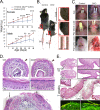
Results: At birth, mutant mice appeared indistinguishable from control littermates; however, their rates of postnatal weight gain were 67% lower than those of controls. After weaning, mutants also exhibited spontaneous skin ulcerations, predominantly in the tail and lower lip, and died 45 to 60 days after birth. The mutant CE displayed an increase in stratal thickness, increased levels of Krt12 in superficial cells, and decreased exfoliation rates. Accordingly, the absence of Myc perturbed protein and mRNA levels of genes modulating differentiation and proliferation processes, including ΔNp63β, Ets1, and two Notch target genes, Hey1 and Maml1. Furthermore, Myc promoted CE wound closure and wound-induced hyperproliferation.
Conclusions: Myc regulates the balance among CE stratification, differentiation, and surface exfoliation and promotes the transition to the hyperproliferative state during wound healing. Its effect on this balance may be exerted through the control of multiple regulators of cell fate, including isoforms of tumor protein p63.
Conflict of interest statement
Disclosure:
Figures


Similar articles
-
Notch signaling promotes the corneal epithelium wound healing.Mol Vis. 2012;18:403-11. Epub 2012 Feb 9. Mol Vis. 2012. PMID: 22355251 Free PMC article.
-
Functional reconstruction of rabbit corneal epithelium by human limbal cells cultured on amniotic membrane.Mol Vis. 2003 Dec 8;9:635-43. Mol Vis. 2003. PMID: 14685149 Free PMC article.
-
dsRNA Induced IFNβ-MMP13 Axis Drives Corneal Wound Healing.Invest Ophthalmol Vis Sci. 2022 Feb 1;63(2):14. doi: 10.1167/iovs.63.2.14. Invest Ophthalmol Vis Sci. 2022. PMID: 35129588 Free PMC article.
-
Endogenous TSG-6 modulates corneal inflammation following chemical injury.Ocul Surf. 2024 Apr;32:26-38. doi: 10.1016/j.jtos.2023.12.007. Epub 2023 Dec 25. Ocul Surf. 2024. PMID: 38151073
-
KLF4 Plays an Essential Role in Corneal Epithelial Homeostasis by Promoting Epithelial Cell Fate and Suppressing Epithelial-Mesenchymal Transition.Invest Ophthalmol Vis Sci. 2017 May 1;58(5):2785-2795. doi: 10.1167/iovs.17-21826. Invest Ophthalmol Vis Sci. 2017. PMID: 28549095 Free PMC article.
Cited by
-
ETS1-HMGA2 Axis Promotes Human Limbal Epithelial Stem Cell Proliferation.Invest Ophthalmol Vis Sci. 2023 Jan 3;64(1):12. doi: 10.1167/iovs.64.1.12. Invest Ophthalmol Vis Sci. 2023. PMID: 36652264 Free PMC article.
-
Corneal regeneration: insights in epithelial stem cell heterogeneity and dynamics.Curr Opin Genet Dev. 2022 Dec;77:101981. doi: 10.1016/j.gde.2022.101981. Epub 2022 Sep 6. Curr Opin Genet Dev. 2022. PMID: 36084496 Free PMC article. Review.
-
YAP1/TAZ Mediates Rumen Epithelial Cell Proliferation but Not Short-Chain Fatty Acid Metabolism In Vitro.Animals (Basel). 2024 Mar 17;14(6):922. doi: 10.3390/ani14060922. Animals (Basel). 2024. PMID: 38540020 Free PMC article.
-
Wnt/β-Catenin Signaling Activation Induces Differentiation in Human Limbal Epithelial Stem Cells Cultured Ex Vivo.Biomedicines. 2023 Jun 26;11(7):1829. doi: 10.3390/biomedicines11071829. Biomedicines. 2023. PMID: 37509479 Free PMC article.
-
The single-cell transcriptomic atlas and RORA-mediated 3D epigenomic remodeling in driving corneal epithelial differentiation.Nat Commun. 2024 Jan 4;15(1):256. doi: 10.1038/s41467-023-44471-w. Nat Commun. 2024. PMID: 38177186 Free PMC article.
References
-
- Marques-Pereira JP, Leblond CP.. Mitosis and differentiation in the stratified squamous epithelium of the rat esophagus. Am J Anat. 1965; 117: 73–87. - PubMed
-
- Rheinwald JG, Green H.. Serial cultivation of strains of human epidermal keratinocytes: the formation of keratinizing colonies from single cells. Cell. 1975; 6(3): 331–343. - PubMed
-
- Lavker RM, Sun TT.. Epithelial stem cells: the eye provides a vision. Eye (Lond). 2003; 17(8): 937–942. - PubMed
-
- Mackenzie IC, Bickenbach JR.. Label-retaining keratinocytes and Langerhans cells in mouse epithelia. Cell Tissue Res. 1985; 242(3): 551–556. - PubMed
-
- Lehrer MS, Sun TT, Lavker RM.. Strategies of epithelial repair: modulation of stem cell and transit amplifying cell proliferation. J Cell Sci. 1998; 111(pt 1): 2867–2875. - PubMed
Publication types
MeSH terms
Substances
Grants and funding
LinkOut - more resources
Full Text Sources
Medical
Molecular Biology Databases
Research Materials
Miscellaneous

