. 2022 Jan 31;12(1):1641.
doi: 10.1038/s41598-022-05580-6.

H84T BanLec has broad spectrum antiviral activity against human herpesviruses in cells, skin, and mice

Affiliations

H84T BanLec has broad spectrum antiviral activity against human herpesviruses in cells, skin, and mice

M G Lloyd et al. Sci Rep. .

Abstract

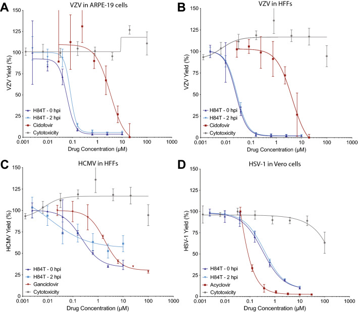
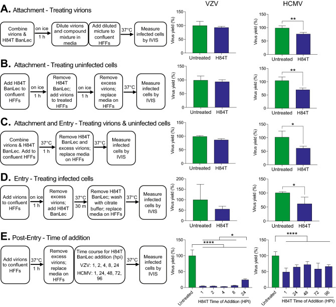
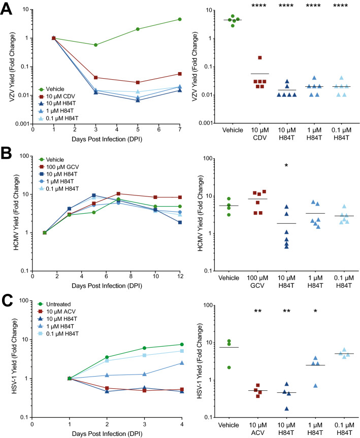
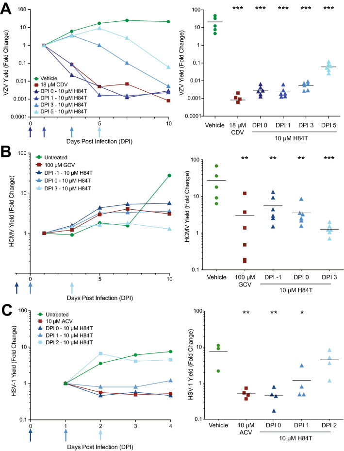
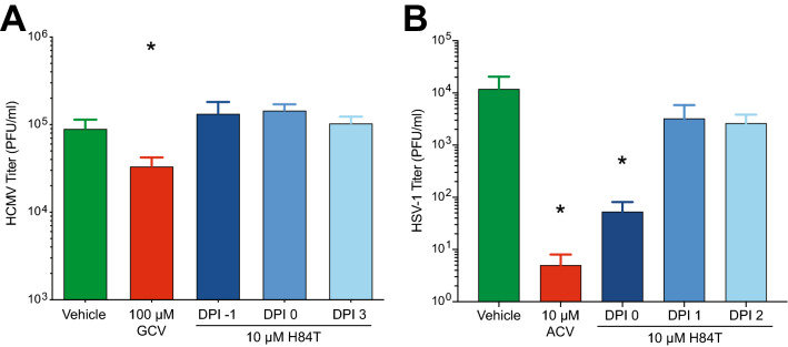
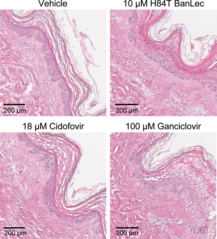
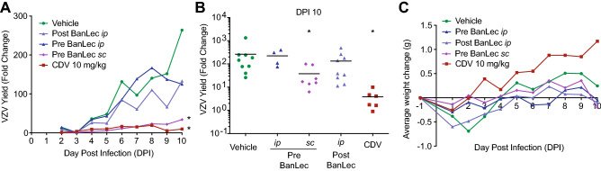
H84T BanLec is a molecularly engineered lectin cloned from bananas with broad-spectrum antiviral activity against several RNA viruses. H84T BanLec dimers bind glycoproteins containing high-mannose N-glycans on the virion envelope, blocking attachment, entry, uncoating, and spread. It was unknown whether H84T BanLec is effective against human herpesviruses varicella-zoster virus (VZV), human cytomegalovirus (HCMV), and herpes simplex virus 1 (HSV-1), which express high-mannose N-linked glycoproteins on their envelopes. We evaluated H84T BanLec against VZV-ORF57-Luc, TB40/E HCMV-fLuc-eGFP, and HSV-1 R8411 in cells, skin organ culture, and mice. The H84T BanLec EC50 was 0.025 µM for VZV (SI50 = 4000) in human foreskin fibroblasts (HFFs), 0.23 µM for HCMV (SI50 = 441) in HFFs, and 0.33 µM for HSV-1 (SI50 = 308) in Vero cells. Human skin was obtained from reduction mammoplasties and prepared for culture. Skin was infected and cultured up to 14 days. H84T BanLec prevented VZV, HCMV and HSV-1 spread in skin at 10 µM in the culture medium, and also exhibited dose-dependent antiviral effects. Additionally, H84T BanLec arrested virus spread when treatment was delayed. Histopathology of HCMV-infected skin showed no overt toxicity when H84T BanLec was present in the media. In athymic nude mice with human skin xenografts (NuSkin mice), H84T BanLec reduced VZV spread when administered subcutaneously prior to intraxenograft virus inoculation. This is the first demonstration of H84T BanLec effectiveness against DNA viruses. H84T BanLec may have additional unexplored activity against other, clinically relevant, glycosylated viruses.


Conflict of interest statement

The authors declare no competing interests.

Figures

Figure 1
Figure 1
H84T BanLec potency against VZV, HCMV, and HSV-1 in cultured cells. Cell-free viruses were added to cell monolayers at 37 °C, and H84T BanLec or positive control antiviral compounds were added at 0 h post-inoculation (HPI) or 2 HPI. The antiviral compounds remained in the culture medium until virus yield was measured by bioluminescence imaging. ARPE-19 cells or HFF cells were infected with VZV for 3 days (A, B), HFF cells were infected with HCMV for 7 days (C), and Vero cells were infected with HSV-1 for 2 days (D). Virus yield was calculated from the average Total Flux of untreated wells divided by the average Total Flux of the treated wells. H84T BanLec cytotoxicity was measured using a neutral red dye uptake assay in Vero cells treated for 48 h (D) and HFFs or ARPE-19 cells treated for 72 h (AC). Each point represents the mean ± SD. N = 3–6 biological replicates.
Figure 2
Figure 2
Effects of H84T BanLec on extracellular VZV and HCMV virions and on infected cells. Each experimental condition tests a different aspect of H84T BanLec’s mechanism of action including attachment (A, B), attachment and entry (C), entry (D), and post-entry steps (E). Cell-free virions and H84T BanLec were added to HFF cell monolayers based on the experimental design for each condition. Cell-free VZV was prepared from a fresh culture of infected HFF cells; the MOI was approximately 0.01. HCMV was prepared from a frozen, titered stock; the MOI was 0.05. H84T BanLec was used at a concentration of 100 nM for VZV and 2 µM for HCMV. (A) Virions were mixed with H84T BanLec on ice for 1 h, and were then diluted 1:50 for VZV and 1:1000 for HCMV prior to adding to HFFs. (AD) Infected cells were measured by bioluminescence imaging 24 h post-infection for VZV, and 7 days post-infection for HCMV. (E) Infected cells were measured by bioluminescence imaging 48 h post-infection for VZV, and 7 days post-infection for HCMV. Virus yield (%) was calculated from the average Total Flux of untreated wells divided by the Total Flux of each well. Bars and error bars represent the mean + SD. N = 4 biological replicates. Asterisks indicate significance [*p < 0.05, **p < 0.01, ****p < 0.0001, p < 0.05, Student’s t-test (AD) or one-way ANOVA with Dunnett’s post hoc test (E)].
Figure 3
Figure 3
H84T BanLec evaluation in skin organ culture. Adult skin explants were inoculated with VZV (A), HCMV (B), or HSV-1 (C) and then placed on NetWells over medium that contained H84T BanLec or positive control antiviral compounds (cidofovir, CDV, 10 µM; ganciclovir, GCV, 100 µM; acyclovir, ACV, 10 µM). Virus spread was measured by bioluminescence imaging starting on 1 DPI. Virus yield on subsequent days was calculated by dividing the average Total Flux (photons/sec/cm2/steradian) by the average Total Flux on 1 DPI. Viral growth kinetics (left panels, each symbol is the average for the group) were analyzed for statistical significance on the last day (right panels), where each symbol represents one piece of skin and the bar is the mean of the group. Asterisks indicate significance between the treated groups and vehicle, [*p < 0.05, **p < 0.01, ****p < 0.0001, p < 0.05, one-way ANOVA, Dunnett’s (A, C) or Dunn’s (B) post hoc test]. N = 4–6 biological replicates.
Figure 4
Figure 4
H84T BanLec addition after infection prevented VZV, HCMV, and HSV-1 spread in skin. Adult skin explants were inoculated with VZV (A), HCMV (B), or HSV-1 (C) and then placed on NetWells over medium that contained H84T BanLec (10 µM) added according to the schedule on the x-axis (blue arrows). Control antiviral compounds (cidofovir, CDV, 18 µM; ganciclovir, GCV, 100 µM; acyclovir, ACV, 10 µM) were added at DPI 0. Virus spread was measured by bioluminescence imaging starting on DPI 1. Virus yield on subsequent days was calculated by dividing the average Total Flux (photons/sec/cm2/steradian) by the average Total Flux on DPI 1. Viral growth kinetics (left panels, each symbol is the average for the group) were analyzed for statistical significance on the last day (right panels), where each symbol represents one piece of skin and the bar is the mean of the group. The Vehicle and ACV groups in (C) are the same as in Fig. 3C, as these experiments were conducted together. Asterisks indicate significance between the treated groups and vehicle (*p < 0.05, **p < 0.01, ***p < 0.001). p < 0.01, one-way ANOVA, Dunnett’s post hoc test. N = 4–6 biological replicates.
Figure 5
Figure 5
H84T BanLec reduces the infectivity of HSV-1 but not HCMV in skin organ culture. HCMV and HSV-1 infected skin pieces from the assay shown in Fig. 4 were collected on the last day and processed for virus titration. (A) HCMV was measured by TCID50 assay in HFF cells (*p < 0.05, one-way ANOVA with Dunnett’s post hoc test). (B) HSV-1 was measured by plaque assay in Vero cells with a methyl cellulose overlay. (*p < 0.05, one-way ANOVA with Dunnett’s post hoc test). Bars and error bars represent the mean + SEM. N = 3–6 infected skin pieces per group.
Figure 6
Figure 6
Effects of H84T BanLec on adult skin. Skin was infected with HCMV, cultured on Netwells, and treated every other day with compounds in the medium. Skin was collected 14 DPI and fixed in 4% paraformaldehyde for H&E staining. The epidermis faces the right side of the image (appears purple) and the dermis is to the left (appears pink). The epidermis appears normal, and the dermal–epidermal junction is intact (HCMV replicates in dermal cells). Images are representative of 2–3 sections per tissue sample from a single tissue donor.
Figure 7
Figure 7
H84T BanLec evaluation in NuSkin mice infected with VZV. NuSkin mice were implanted with a single subcutaneous xenograft of adult human skin and xenografts were inoculated 3–4 weeks later with VZV by intraxenograft injection and scarification. Mice were treated with 50 mg/kg/day H84T BanLec by subcutaneous (sc) or intraperitoneal (ip) routes given 6 h pre-inoculation (−6 HPI) and on DPI 4 and 8 to the Pre-BanLec groups, or by the ip route on DPI 3 and 7 to the Post-BanLec group. Cidofovir was administered daily by ip injection from DPI 3–9. (A) VZV yield was measured by bioluminescence and the fold change calculated as the Total Flux on each day divided by the Total Flux on DPI 2 or 3 (the lowest value for that mouse). Lines represent the average VZV yield per group. (B) VZV yield on DPI 10 was analyzed for statistical significance where each symbol represents one mouse, and the bar is the mean of the group. (*) Signifies statistical significance between the vehicle and a treatment group (p < 0.05, one-way ANOVA, Dunnett’s post hoc test). (C) Mouse weights were measured daily, and the lines show the average weight change for each group from the onset to conclusion of the study. N = 4–12 mice per group.

Similar articles

Cited by

References

    1. Payne S. Chapter 34: Family Herpesviridae. Elsevier Science; 2017.
    1. Lan K, Luo MH. Herpesviruses: epidemiology, pathogenesis, and interventions. Virol. Sin. 2017;32:347–348. doi: 10.1007/s12250-017-4108-2. - DOI - PMC - PubMed
    1. Anonymous. Prevention and control of herpesvirus diseases. Part 1. Clinical and laboratory diagnosis and chemotherapy. A WHO meeting. Bull. World Health Org. 1985;63:185–201. - PMC - PubMed
    1. McQuillan, G., Kruszon-Moran, D., Flagg, E. W., Paulose-Ram, R. Prevalence of herpes simplex virus type 1 and type 2 in persons aged 14–49: United States, 2015–2016. NCHS Data Brief 1–8 (2018). - PubMed
    1. Lopez AS, Zhang J, Marin M. Epidemiology of varicella during the 2-dose varicella vaccination program—United States, 2005–2014. MMWR Morb. Mortal. Wkly. Rep. 2016;65:902–905. doi: 10.15585/mmwr.mm6534a4. - DOI - PubMed

Publication types

MeSH terms