. 2022 Jan 14:12:783213.
doi: 10.3389/fphar.2021.783213. eCollection 2021.

MicroRNA-92a-3p Enhances Cisplatin Resistance by Regulating Krüppel-Like Factor 4-Mediated Cell Apoptosis and Epithelial-to-Mesenchymal Transition in Cervical Cancer

Affiliations

MicroRNA-92a-3p Enhances Cisplatin Resistance by Regulating Krüppel-Like Factor 4-Mediated Cell Apoptosis and Epithelial-to-Mesenchymal Transition in Cervical Cancer

Jing Yang et al. Front Pharmacol. .

Abstract

Recent studies have confirmed the existence and key roles of microRNA (miRNAs) in cancer drug resistance, including cervical cancer (CC). The present study aims to establish a novel role for miR-92a-3p and its associated gene networks in cisplatin (DDP) resistance of CC. First, the disparities in miRNA expression between CC tissues and adjacent normal tissues were screened based on GSE19611 microarray data that retrieved from Gene Expression Omnibus (GEO), and we identified several miRs that were significantly downregulated or upregulated in CC tissues including miR-92a-3p. Moreover, miR-92a-3p was significantly up-regulated in DDP-resistant cells and was the most differently expressed miRNA. Functionally, knockdown of miR-92a-3p increased the sensitivity of DDP-resistant cells to DDP via inhibiting cell proliferation, migration and invasion, and promoting apoptosis. Conversely, overexpression of miR-92a-3p significantly induced DDP resistance in CC parental cells including HeLa and SiHa cells. Moreover, Krüppel-like factor 4 (KLF4) was identified as a direct target of miR-92a-3p, and an obvious inverse correlation was observed between the expression of miR-92a-3p and KLF4 in 40 pairs of cancer tissues. Furthermore, KLF4 knockdown reversed the promoting effect of miR-92a-3p inhibition on DDP sensitivity in DDP-resistant CC cells. Besides, high expression of miR-92a-3p was associated with DDP resistance, as well as a short overall survival in clinic. Taken together, these findings provide important evidence that miR-92a-3p targets KLF4 and is significant in DDP resistance in CC, indicating that miR-92a-3p may be an attractive target to increase DDP sensitivity in clinical CC treatment.

Keywords: KLF4; apoptosis; cervical cancer; cisplatin resistance; miR-92a-3p.


Conflict of interest statement

The authors declare that the research was conducted in the absence of any commercial or financial relationships that could be construed as a potential conflict of interest.

Figures

FIGURE 1
FIGURE 1
The expression of miRNAs in cisplatin-sensitive and -resistant cervical cancer cells. Effects of cisplatin at different concentrations on cell viability were analyzed using CCK-8 assay in SiHa and SiHa/DDP (A), and in HeLa and HeLa/DDP (B). (C) Differentially expressed miRNAs were analyzed between cervical cancer tissues and the adjacent normal tissues group. Data were retrieved from Gene Expression Omnibus (GEO) dataset, with the accession number GSE19611. The color code in the heat map is linear and the expression levels of miRNAs that were upregulated are shown in green to red, whereas the miRNAs that were downregulated are shown from red to green. (D, E) miR-92a-3p, miR-221-3p, miR-21, miR-146a, miR-196a, miR-383, miR-206 and miR-497 were further analyzed using qRT-PCR in SiHa/DDP and HeLa/DDP cells. Data are presented as means ± SD of three individual experiments. *p < 0.05, **p < 0.01 vs. SiHa or HeLa cells.
FIGURE 2
FIGURE 2
Knockdown of miR-92a-3p increased the sensitivity of DDP resistant cells to DDP. DDP resistant cells (HeLa/DDP and SiHa/DDP) were treated with miR-92a-3p inhibitor or inhibitor NC for 24 h, followed by treatment with 10 μM DDP for 48 h. (A) miR-92a-3p expression was detected by qRT-PCR. (B–D) Effects of 10 μM DDP on cell viability were analyzed using CCK-8 assay in miR-92a-3p inhibitor transfected SiHa/DDP and HeLa/DDP cells. (E) The activity of caspase 3 was measured by a Caspase-3 Activity kit. (F, G) The apoptosis rates were analyzed by Flow Cytometry in HeLa/DDP and SiHa/DDP cells. (H) The protein levels of cleaved caspase 3 was detected by IFA. Data are presented as means ± SD of three individual experiments. **p < 0.01 vs. Inhibitor NC group; ##p < 0.01 vs. Inhibitor NC + DDP group.
FIGURE 3
FIGURE 3
Overexpression of miR-92a-3p induced DDP resistance of cervical cancer cells. Parental HeLa and SiHa cells were treated with miR-92a-3p mimics or mimics NC for 24 h, followed by the treatment with 2 μM DDP for 48 h. (A) miR-92a-3p expression was detected by qRT-PCR. (B–D) Effects of 2 μM DDP on cell viability was analyzed using CCK-8 assay in miR-92a-3p mimics transfected SiHa and HeLa cells. (E) The activity of caspase 3 was measured by a Caspase-3 Activity kit. (F, G) The apoptosis rates were analyzed by Flow Cytometry. (H) The protein levels of cleaved caspase 3 was detected by IFA. Data are presented as means ± SD of three individual experiments. *p < 0.05, **p < 0.01 vs. mimics NC group; ##p < 0.01 vs. mimics NC + DDP group.
FIGURE 4
FIGURE 4
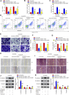
The effects of DDP on the migration, invasion and EMT process in miR-92a-3p inhibitor transfected DDP-resistant cervical cancer cells. DDP resistant cells (HeLa/DDP and SiHa/DDP) were treated with miR-92a-3p inhibitor or inhibitor NC for 24 h, followed by treatment with 10 μM DDP for 48 h. (A, B) Effects of 10 μM DDP on cell invasion was analyzed using Transwell assay in miR-92a-3p inhibitor transfected SiHa/DDP and HeLa/DDP cells. (C, D, E) Effects of 10 μM DDP on the migration was detected by wound healing assay in miR-92a-3p inhibitor transfected HeLa/DDP and SiHa/DDP cells. (F, G) The epithelial protein (E-cadherin) and mesenchymal proteins (Vimentin and N-cadherin) were measured by western blot assay. Data are presented as means ± SD of three individual experiments. **p < 0.01 vs. Inhibitor NC group; ##p < 0.01 vs. Inhibitor NC + DDP group.
FIGURE 5
FIGURE 5
Overexpression of miR-92a-3p promoted DDP resistance in CC parental cells. Parental HeLa and SiHa cells were treated with miR-92a-3p mimics or mimics NC for 24 h, followed by the treatment with 2 μM DDP for 48 h. (A, B) Effects of 2 μM DDP on cell invasion was analyzed using Transwell assay in miR-92a-3p mimics transfected SiHa and HeLa cells. (C, D, E) Effects of 2 μM DDP on the migration was detected by wound healing assay in miR-92a-3p mimics transfected HeLa and SiHa cells. (F, G) The epithelial protein (E-cadherin) and mesenchymal proteins (Vimentin and N-cadherin) were measured by western blot assay. Data are presented as means ± SD of three individual experiments. **p < 0.01 vs. mimics NC group; ##p < 0.01 vs. mimics NC + DDP group.
FIGURE 6
FIGURE 6
KLF4 was a direct target of miR-92a-3p. (A) miR-92a-3p sequence is shown to be highly conserved among species. (B) The predicted complementary sequences for miR-92a-3p in the 3′UTR of KLF4 and the mutations are shown in the seed region of miR-92a-3p. (C, D) The SiHa and HeLa cells were co-transfected with either pGLO-KLF4-3ʹ-UTR or pGLO-KLF4-mut-3ʹ-UTR, and miR-92a-3p mimics or corresponding NC and the relative luciferase activity were measured. **p < 0.01 vs mimics NC. (E, F) The SiHa and HeLa cells were transfected with miR-92a-3p mimics/inhibitor or corresponding NC, and the KLF4 protein level was measured using western blot analysis. (G) The KLF4 protein level was measured in SiHa/DDP and HeLa/DDP cells.
FIGURE 7
FIGURE 7
KLF4 is responsible for miR-92a-3p-mediated DDP resistance. HeLa/DDP and SiHa/DDP cells were co-transfected with miR-92a-3p inhibitor and si-KLF4, followed by the treatment with 10 μM DDP for 48 h. (A) Effects of 10 μM DDP on cell viability was analyzed using CCK-8 assay in miR-92a-3p inhibitor and si-KLF4 cotransfected SiHa/DDP and HeLa/DDP cells. (B) The activity of caspase 3 was measured by a Caspase-3 Activity kit. (C, D) The apoptosis rates were analyzed by Flow Cytometry. (E, F) Effects of 10 μM DDP on cell invasion was analyzed using Transwell assay in miR-92a-3p inhibitor and si-KLF4 cotransfected SiHa/DDP and HeLa/DDP cells. (G–I) Effects of 10 μM DDP on the migration was detected by wound healing assay in miR-92a-3p inhibitor and si-KLF4 cotransfected SiHa/DDP and HeLa/DDP cells. (J, K) E-cadherin, Vimentin and N-cadherin protein expression levels were measured by western blot assay in miR-92a-3p inhibitor and si-KLF4 cotransfected SiHa/DDP and HeLa/DDP cells. Data are presented as means ± SD of three individual experiments. *p < 0.05, **p < 0.01 vs. si-Scramble group; ##p < 0.01 vs. miR-92a-3p inhibitor + DDP group.
FIGURE 8
FIGURE 8
High miR-92a-3p expression is associated with poor prognosis of cervical cancer patients receiving DDP therapy. (A) miR-92a-3p levels in 40 patients (19 DDP-sensitive and 21 DDP-resistant cervical cancer tissues) were analyzed using qRT-PCR. **p < 0.01. vs. Adjacent normal tissues, ##p < 0.01. vs. Chemosensitive CC tissues. (B) Cervical cancer patients were separated into groups based on low or high miR-92a-3p expression. Kaplan-Meier survival curves were used to compare the overall survival rate between the two groups. **p < 0.01. (C) KLF4 levels in 40 patients who responded to DDP (n = 19) and who did not respond (n = 21) to DDP were analyzed using qRT-PCR. **p < 0.01. vs. Adjacent normal tissues, ##p < 0.01. vs. Chemosensitive CC tissues. (D) Cervical cancer patients were separated into groups based on low or high KLF4 expression. Kaplan-Meier survival curves were used to compare the overall survival rate between the two groups. **p < 0.01. (E) The correlation between miR-92a-3p and KLF4 was analyzed in cervical cancer tissues. p < 0.01.

Similar articles

Cited by

References

    1. Amable L. (2016). Cisplatin Resistance and Opportunities for Precision Medicine. Pharmacol. Res. 106, 27–36. 10.1016/j.phrs.2016.01.001 - DOI - PubMed
    1. Bartel D. P. (2009). MicroRNAs: Target Recognition and Regulatory Functions. Cell 136 (2), 215–233. 10.1016/j.cell.2009.01.002 - DOI - PMC - PubMed
    1. Cao W., Yang W., Fan R., Li H., Jiang J., Geng M., et al. (2014). miR-34a Regulates Cisplatin-Induce Gastric Cancer Cell Death by Modulating PI3K/AKT/survivin Pathway. Tumour Biol. 35 (2), 1287–1295. 10.1007/s13277-013-1171-7 - DOI - PubMed
    1. Chatterjee K., Mukherjee S., Vanmanen J., Banerjee P., Fata J. E. (2019). Dietary Polyphenols, Resveratrol and Pterostilbene Exhibit Antitumor Activity on an HPV E6-Positive Cervical Cancer Model: An In Vitro and In Vivo Analysis. Front. Oncol. 9, 352. 10.3389/fonc.2019.00352 - DOI - PMC - PubMed
    1. Chen C., Ma Z., Zhang H., Liu X., Yu Z. (2017). Krüppel-Like Factor 4 Enhances Sensitivity of Cisplatin to Esophageal Squamous Cell Carcinoma (ESCC) Cells. Med. Sci. Monit. 23, 3353–3359. 10.12659/msm.902583 - DOI - PMC - PubMed

LinkOut - more resources