. 2022 Apr 1;322(4):H549-H567.
doi: 10.1152/ajpheart.00460.2021. Epub 2022 Jan 28.

Electronic cigarette exposure causes vascular endothelial dysfunction due to NADPH oxidase activation and eNOS uncoupling

Affiliations

Electronic cigarette exposure causes vascular endothelial dysfunction due to NADPH oxidase activation and eNOS uncoupling

Mohamed A El-Mahdy et al. Am J Physiol Heart Circ Physiol. .

Abstract

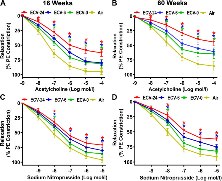
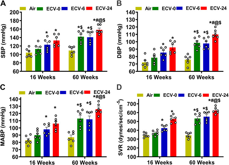
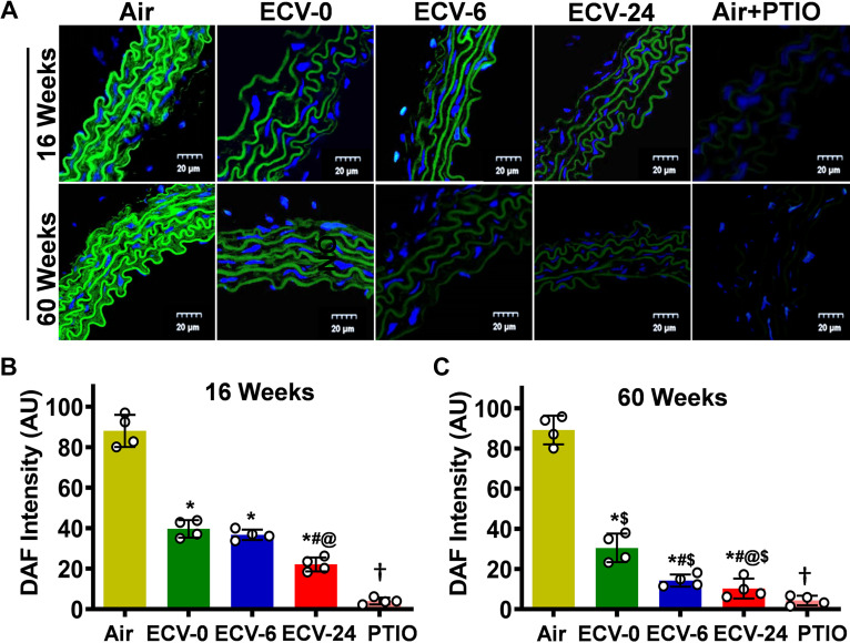
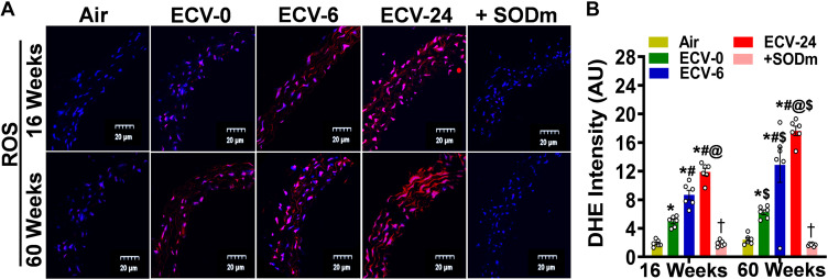
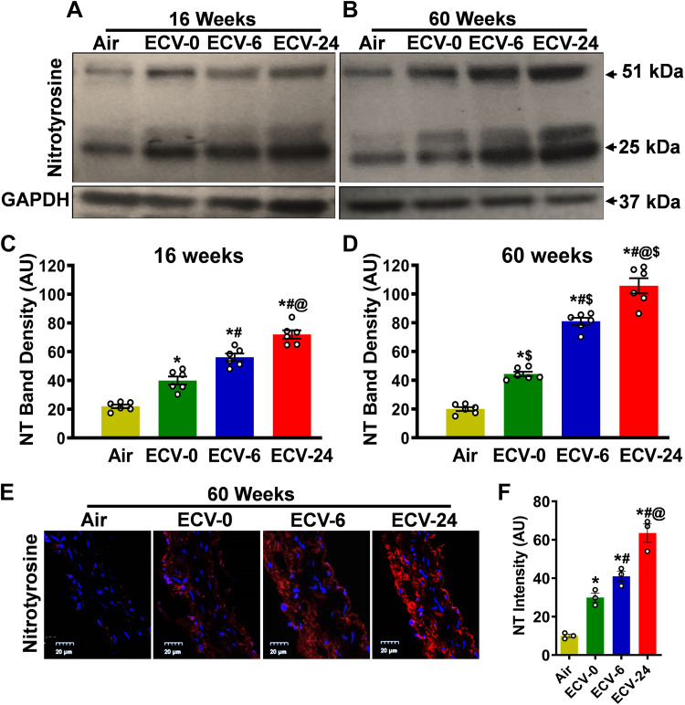
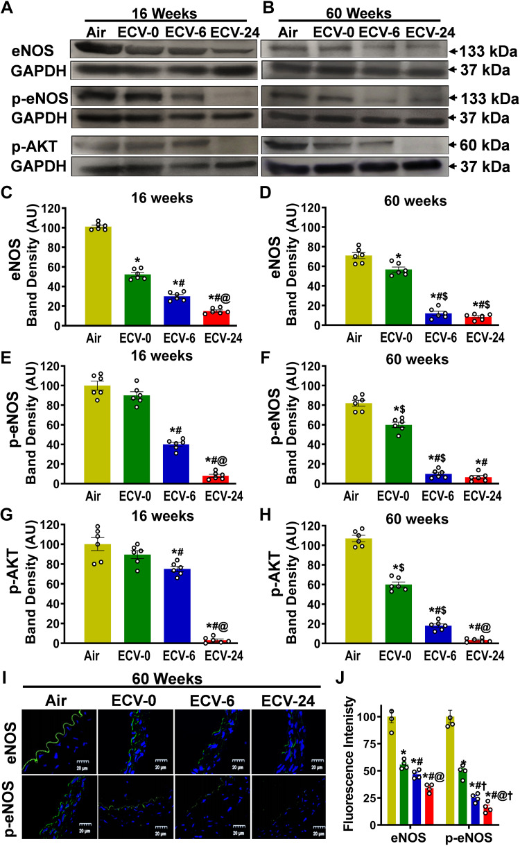
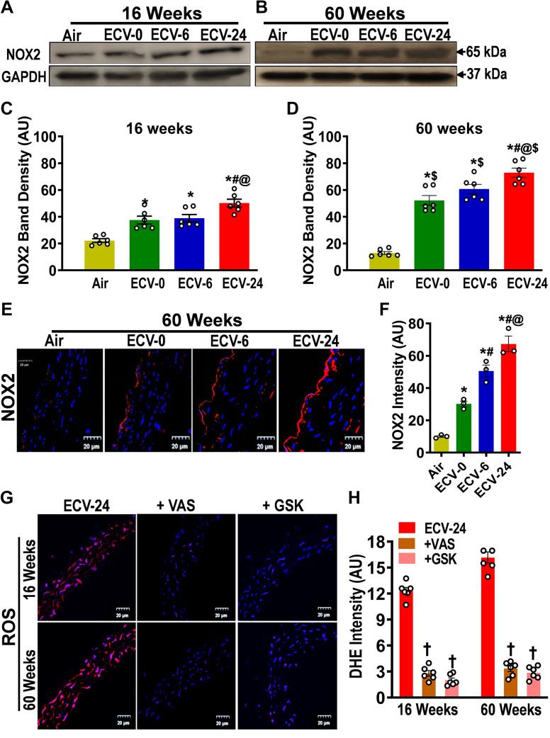
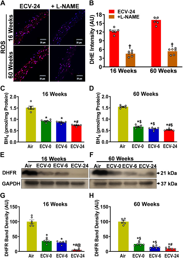
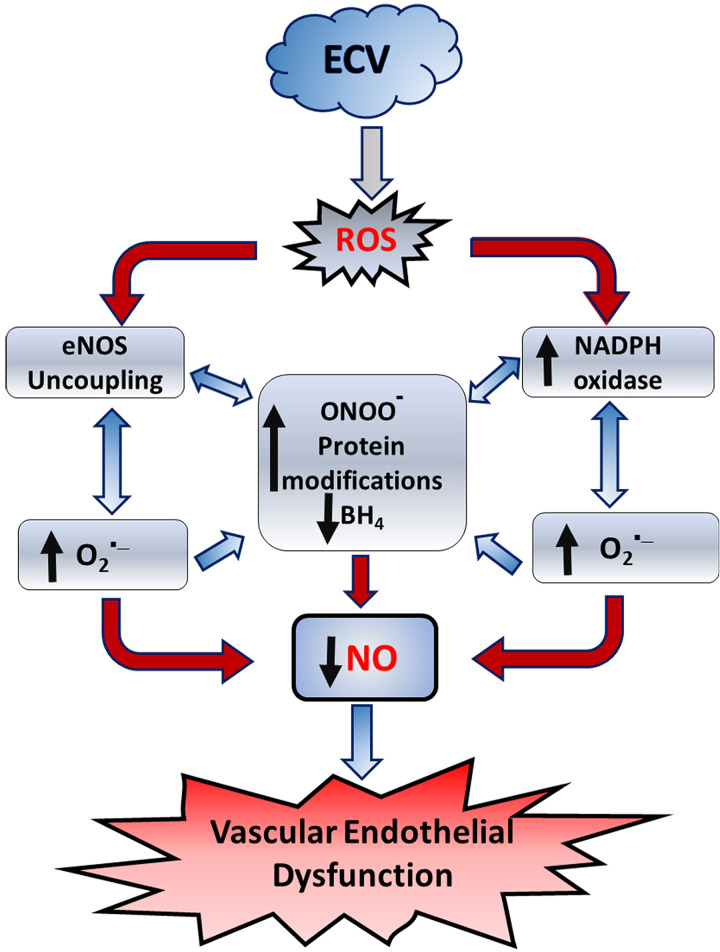
We recently reported a mouse model of chronic electronic cigarette (e-cig) exposure-induced cardiovascular pathology, where long-term exposure to e-cig vape (ECV) induces cardiac abnormalities, impairment of endothelial function, and systemic hypertension. Here, we delineate the underlying mechanisms of ECV-induced vascular endothelial dysfunction (VED), a central trigger of cardiovascular disease. C57/BL6 male mice were exposed to ECV generated from e-cig liquid containing 0, 6, or 24 mg/mL nicotine for 16 and 60 wk. Time-dependent elevation in blood pressure and systemic vascular resistance were observed, along with an impairment of acetylcholine-induced aortic relaxation in ECV-exposed mice, compared with air-exposed control. Decreased intravascular nitric oxide (NO) levels and increased superoxide generation with elevated 3-nitrotyrosine levels in the aorta of ECV-exposed mice were observed, indicating that ECV-induced superoxide reacts with NO to generate cytotoxic peroxynitrite. Exposure increased NADPH oxidase expression, supporting its role in ECV-induced superoxide generation. Downregulation of endothelial nitric oxide synthase (eNOS) expression and Akt-dependent eNOS phosphorylation occurred in the aorta of ECV-exposed mice, indicating that exposure inhibited de novo NO synthesis. Following ECV exposure, the critical NOS cofactor tetrahydrobiopterin was decreased, with a concomitant loss of its salvage enzyme, dihydrofolate reductase. NADPH oxidase and NOS inhibitors abrogated ECV-induced superoxide generation in the aorta of ECV-exposed mice. Together, our data demonstrate that ECV exposure activates NADPH oxidase and uncouples eNOS, causing a vicious cycle of superoxide generation and vascular oxidant stress that triggers VED and hypertension with predisposition to other cardiovascular disease.NEW & NOTEWORTHY Underlying mechanisms of e-cig-induced vascular endothelial dysfunction are delineated. e-cig exposure activates and increases expression of NADPH oxidase and disrupts activation and coupling of eNOS, leading to a vicious cycle of superoxide generation and peroxynitrite formation, with tetrahydrobiopterin depletion, causing loss of NO that triggers vascular endothelial dysfunction. This process is progressive, increasing with the duration of e-cig exposure, and is more severe in the presence of nicotine, but observed even with nicotine-free vaping.

Keywords: NADPH oxidase; e-cigarettes; eNOS uncoupling; tetrahydrobiopterin; vascular endothelial dysfunction.


Conflict of interest statement

No conflicts of interest, financial or otherwise, are declared by the authors.

Figures

Figure 1.
Figure 1.
Endothelium-dependent and -independent vascular relaxation. Male C57BL/6J mice were exposed to 16 or 60 wk of either air or electronic cigarette (e-cig) vape generated from e-cig liquid containing nicotine (NIC) 0 mg/mL (ECV-0), 6 mg/mL (ECV-6), or 24 mg/mL (ECV-24). For endothelium-dependent function, aortic rings from 16-wk-exposed (A) and 60-wk-exposed (B) mice were mounted in a wire myograph, constricted by 1 µM phenylephrine (PE), and acetylcholine response curve was derived. For endothelium-independent function, aortic rings from 16-wk-exposed (C) and 60-wk-exposed (D) mice were constricted by 1 µM PE, and sodium nitroprusside response curve was derived. Data are presented as means ± SE of 6 mice. Analysis was done using repeated-measures two-way ANOVA. The differences were considered statistically significant at P ≤ 0.05. *Significant difference from relative air-exposed controls.
Figure 2.
Figure 2.
Blood pressure and vascular resistance. Male C57BL/6J mice were exposed to 16 or 60 wk of either air or electronic cigarette (e-cig) vape generated from e-cig liquid containing nicotine (NIC) 0 mg/mL (ECV-0), 6 mg/mL (ECV-6), or 24 mg/mL (ECV-24). A: systolic blood pressure (SBP). B: diastolic blood pressure (DBP). C: mean arterial blood pressure (MABP). D: systemic vascular resistance (SVR). ECV exposure resulted in a NIC-and an exposure time-dependent vascular endothelial dysfunction with elevated MABP and SVR. Data are presented as means ± SE of 6 mice. Each point represents the average of at least 10 measurements per animal. Analysis was done using two-way ANOVA followed by Bonferroni multiple-comparisons test. The differences were considered statistically significant at P ≤ 0.05. *Significant from air-exposed controls at P < 0.05; #significant from ECV-0 at P < 0.05; @significant from ECV-6 at P < 0.05; $significant from the same exposure at 16 wk at P < 0.05.
Figure 3.
Figure 3.
Nitric oxide (NO) bioavailability in the aorta. Aortic sections were studied from mice exposed to 16 and 60 wk of either air or electronic cigarette (e-cig) vape generated from e-cig liquid containing nicotine (NIC) 0 mg/mL (ECV-0), 6 mg/mL (ECV-6), or 24 mg/mL (ECV-24). A: sections were treated with the fluorescent nitric oxide (NO) probe diaminofluorescein-FM (DAF; green) and the nuclear fluorescent stain diamidino-2-phenylindole (DAPI; blue), with (air-exposed) or without the NO trap 2-(4-carboxyphenyl)-4,4,5,5-tetramethylimidazoline-1-oxyl-3-oxide (PTIO treated), and visualized by confocal fluorescence microscopy. A 20-µm scale bar is shown toward the lower right corner of each figure. B and C: quantitation of the green fluorescence in A. ECV exposure decreased NO levels in the aorta in an exposure time- and a NIC-dependent manner. Data show the means ± SE of aortic sections from 4 mice. Analysis was done using two-way ANOVA followed by Bonferroni multiple-comparisons test. The differences were considered statistically significant at P ≤ 0.05. *Significant from air-exposed controls at P < 0.05; #significant from ECV-0 at P < 0.05; @significant from ECV-6 at P < 0.05; $significant from the same exposure at 16 wk at P < 0.05; †significant difference from ECV-24 in the absence of PTIO at P < 0.05.
Figure 4.
Figure 4.
Superoxide radical generation in aorta. Aortic sections from mice exposed to 16 and 60 wk of either air or electronic cigarette (e-cig) vape generated from e-cig liquid containing nicotine (NIC) 0 mg/mL (ECV-0), 6 mg/mL (ECV-6), or 24 mg/mL (ECV-24) were studied. A: incubated with the superoxide probe dihydroethidium (DHE; red) and the nuclear fluorescent stain diamidino-2-phenylindole (DAPI; blue), with or without the superoxide dismutase mimetic Mn(III)tetrakis(4-benzoic acid)porphyrin chloride (+SODm), and imaged with confocal fluoroscopy. B: quantitation of the red fluorescence in A, showing that ECV exposure induced superoxide generation that was quenched by the SODm. Thus, ECV exposure results in vascular superoxide production in an exposure time- and a NIC-dependent manner. Data represent means ± SE of aortic sections from 6 mice. Analysis was done using two-way ANOVA followed by Bonferroni multiple-comparisons test. The differences were considered statistically significant at P ≤ 0.05. *Significant from air-exposed controls at P < 0.05; #significant from ECV-0 at P < 0.05; @significant from ECV-6 at P < 0.05; $significant from the same exposure at 16 wk at P < 0.05; †significant from ECV-24 in the absence of the SODm at P < 0.05. ROS, reactive oxygen species.
Figure 5.
Figure 5.
Nitrotyrosine formation in aorta. Nitrotyrosine (NT) levels were measured by Western blotting in aortic homogenates (AD) and by immunofluorescence in aortic sections (E and F) from mice exposed to 16 or 60 wk of either air or electronic cigarette (e-cig) vape generated from e-cig liquid containing nicotine (NIC) 0 mg/mL (ECV-0), 6 mg/mL (ECV-6), or 24 mg/mL (ECV-24). A and B: immunoblots of NT levels. C and D: quantitation of NT bands in A and B, respectively, showing increased NT in a time- and NIC-dependent manner. E: aortic sections were incubated with primary antibody against NT followed by corresponding fluorescent labeled secondary antibody (red). The fluorescent dye diamidino-2-phenylindole (DAPI; blue) was used as a nuclear stain. F: quantitation of red fluorescence in E, showing similar effects as in C and D. These data indicate that ECV-generated superoxide scavenges nitric oxide (NO) producing cytotoxic peroxynitrite, with loss of NO bioavailability and ECV-induced VED. For C and D, data are presented as means ± SE with measurements performed in 6 mice. In F, each point is derived from measurements in 3 mice. Analysis was done using two-way ANOVA followed by Bonferroni multiple-comparisons test. The differences were considered statistically significant at P ≤ 0.05. *Significant from air-exposed controls at P < 0.05; #significant from ECV-0 at P < 0.05; @significant from ECV-6 at P < 0.05; $significant from the same exposure at 16 wk at P < 0.05.
Figure 6.
Figure 6.
Endothelial nitric oxide (NO) synthase expression and phosphorylation in aorta. A and B: Western blotting was used to measure the expression and phosphorylation of endothelial nitric oxide synthase (eNOS) and phosphorylation of AKT (p-AKT) in the aorta from mice exposed to 16 and 60 wk of either air or electronic cigarette (e-cig) vape generated from liquid containing nicotine (NIC) 0 mg/mL (ECV-0), 6 mg/mL (ECV-6), or 24 mg/mL (ECV-24). C and D: quantitation of eNOS bands in A and B, respectively, showing downregulation of e-NOS in an exposure time- and NIC-dependent manner. E and F: quantitation of p-eNOS bands in A and B, respectively, showing similar decreases as in C and D. G and H: quantitation of p-AKT bands in A and B, respectively, also exhibiting similar decreases. I: frozen aortic sections were incubated with primary antibody against eNOS and p-eNOS followed by corresponding secondary fluorescence antibody (green) and the nuclear stain DAPI (blue). J: quantitation of green fluorescence of eNOS and p-eNOS. ECV exposure downregulated eNOS expression and decreased Akt-dependent eNOS phosphorylation, contributing to the decline of NO synthesis and onset of VED. Data are presented as means ± SE of 6 experiments. For CH, data are presented as means ± SE of independent experiments in 6 mice, whereas for J, each point is based on independent experiments in 4 mice. Analysis was done using two-way ANOVA followed by Bonferroni multiple-comparisons test. The differences were considered statistically significant at P ≤ 0.05. *Significant from air-exposed controls at P < 0.05; #significant from ECV-0 at P < 0.05; @significant from ECV-6 at P < 0.05; $significant from the same exposure at 16 wk at P < 0.05. †significant different from eNOS expression at P < 0.05.
Figure 7.
Figure 7.
Expression of NADPH oxidase 2 in the aorta. NADPH oxidase 2 (NOX2) levels were measured by Western blotting (AD) and immunofluorescence (E and F) from mice exposed to 16 and 60 wk of either air or electronic cigarette (e-cig) vape generated from liquid containing nicotine (NIC) 0 mg/mL (ECV-0), 6 mg/mL (ECV-6), or 24 mg/mL (ECV-24). A and B: immunoblots of NOX2. C and D: quantitation of NOX2 band density in A and B, respectively, showing exposure time- and NIC-dependent increases in NOX2 expression. E: aortic sections were incubated with primary antibody against NOX2 followed by corresponding secondary fluorescent tagged antibody (red), with DAPI nuclear stain (blue). F: quantitation of red fluorescence in E, showing a similar response as in D. G: aortic sections from ECV-24-exposed mice were incubated with the superoxide probe dihydroethidium (DHE; red) and DAPI (blue), with or without NADPH oxidase inhibitors 3-benzyl-7-(2-benzoxazolyl)thio-1,2,3-triazolo[4,5-d]pyrimidine (+VAS) or GSK2795039 (+GSK), and visualized by confocal microscopy. H: quantitation of the superoxide-derived red fluorescence in G, showing that NOX2 inhibitors decreased superoxide in ECV-24-exposed aortic sections. Thus, induction of NOX2 in the endothelium is a major source of the ECV-stimulated superoxide seen in all ECV-exposed groups, and its induction increased with exposure duration and NIC content. For C and D, data are presented as means ± SE of 6 mice, whereas for F and H, n = 3 or 6 mice, respectively. Analysis was done using two-way ANOVA followed by Bonferroni multiple-comparisons test. The differences were considered statistically significant at P ≤ 0.05. *Significant from air-exposed controls at P < 0.05; #significant from ECV-0 at P < 0.05; @significant from ECV-6 at P < 0.05; $significant from the same exposure at 16 wk at P < 0.05. †significant different from ECV-24-exposed sections stained with DHE.
Figure 8.
Figure 8.
Nitric oxide synthase uncoupling in the aorta. A: aortic sections from ECV-24-exposed mice were incubated with the superoxide probe dihydroethidium (DHE; red) and DAPI (blue), with or without nitric oxide synthase inhibitor l-NAME. B: quantitation of fluorescence in A, showing that l-NAME decreased the DHE fluorescence in ECV-24-exposed aortic sections. C and D: ELISA quantitation of aortic tetrahydrobiopterin (BH4) following either air or electronic cigarette (e-cig) vape generated from e-cig liquid containing nicotine (NIC) 0 mg/mL (ECV-0), 6 mg/mL (ECV-6), or 24 mg/mL (ECV-24). E and F: immunoblots of DHFR expression. G and H: quantitation of band density in E and F, respectively, showing exposure time- and NIC-dependent downregulation of DHFR. Thus, NOS is a source of ECV-induced superoxide with ECV depleting BH4 and DHFR, leading to eNOS uncoupling. For B, C, D, G, and H, data are presented as means ± SE of values from 6 mice. Analysis was done using two-way ANOVA followed by Bonferroni’s multiple-comparisons test. The differences were considered statistically significant at P ≤ 0.05. *Significant from air-exposed controls at P < 0.05; #significant from ECV-0 at P < 0.05; @significant from ECV-6 at P < 0.05; $significant from the same exposure at 16 wk at P < 0.05. †significant different from ECV-24 sections in the absence of l-NAME. DHFR, dihydrofolate reductase enzyme; enos, endothelial nitric oxide synthase; l-NAME, NG-nitro-l-arginine methyl ester; NOS, nitric oxide synthase.
Figure 9.
Figure 9.
Process by which electronic cigarette vape (ECV) exposure triggers endothelial reactive oxygen species (ROS) generation and vascular endothelial dysfunction. ECV exposure generates ROS that increase the activation and expression of NADPH oxidase and trigger nitric oxide (NO) synthase (eNOS) uncoupling, leading to a vicious cycle of superoxide production and peroxynitrite formation, with protein modifications, and tetrahydrobiopterin (BH4) depletion. Together these cause decreased NO synthesis and bioavailability, leading to endothelial dysfunction.

Similar articles

Cited by

References

    1. Galley HF, Webster NR. Physiology of the endothelium. Br J Anaesth 93: 105–113, 2004. doi:10.1093/bja/aeh163. - DOI - PubMed
    1. Palmer RM, Ferrige AG, Moncada S. Nitric oxide release accounts for the biological activity of endothelium-derived relaxing factor. Nature 327: 524–526, 1987. doi:10.1038/327524a0. - DOI - PubMed
    1. Moncada S, Palmer RMJ, Higgs EA. Nitric oxide: physiology, pathophysiology, and pharmacology. Pharmacol Rev 43: 109–142, 1991. - PubMed
    1. White KA, Marletta MA. Nitric oxide synthase is a cytochrome-P-450 type hemoprotein. Biochemistry 31: 6627–6631, 1992. doi:10.1021/bi00144a001. - DOI - PubMed
    1. Zweier JL, Ilangovan G. Regulation of nitric oxide metabolism and vascular tone by cytoglobin. Antioxid Redox Signal 32: 1172–1187, 2020. doi:10.1089/ars.2019.7881. - DOI - PMC - PubMed

Publication types