Review
. 2022 Jan 11:8:814496.
doi: 10.3389/fmed.2021.814496. eCollection 2021.

Liver Fibrosis-From Mechanisms of Injury to Modulation of Disease

Affiliations
Review

Liver Fibrosis-From Mechanisms of Injury to Modulation of Disease

Christian Liedtke et al. Front Med (Lausanne). .

Abstract

The Transregional Collaborative Research Center "Organ Fibrosis: From Mechanisms of Injury to Modulation of Disease" (referred to as SFB/TRR57) was funded for 13 years (2009-2021) by the German Research Council (DFG). This consortium was hosted by the Medical Schools of the RWTH Aachen University and Bonn University in Germany. The SFB/TRR57 implemented combined basic and clinical research to achieve detailed knowledge in three selected key questions: (i) What are the relevant mechanisms and signal pathways required for initiating organ fibrosis? (ii) Which immunological mechanisms and molecules contribute to organ fibrosis? and (iii) How can organ fibrosis be modulated, e.g., by interventional strategies including imaging and pharmacological approaches? In this review we will summarize the liver-related key findings of this consortium gained within the last 12 years on these three aspects of liver fibrogenesis. We will highlight the role of cell death and cell cycle pathways as well as nutritional and iron-related mechanisms for liver fibrosis initiation. Moreover, we will define and characterize the major immune cell compartments relevant for liver fibrogenesis, and finally point to potential signaling pathways and pharmacological targets that turned out to be suitable to develop novel approaches for improved therapy and diagnosis of liver fibrosis. In summary, this review will provide a comprehensive overview about the knowledge on liver fibrogenesis and its potential therapy gained by the SFB/TRR57 consortium within the last decade. The kidney-related research results obtained by the same consortium are highlighted in an article published back-to-back in Frontiers in Medicine.

Keywords: chemokines; cirrhosis; cytokines; extracellular matrix; hepatic stellate cell; hepatocytes; inflammation; resolution.


Conflict of interest statement

The authors declare that the research was conducted in the absence of any commercial or financial relationships that could be construed as a potential conflict of interest.

Figures

Figure 1
Figure 1
Relevant mechanisms of liver fibrosis initiation reviewed in this article. Four liver-related aspects will be reviewed which aimed to characterize and prevent fibrosis initiation at the level of hepatocyte injury or myofibroblast activation. Hepatocyte injury can be mediated by a variety of mechanisms such as alcohol, NASH, RIPK3-mediated necroptosis, impaired c-Met signaling, iron overload and AAT. In any case, death of hepatocytes triggers activation of HSCs and their transdifferentiation and proliferation of Myofibroblasts. The latter process depends on the cell cycle regulator Cyclin E1 and its interacting kinase Cdk2. It is hypothesized that fibrosis can be halted by inducing Caspase-mediated MFB cell death. AAT, α1-antitrypsin; AAT-d, α1-antitrypsin-deficiency; DAMPs, Danger-associated molecular patterns; Fe, Iron overload; NASH, non-alcoholic steatohepatitis.
Figure 2
Figure 2
Role of cell cycle mediators for liver fibrosis initiation. Initiation of liver fibrogenesis is associated with increased hepatic cell cycle activity in HSCs. Important cell cycle mediators in these processes are c-myc and the Cyclin-dependent kinase 2 (Cdk2) in complex with its regulatory subunit Cyclin E1. In general, c-myc is involved in activation of Cyclin E1/Cdk2. Moreover, over-expression of c-myc in hepatocytes triggers activation of HSCs and is observed in patients and mice with liver fibrosis. Cyclin E1 is indispensable for initiation of liver fibrosis and also essential for proliferation, differentiation and survival of HSCs. Direct targeting of Cyclin E1 is currently feasible by using RNA interference (siRNA) in vivo. Anti-fibrotic effects in HSCs can also be triggered by the use of pharmacological inhibition of Cdk-activity.
Figure 3
Figure 3
Summary of the involvement of the necroptosis-associated proteins RIPK1, RIPK3 and MLKL in obese/NASH patients and in murine NASH models. The necroptosis-associated proteins RIPK1, RIPK3, and MLKL are overexpressed in the liver and/or adipose tissue of NASH patients. Murine NASH studies with distinct knock-in and knock-out mouse models confirmed an essential function of RIPK1, RIPK3, and MLKL in the transition from NAFLD to NASH and NASH-fibrosis, including both protective and inducing mechanisms. While RIPK3 deletion led to a decrease in fibrosis in the MCD model, NASH progression was observed in the HFD and CD-HFD models. Deletion of RIPK1 and MLKL was predominantly protective in the MCD and HFD models.
Figure 4
Figure 4
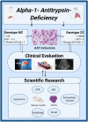
Clinical and scientific insights in alpha1-antitrypsin deficiency. Genotype MZ/ZZ refers to a presence of a heterozygous/homozygous Pi*Z mutation in alpha1-antitrypsin (AAT) gene. The upper boxes describe the frequency of both genotypes in Caucasians, the resulting serum AAT levels and their risk to develop advanced liver fibrosis compared to individuals without AAT mutations. Non-invasive techniques such as liver stiffness measurement by transient elastography or determination of aspartate aminotransferase to platelet ratio index (APRI) are suitable to estimate the amount of histological liver fibrosis. Liver ultrasounds are recommended for individuals with advanced liver fibrosis as a method of HCC surveillance. Several molecular pathways (NF-κB, JNK), protein degradation machineries (UPR, autophagy) as well as epigenetic modifications or appearance of extracellular vesicles with pro-fibrogenic cargo may contribute to disease developments. Created with BioRender.com. iPSCs, induced pluripotent stem cells; JNK, C-Jun N-terminal kinase; NF-κB, nuclear factor 'kappa-light-chain-enhancer' of activated B-cells; UPR, unfolded protein response.
Figure 5
Figure 5
Immunological mechanisms in liver fibrogenesis. Selected potential signaling pathways and proposed cell-cell interactions discussed in this article are shown schematically. Mediators and triggers are depicted in smaller font size as follows: NASH, non-alcoholic steatohepatitis; CXCR7, CXC chemokine receptor type 7; HCV, hepatitis C virus; ILCs, Innate lymphoid cells; NKT, Natural killer T-cells; MIF, Macrophage migration inhibitory factor. In the center of this chapter is the potential crosstalk of monocytes, macrophages, hepatic stellate cells and ILCs, which can be modulated by distinct triggers such as NASH, toxins, inflammasome activation or HCV infection. Crosstalks may eventually lead to modulation of stellate cell activation and subsequent liver fibrogenesis.
Figure 6
Figure 6
Macrophage migration inhibitory factor in chronic liver injury and fibrogenesis. In the liver, the atypical chemokine Macrophage migration inhibitory factor (MIF) is mostly expressed by infiltrating immune cells as well as hepatocytes especially under stress condition e.g., fatty degeneration or inflammation during NASH, ASH and viral hepatitis. In addition, it is most likely that MIF is involved in the pathogenesis of hepatic autoimmune disorders because this cytokine was previously shown to be associated with many autoimmune disorders including rheumatoid arthritis, inflammatory arthritis, inflammatory bowel disease, multiple sclerosis, autoimmune uveitis., autoimmune glomerulonephritis, systemic lupus erythematosus, sarcoidosis and many other autoimmune inflammatory disorder [for review see (85, 100)]. MIF engages high-affinity non-cognate interactions with three different surface receptors expressed by liver resident cells as well as infiltrating cells, the CXC chemokine receptors CXCR2 and CXCR4 as well as with CD74, the membrane form of invariant chain (Ii). While MIF engagement with the CD74 receptor on hepatocytes and hepatic stellate cells exert protective effect via AMPK signaling during chronic liver injury, MIF also mediates pro-inflammatory effects orchestrating the intrahepatic recruitment and activation of inflammatory immune cells via engagement of CXCR2 and CXCR4. The balance between these opposing, regulatory roles of the MIF/receptor network is crucial for the overall impact of MIF on severity and progression of chronic liver disease in distinct settings and has to be considered when designing MIF-directed therapeutic strategies. Blue arrow indicates MIF release. Red arrow marks pro-inflammatory properties and green arrow protective features of MIF on respective pathways.
Figure 7
Figure 7
The dual role of inflammatory cells in liver fibrosis. While the chemokine-axes CCL2/CCR2, CCL1/CCR8, and CXCL16/CXCR6 have been shown to play a role in disease propagation through induction of a pro-inflammatory and pro-fibrogenic environment, the axes CX3CL1/CX3CR1, and CCL20/CCR6 were identified to be essential to obtain amelioration of liver function after acute or chronic damage. Besides its effects on inflammatory cell recruitment, the liver environment also influences the function of immune cells, such as through secretion of histidine-rich glycoprotein (HRG), promoting the polarization of macrophages toward an inflammatory phenotype. While therapeutic agents targeting the inflammatory system may be a promising strategy, their potential off-target effects limit their future use. Cell-specific nano-scale delivery systems such as liposomes, polymers and microbubbles may therefore aid in the development of such inflammatory-specific therapeutic tools.
Figure 8
Figure 8
Multitudes of metabolic products like alcohol and free fatty acids can lead to enhanced intestinal permeability by disrupting tight junctions of intestinal epithelial cells. PAMPs and DAMPs that enter the liver initiate gene transcription of pro-IL-1β, pro-IL-18, and NLRP3 itself by binding to an appropriate receptor (here TLR4; signal 1). Injured and dying hepatocytes release DAMPs including endogenous ATP or uric acid that promote the assembly of the three main effectors NLRP3, ASC and procaspase 1 to form the inflammasome. The active caspase 1 cleaves pro-IL-1β and pro-IL-18 as well as Gasdermin (GSDMD) into their mature forms. The N-terminal GSDMD fragments form a membrane pore to enable the release of IL-1β and IL-18 in order to attract further immune cells (Figure was partly created by Biorender).
Figure 9
Figure 9
Concepts for the repair and modulation of liver fibrosis. Detailed explanations are given in the main text. CCN1, Cellular Communication network factor 1; PDGF, Platelet-derived growth factor.
Figure 10
Figure 10
CCN1 in liver homeostasis and disease. CCN1 consists of a secretory signal (SP), an insulin-like growth factor-binding protein domain (IGFBP), a von Willebrand type C domain (VWC), a thrombospondin-1 domain (TSP-1), and a cysteine knot (CT). The biological activities of CCN proteins manifest during liver injury. They stimulate the activation and transdifferentiation of hepatic stellate cells (HSC) to matrix-producing myofibroblasts (MFB), modulate cytokine activity, regulate apoptosis/necrosis, and fine-tune mechanisms involved in control of cell-cell contacts, cell renewal, epithelial-to-mesenchymal transition (EMT), and (neo-)angiogenesis. Large quantities induce endoplasmic reticulum stress and unfolded protein response. Moreover, they can bind cytokines such as TGF-β, thereby modulating their activities and pathways.
Figure 11
Figure 11
Renin-angiotensin-system (RAS) in liver fibrosis. Angiotensinogen is cleaved by renin into Angiotensin I (Ang I), which is further converted to Angiotensin II (Ang II) by Angiontensin-Coverting-Enzyme (ACE). Ang II is the agonist of AT1R, which signals G-protein dependent via Janus-kinase 2 (JAK2), Argef1/RhoA/Rho-kinase. This constitutes the classical RAS, which is known to lead to activation of hepatic stellate cells (HSC) and thereby to fibrosis and their contraction. The G-protein coupled pathway is terminated by beta-arrestin-2 binding to AT1R, which may terminate the contraction, but still may induce fibrosis via ERK-activation. Ang II may be further metabolized to Ang1-7 by ACE2, which represents the alternative RAS-pathway. The alternative RAS-pathway may block contraction via mas-receptor (masR) stimulation. The role of masR and beta-arrestin-2 are still under investigation and be crucial to elucidate the mechanisms in HSC, but also may offer therapeutic options for liver fibrosis and portal hypertension.
Figure 12
Figure 12
Imaging liver fibrosis. (A) Contrast-enhanced in vivo and ex vivo micro-CT imaging reveals pathological angiogenesis in CCl4-induced liver fibrosis, as well as inhibition of fibrosis-associated angiogenesis upon anti-CCL2 RNA aptamer therapy. (B,C) Micro-CT-based assessment of the antitumor and anti-angiogenic effect of anti-CCL2 RNA aptamer therapy in the DEN-CCl4 fibrosis-HCC mouse model. (D) ESMA-enhanced molecular MRI of perivascular elastin deposition in CCl4-induced liver fibrosis in mice. (E,F) Multimodal optical imaging was employed to demonstrate that CCl4-induced liver fibrosis affects the organ distribution and cellular accumulation of prototypic drug delivery systems in the liver. Images reproduced, with permission, from (118, 120, 196, 198, 199).

Similar articles

Cited by

References

    1. Rockey DC, Bell PD, Hill JA. Fibrosis–a common pathway to organ injury and failure. N Engl J Med. (2015) 373:96. 10.1056/NEJMra1300575 - DOI - PubMed
    1. Nevzorova YA, Tschaharganeh D, Gassler N, Geng Y, Weiskirchen R, Sicinski P, et al. . Aberrant cell cycle progression and endoreplication in regenerating livers of mice that lack a single E-type cyclin. Gastroenterology. (2009) 137:691–703.e6. 10.1053/j.gastro.2009.05.003 - DOI - PMC - PubMed
    1. Hu W, Nevzorova YA, Haas U, Moro N, Sicinski P, Geng Y, et al. . Concurrent deletion of cyclin E1 and cyclin-dependent kinase 2 in hepatocytes inhibits DNA replication and liver regeneration in mice. Hepatology. (2014) 59:651–60. 10.1002/hep.26584 - DOI - PMC - PubMed
    1. Zheng K, Cubero FJ, Nevzorova YA. c-MYC-Making Liver sick: role of c-MYC in hepatic cell function, homeostasis and disease. Genes. (2017) 8:123. 10.3390/genes8040123 - DOI - PMC - PubMed
    1. Nevzorova YA, Hu W, Cubero FJ, Haas U, Freimuth J, Tacke F, et al. . Overexpression of c-myc in hepatocytes promotes activation of hepatic stellate cells and facilitates the onset of liver fibrosis. Biochim Biophys Acta. (2013) 1832:1765–75. 10.1016/j.bbadis.2013.06.001 - DOI - PubMed

LinkOut - more resources