. 2022 Mar 1;322(3):C395-C409.
doi: 10.1152/ajpcell.00397.2021. Epub 2022 Jan 26.

Chloride channels with ClC-1-like properties differentially regulate the excitability of dopamine receptor D1- and D2-expressing striatal medium spiny neurons

Affiliations

Chloride channels with ClC-1-like properties differentially regulate the excitability of dopamine receptor D1- and D2-expressing striatal medium spiny neurons

Viktor Yarotskyy et al. Am J Physiol Cell Physiol. .

Abstract

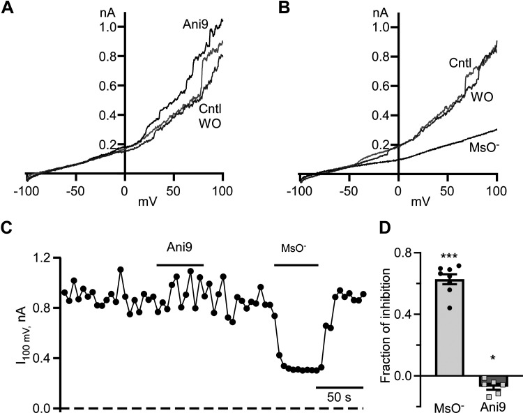
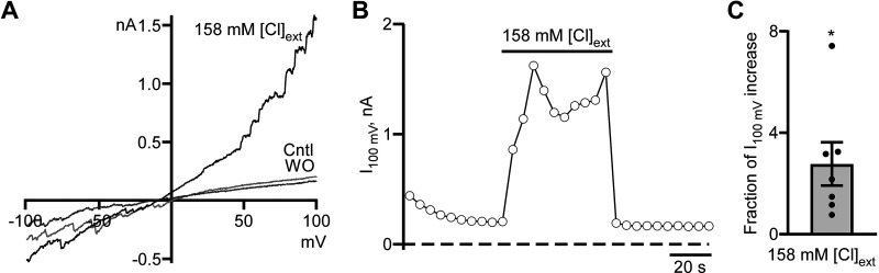
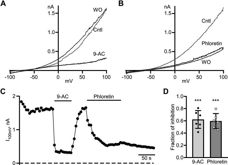
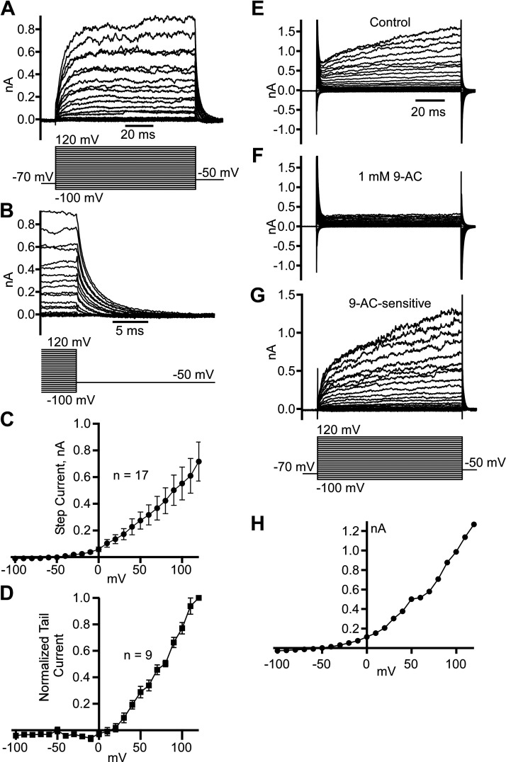
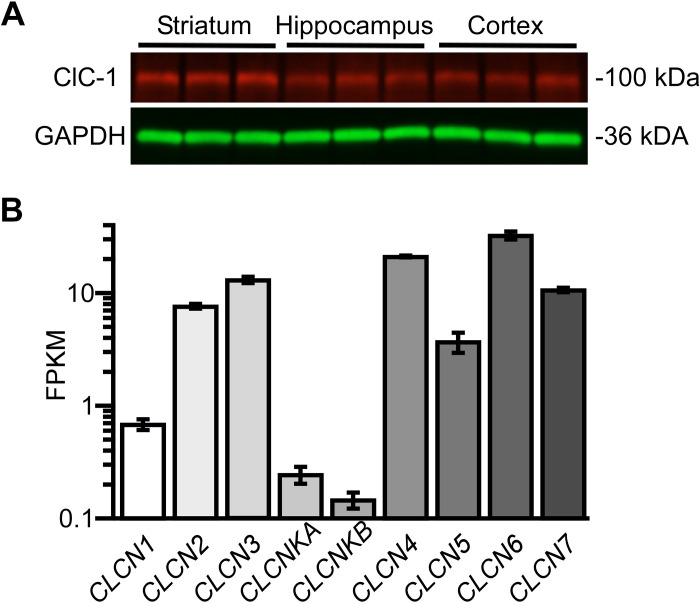
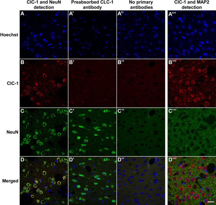
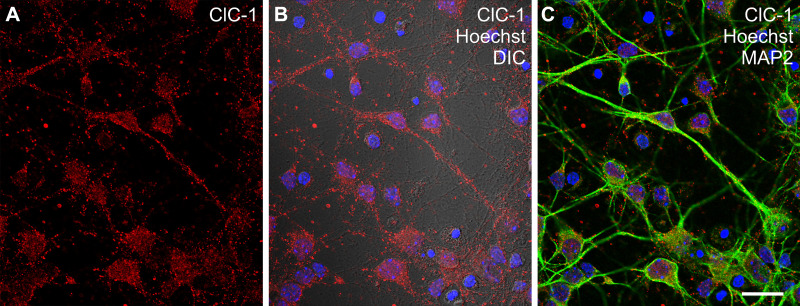
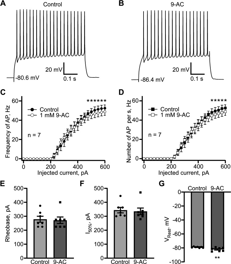
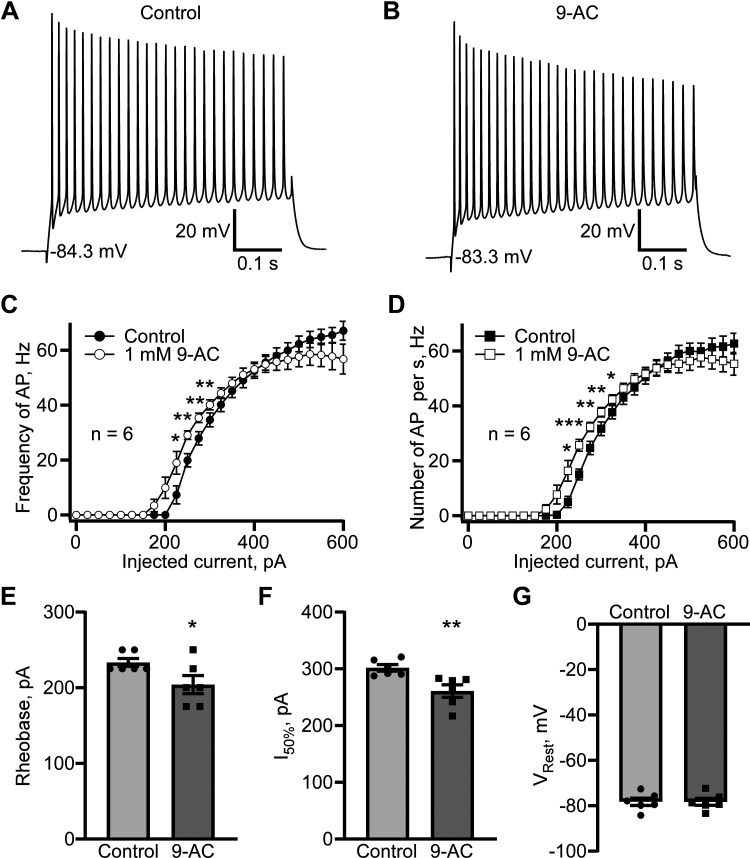
Dynamic chloride (Cl-) regulation is critical for synaptic inhibition. In mature neurons, Cl- influx and extrusion are primarily controlled by ligand-gated anion channels (GABAA and glycine receptors) and the potassium chloride cotransporter K+-Cl- cotransporter 2 (KCC2), respectively. Here, we report for the first time, to our knowledge, a presence of a new source of Cl- influx in striatal neurons with properties similar to chloride voltage-gated channel 1 (ClC-1). Using whole cell patch-clamp recordings, we detected an outwardly rectifying voltage-dependent current that was impermeable to the large anion methanesulfonate (MsO-). The anionic current was sensitive to the ClC-1 inhibitor 9-anthracenecarboxylic acid (9-AC) and the nonspecific blocker phloretin. The mean fractions of anionic current inhibition by MsO-, 9-AC, and phloretin were not significantly different, indicating that anionic current was caused by active ClC-1-like channels. In addition, we found that Cl- current was not sensitive to the transmembrane protein 16A (TMEM16A; Ano1) inhibitor Ani9 and that the outward Cl- rectification was preserved even at a very high intracellular Ca2+ concentration (2 mM), indicating that TMEM16B (Ano2) did not contribute to the total current. Western blotting and immunohistochemical analyses confirmed the presence of ClC-1 channels in the striatum mainly localized to the somata of striatal neurons. Finally, we found that 9-AC decreased action potential firing frequencies and increased excitability in medium spiny neurons (MSNs) expressing dopamine type 1 (D1) and type 2 (D2) receptors in the brain slices, respectively. We conclude that ClC-1-like channels are preferentially located at the somata of MSNs, are functional, and can modulate neuronal excitability.

Keywords: ClC-1, chloride voltage-gated channel 1; TMEM16A, transmembrane protein 16A (Ano1); chloride homeostasis; dopamine type 1 and type 2 receptors; striatal medium spiny neurons.


Conflict of interest statement

No conflicts of interest, financial or otherwise, are declared by the authors.

Figures

Figure 1.
Figure 1.
Cl currents in striatal neurons in mixed neuronal-glial coculture. A: representative voltage ramp-evoked current traces were recorded in control (Cntl), at the end of 10 µM Ani9 application (Ani9), and after Ani9 washout (WO). B: typical voltage ramp-evoked current traces were recorded in controls (Cntl), at the end of extracellular Cl replacement with methanesulfonate (MsO), and after MsO washout (WO). C: time course of a current measured at the end of voltage ramp at +100 mV (I100 mV). Durations of Ani9 application and Cl replacement with MsO are shown as horizontal lines. Currents were measured every 5 s. D: fraction of inhibition of I100 mV in the presence of MsO and Ani9. ***P < 0.001, n = 8, one-sample t test, *P < 0.05, n = 6, one-sample t test.
Figure 2.
Figure 2.
Cl current in striatal neurons in mixed neuronal-glial coculture is voltage dependent. A: Representative voltage ramp-evoked current traces were recorded in control (Cntl), at the end of symmetrical Cl solution application (158 mM [Cl]ext), and washout (WO). B: time course of Cl current measured at the end of voltage ramp at +100 mV (I100 mV). The duration of symmetrical Cl solution (158 mM [Cl]ext) application is shown as a horizontal line. Currents were measured every 5 s. C: the fraction of I100 mV increase in symmetrical Cl solution. *P < 0.05, n = 7, one-sample t test.
Figure 3.
Figure 3.
Clcurrent is sensitive to ClC-1 inhibitor 9-AC and nonspecific blocker phloretin. A: representative voltage ramp-evoked current traces were recorded in control (Cntl), at the end 1 mM 9-AC application (9-AC), and washout (WO). B: typical voltage ramp-evoked current traces were recorded in control (Cntl), at the end of extracellular 0.3 mM phloretin application (phloretin), and washout (WO). C: time course of a current measured at the end of voltage ramp at +100 mV (I100 mV). Durations of 9-AC and phloretin application are shown as horizontal lines. Currents were measured every 5 s. D: fraction of inhibition of I100 mV in the presence of 9-AC and phloretin. ***P < 0.001, n = 6, one-sample t test.
Figure 4.
Figure 4.
Leak-subtracted voltage-dependent Cl current demonstrates a predicted reversal potential below −50 mV. A: examples of leak-subtracted (P/8 method) step currents caused by voltage steps in a range from −100 to +120 mV. B: the same as in A recordings magnify the last 5 ms of step currents and outward tail currents caused by the −50 mV step. C: step current voltage dependence (means ± SE) obtained from 17 neurons. D: normalized tail current voltage dependence (means ± SE) obtained from 9 neurons. Examples of step current traces obtained at conditions of no leak subtraction in control (E), after 1 mM 9-AC application (F), and 9-AC sensitive (G) obtained by subtracting corresponding traces in F from E. H: 9-AC-sensitive step current voltage dependence measured at the end of the step currents in G.
Figure 5.
Figure 5.
ClC-1 channel antigenicity and mRNA were detected in murine striatum. A: CLC-1 channel antigenicity was detected at ∼100 kDa in the striatum, hippocampus, and cerebral cortex by immunoblotting. CLC-1 channel antigenicity was detected at ∼100 kDa in the striatum with anti-ClC-1 primary and Alexa Fluor 647-conjugated secondary antibodies, whereas the loading control (GAPDH) is detected by an Alexa Fluor 488-conjugated secondary antibody at ∼36 kDa. B: next-generation sequencing shows that ClC family gene (ClCN1, ClCN 2, ClCN3, ClCNKA, ClCNKB, ClCN4, ClCN5, ClCN6, ClCN7) transcripts were detected in the striatum. FPKM, fragments per kilobase of transcript per million mapped reads.
Figure 6.
Figure 6.
Pronounced immunohistochemical detection of ClC-1 channel expression in the striatum. Columns show the detection of primary ClC-1 and NeuN immunofluorescence (A), controls in which primary anti-ClC-1 antibodies were preabsorbed with ClC-1 blocking peptide (A′), controls in which primary anti-ClCN1 and NeuN antibodies were excluded (A′′), and the detection of primary ClC-1 and MAP2 immunofluorescence (A′′′). Sections were counterstained with Hoechst 33342 to visualize cell nuclei in the striatum. B, B′, B′′, and B′′′: ClC-1-specific immunofluorescence was preferentially associated with the cell bodies of MSNs; ClC-1 antigenicity was detected in dendrites and perhaps elsewhere confirming its presence in the striatum. NeuN (C, C′, and C′′) and MAP2 (C′′′) markers localize immunofluorescence to soma and dendrites, respectively. D, D′, D′′, and D′′′: merged images confirm soma localization of neuronal ClC-1 channels. Scale bar = 20 µm. MSNs, medium spiny neurons.
Figure 7.
Figure 7.
Immunocytochemical detection of ClC-1-like channel antigenicity in dissociated striatal neurons. A: ClC-1 channel immunofluorescence (red) was readily detected in MSNs in vitro. B: Differential interference contrast (DIC) microscopy revealed ClC-1 immunofluorescence in association with both large- and fine-diameter neuritic processes. C: ClC-1 antigenicity was colocalized with MAP2 (green fluorescence) immunoreactive perikarya and dendrites of MSNs, and the cell cultures were additionally counterstained with Hoechst 33342 to reveal cell nuclei (blue fluorescence). Scale bar = 20 μm. MSNs, medium spiny neurons.
Figure 8.
Figure 8.
ClC-1 inhibitor 9-AC causes a reduction in action potential firing in striatal D1 MSN in the brain slices. Examples of action potential firing evoked by 600 pA current injection in control (A) and after 5 min perfusion with 9-AC (1 mM; B). Action potential firing evaluated as a firing frequency measured as a reciprocal value of mean interaction potential duration (C) and as several action potentials normalized to the stimulus duration (D). *P < 0.05, n = 7, paired t test. E: rheobase (means ± SE) obtained in control and in the presence of 9-AC (1 mM), P > 0.05, not significantly different, paired t test. F: current causing 50% of action potential firing (I50%, pA, means ± SE) calculated in control and in the presence of 9-AC (1 mM), P > 0.05, not significantly different, paired t test. G: resting membrane potential (Vrest, mV, means ± SE) obtained in control and in the presence of 1 mM 9-AC, P < 0.01, n = 7, paired t test. MSN, medium spiny neuron.
Figure 9.
Figure 9.
The ClC-1 inhibitor 9-AC causes a leftward shift in action potential firing vs. injected current dependence in striatal D2 MSN in the brain slices. Examples of action potential firing evoked by 600 pA current injection in control (A) and after perfusing 5 min with 9-AC (1 mM; B). Action potential firing evaluated as a firing frequency measured as a reciprocal value of mean interaction potential duration (C) and as several action potentials normalized to the stimulus duration (D). Total of n = 6 recordings analyzed. Statistically significant differences at *P < 0.05, **P < 0.01, and ***P < 0.001 were obtained using Student’s paired t test analysis. E: rheobase (means ± SE) obtained in control and in the presence of 1 mm 9-AC is significantly different (n = 6, *P <0.05, paired t test). F: current causing 50% of action potential firing (I50%, pA, means ± SE) calculated in control and in the presence of 1 mm 9-AC is significantly different (n = 6, **P <0.01, paired t test). G: resting membrane potential (Vrest, mV, means ± SE) obtained in control and in the presence of 1 mM 9-AC not significantly different (n = 6, P > 0.05, paired t test). MSN, medium spiny neuron.

Similar articles

Cited by

References

    1. Ebihara S, Shirato K, Harata N, Akaike N. Gramicidin-perforated patch recording: GABA response in mammalian neurones with intact intracellular chloride. J Physiol 484: 77–86, 1995. doi:10.1113/jphysiol.1995.sp020649. - DOI - PMC - PubMed
    1. Doyon N, Vinay L, Prescott SA, De Koninck Y. Chloride regulation: a dynamic equilibrium crucial for synaptic inhibition. Neuron 89: 1157–1172, 2016. doi:10.1016/j.neuron.2016.02.030. - DOI - PubMed
    1. Payne JA, Stevenson TJ, Donaldson LF. Molecular characterization of a putative K-Cl cotransporter in rat brain. A neuronal-specific isoform. J Biol Chem 271: 16245–16252, 1996. doi:10.1074/jbc.271.27.16245. - DOI - PubMed
    1. Payne JA. Functional characterization of the neuronal-specific K-Cl cotransporter: implications for [K+]o regulation. Am J Physiol Cell Physiol 273: C1516–C1525, 1997. doi:10.1152/ajpcell.1997.273.5.C1516. - DOI - PubMed
    1. Johansson S, Yelhekar TD, Druzin M. Commentary: chloride regulation: a dynamic equilibrium crucial for synaptic inhibition. Front Cell Neurosci 10: 182, 2016. doi:10.3389/fncel.2016.00182. - DOI - PMC - PubMed

Publication types

MeSH terms

LinkOut - more resources