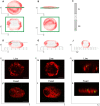
Evaluation of chromatin mesoscale organization
- PMID: 35071965
- PMCID: PMC8758204
- DOI: 10.1063/5.0069286
Evaluation of chromatin mesoscale organization
Abstract
Chromatin organization in the nucleus represents an important aspect of transcription regulation. Most of the studies so far focused on the chromatin structure in cultured cells or in fixed tissue preparations. Here, we discuss the various approaches for deciphering chromatin 3D organization with an emphasis on the advantages of live imaging approaches.
© 2022 Author(s).
Figures

Similar articles
-
Implications of liquid-liquid phase separation in plant chromatin organization and transcriptional control.Curr Opin Genet Dev. 2019 Apr;55:59-65. doi: 10.1016/j.gde.2019.06.003. Epub 2019 Jul 12. Curr Opin Genet Dev. 2019. PMID: 31306885 Review.
-
Plant 3D genomics: the exploration and application of chromatin organization.New Phytol. 2021 Jun;230(5):1772-1786. doi: 10.1111/nph.17262. Epub 2021 Mar 4. New Phytol. 2021. PMID: 33560539 Free PMC article. Review.
-
Intranuclear mesoscale viscoelastic changes during osteoblastic differentiation of human mesenchymal stem cells.FASEB J. 2021 Dec;35(12):e22071. doi: 10.1096/fj.202100536RR. FASEB J. 2021. PMID: 34820910
-
Deciphering Hi-C: from 3D genome to function.Cell Biol Toxicol. 2019 Feb;35(1):15-32. doi: 10.1007/s10565-018-09456-2. Epub 2019 Jan 4. Cell Biol Toxicol. 2019. PMID: 30610495 Review.
-
Understanding 3D Genome Organization and Its Effect on Transcriptional Gene Regulation Under Environmental Stress in Plant: A Chromatin Perspective.Front Cell Dev Biol. 2021 Dec 8;9:774719. doi: 10.3389/fcell.2021.774719. eCollection 2021. Front Cell Dev Biol. 2021. PMID: 34957106 Free PMC article. Review.
Cited by
-
Analysis of Fluorescent Proteins for Observing Single Gene Locus in a Live and Fixed Escherichia coli Cell.J Phys Chem B. 2024 Jul 18;128(28):6730-6741. doi: 10.1021/acs.jpcb.4c01816. Epub 2024 Jul 5. J Phys Chem B. 2024. PMID: 38968413 Free PMC article.
-
Mechanobiology of the cell nucleus.APL Bioeng. 2022 Dec 16;6(4):040401. doi: 10.1063/5.0135299. eCollection 2022 Dec. APL Bioeng. 2022. PMID: 36536804 Free PMC article. No abstract available.
-
Fixation can change the appearance of phase separation in living cells.Elife. 2022 Nov 29;11:e79903. doi: 10.7554/eLife.79903. Elife. 2022. PMID: 36444977 Free PMC article.
-
Chromatin Liquid-Liquid Phase Separation (LLPS) Is Regulated by Ionic Conditions and Fiber Length.Cells. 2022 Oct 6;11(19):3145. doi: 10.3390/cells11193145. Cells. 2022. PMID: 36231107 Free PMC article.
References
LinkOut - more resources
Full Text Sources

