

A novel animal model for vulnerable atherosclerotic plaque: dehydrated ethanol lavage in the carotid artery of rabbits fed a Western diet
- PMID: 35070793
- PMCID: PMC8748485
- DOI: 10.21037/cdt-21-291
A novel animal model for vulnerable atherosclerotic plaque: dehydrated ethanol lavage in the carotid artery of rabbits fed a Western diet
Abstract
Background: The study of unstable atherosclerotic plaques is limited by the absence of ideal animal models to reproduce the plaque instability observed in humans. In this study, we attempted to develop a novel animal model for vulnerable atherosclerotic plaques using dehydrated ethanol lavage in rabbits fed a Western diet (WD).
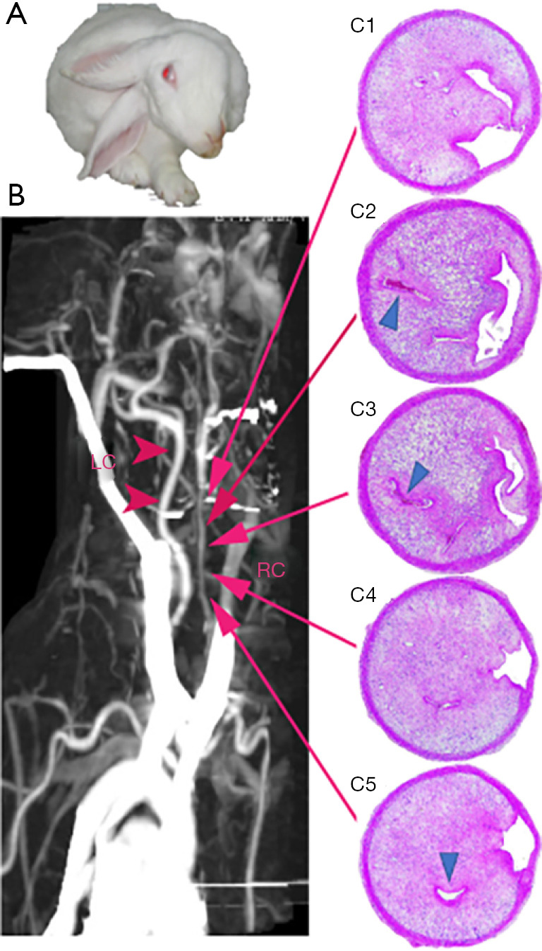
Methods: A total of 30 New Zealand White (NZW) rabbits were randomized to 5 groups, including a control group with or without WD, a balloon injury with WD group, and an ethanol injury with or without WD group. Operations were conducted using the right common carotid artery as the target vessel. All animals were followed up for 3 months unless a vascular event occurred. Blood samples and carotid artery specimens were ultimately collected for analysis of atherogenesis.
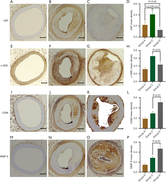
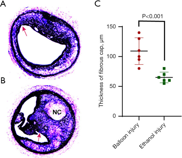
Results: Compared to rabbits in which lesions were induced by balloon injury, those subjected to an ethanol lavage with high cholesterol diet showed progressive atherosclerotic lesions in all carotid artery segments, which were characterized by greater plaque burden, smaller minimum lumen area (MLA), and increased vulnerability as indicated by abundant macrophages, scattered smooth muscle cell (SMC) composition, higher matrix metalloproteinase-9 (MMP-9) expression in plaques, thinner fibrous cap thickness, and higher possibility of stroke event (50% vs. 0%). Meanwhile, the serum interleukin-1β (IL-1β) and monocyte chemoattractant protein-1 (MCP-1) levels in the ethanol injury group with a high-cholesterol diet were significantly higher than those in the balloon injury group after 3 months (all P<0.01).
Conclusions: We successfully established a novel animal model for vulnerable atherosclerosis by ethanol exposure of the carotid segment that has a higher predictive value for the probability of ischemic events than the balloon injury model. Therefore, it may represent a promising animal model for investigating new therapeutic approaches, novel imaging modalities, and underlying mechanisms for vulnerable atherosclerotic plaque.
Keywords: Animal model; endothelial cell (EC) injury; inflammatory marker; vulnerable atherosclerotic plaque.
2021 Cardiovascular Diagnosis and Therapy. All rights reserved.
Conflict of interest statement
Conflicts of Interest: All authors have completed the ICMJE uniform disclosure form (available at https://dx.doi.org/10.21037/cdt-21-291). HC reports that the study was supported by grant of Zhejiang Provincial Natural Science Foundation, China (grant No. LY12H02001) and Zhejiang Provincial Key Research Project (grant No. 2021C03096). HY reports that the study was partly supported by Ningbo Social Development Research Project (Grant No. 2011C51003 to HY). The other authors have no conflicts of interest to declare.
Figures

Similar articles
-
A novel rabbit model of atherosclerotic vulnerable plaque established by cryofluid-induced endothelial injury.Sci Rep. 2024 Apr 24;14(1):9447. doi: 10.1038/s41598-024-60287-0. Sci Rep. 2024. PMID: 38658774 Free PMC article.
-
Mesenchymal Stem Cells Stabilize Atherosclerotic Vulnerable Plaque by Anti-Inflammatory Properties.PLoS One. 2015 Aug 19;10(8):e0136026. doi: 10.1371/journal.pone.0136026. eCollection 2015. PLoS One. 2015. PMID: 26288013 Free PMC article.
-
[Effect of local unstable atherosclerotic plaque on plaque formation in the carotid artery and abdominal aorta of rabbits].Nan Fang Yi Ke Da Xue Xue Bao. 2023 Jan 20;43(1):117-121. doi: 10.12122/j.issn.1673-4254.2023.01.16. Nan Fang Yi Ke Da Xue Xue Bao. 2023. PMID: 36856219 Free PMC article. Chinese.
-
Atherosclerotic plaques composed of a large core of foam cells covered with thin fibrous caps in twice-injured carotid arterial specimens obtained from high cholesterol diet-fed rabbits.J Atheroscler Thromb. 2000;7(2):83-90. doi: 10.5551/jat1994.7.83. J Atheroscler Thromb. 2000. PMID: 11426587
-
Triggering of plaque disruption and arterial thrombosis in an atherosclerotic rabbit model.Circulation. 1995 Feb 1;91(3):776-84. doi: 10.1161/01.cir.91.3.776. Circulation. 1995. PMID: 7828306
References
LinkOut - more resources
Full Text Sources
Research Materials
Miscellaneous