. 2022 Jan 11;14(1):125.
doi: 10.3390/v14010125.

Novel Neutralizing Epitope of PEDV S1 Protein Identified by IgM Monoclonal Antibody

Affiliations

Novel Neutralizing Epitope of PEDV S1 Protein Identified by IgM Monoclonal Antibody

Techit Thavorasak et al. Viruses. .

Abstract

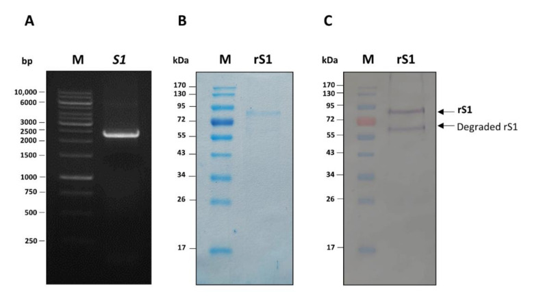
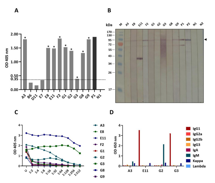
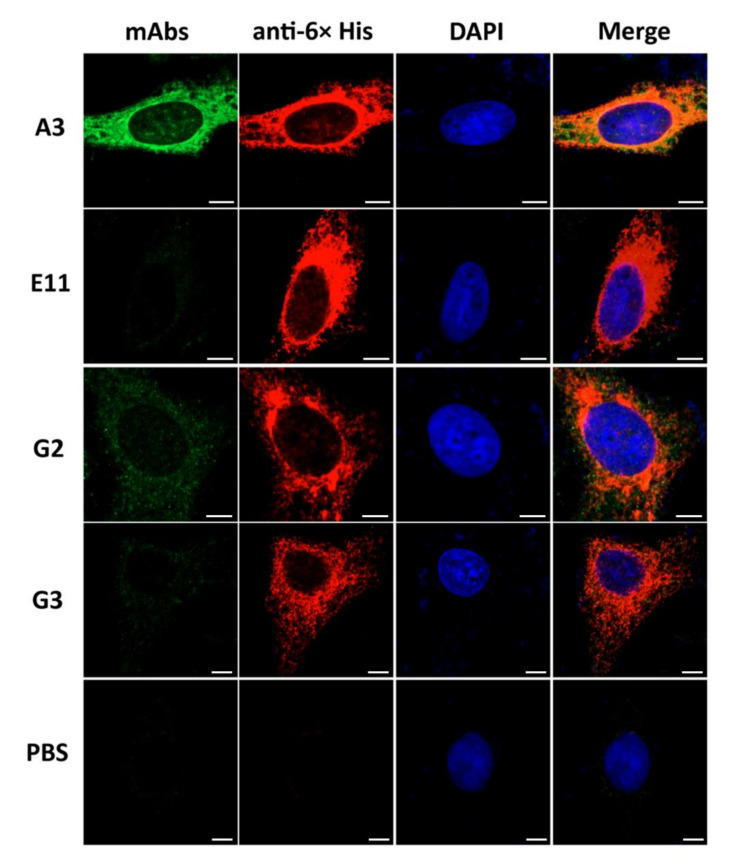
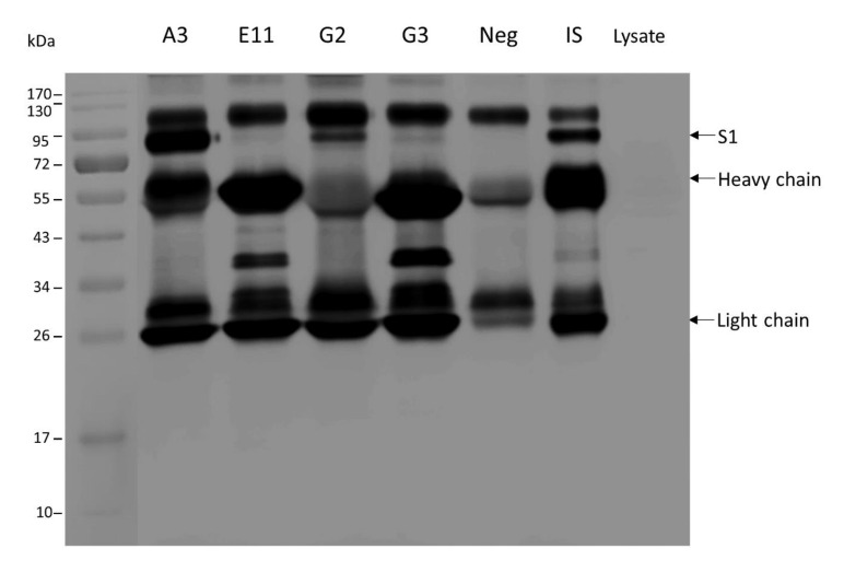
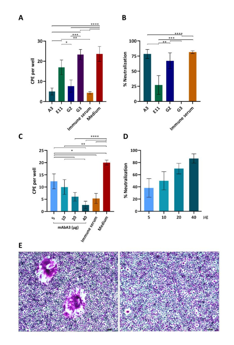
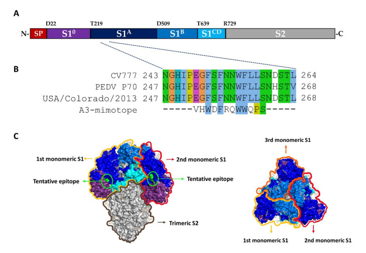
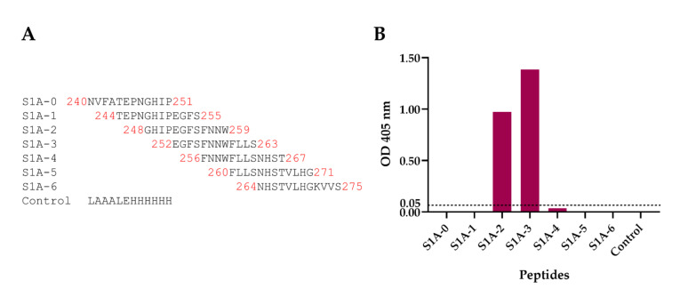
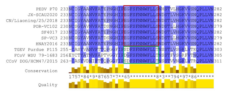
Porcine epidemic diarrhea virus (PEDV) causes devastating enteric disease that inflicts huge economic damage on the swine industry worldwide. A safe and highly effective PEDV vaccine that contains only the virus-neutralizing epitopes (not enhancing epitope), as well as a ready-to-use PEDV neutralizing antibody for the passive immunization of PEDV vulnerable piglets (during the first week of life) are needed, particularly for PEDV-endemic farms. In this study, we generated monoclonal antibodies (mAbs) to the recombinant S1 domain of PEDV spike (S) protein and tested their PEDV neutralizing activity by CPE-reduction assay. The mAb secreted by one hybrodoma clone (A3), that also bound to the native S1 counterpart from PEDV-infected cells (tested by combined co-immunoprecipitation and Western blotting), neutralized PEDV infectivity. Epitope of the neutralizing mAb (mAbA3) locates in the S1A subdomain of the spike protein, as identified by phage mimotope search and multiple sequence alignment, and peptide binding-ELISA. The newly identified epitope is shared by PEDV G1 and G2 strains and other alphacoronaviruses. In summary, mAbA3 may be useful as a ready-to-use antibody for passive immunization of PEDV-susceptible piglets, while the novel neutralizing epitope, together with other, previously known protective epitopes, have potential as an immunogenic cocktail for a safe, next-generation PEDV vaccine.

Keywords: monoclonal antibody; neutralization assay; neutralizing antibody; phage mimotope; porcine epidemic diarrhea virus (PEDV); spike (S) protein.


Conflict of interest statement

No potential conflict of interest was reported by the authors.

Figures

Figure 1
Figure 1
Production of recombinant S1 (rS1) of porcine epidemic diarrhea virus (PEDV). (A) Polymerase chain reaction (PCR) amplificon of PEDV S1 gene (S1). Lane M, 1 kilobase (kb) DNA ladder. Numbers on the left are DNA sizes in base pairs (bp). (B) Sodium dodecyl sulfate-polyacrylamide gel electrophoresis (SDS-PAGE) separated pattern of the purified rS1 protein after staining with Coomassie Brilliant Blue G-250 (CBB) dye. (C) Western blot pattern of the rS1 as detected by anti-His antibody. Lanes M and numbers on the left of (B) and (C) are protein molecular mass marker and protein molecular masses in kilo-Daltons (kDa), respectively.
Figure 2
Figure 2
Binding of monoclonal antibodies (mAbs) in culture fluids of different hybridoma clones to rS1 and titers and isotypes of the mAbs secreted by the selected hybridomas. (A) Indirect ELISA OD 405 nm of mAbs in individual hybridoma culture fluids to the rS1. *, the clone that gave OD 405 nm ≥ 0.35 (arbitrarily cut-off level), which was selected for further testing. P1, mouse immune serum (1:50,000) to rS1 (IS) as positive binding control; N1, SP2/0-Ag14 spent medium as negative binding control. Broken line, cut-off OD. (B) Binding of the mAbs of the selected hybridoma clones to SDS-PAGE-separated rS1 (arrowhead) by Western blotting. P1 is mouse immune serum to rS1 (IS) (1:10,000) and P2 is anti-His antibody, both served as positive controls for binding to 8× His tagged-rS1. N1 and N2 are SP2/0-Ag14 spent medium and PBS, respectively, which served as negative binding controls. Lane M, protein molecular mass marker. Numbers on the left are protein molecular masses in kDa. The prominent band at about 40 kDa (lane E11) should be N-terminal portion of degraded rS1 which contained epitope of the mAbE11; this band was not bound by the anti-His (P2) as the recombinant S1 had the 8× His tag at the C-terminus; the band was not recognized by the mouse immune serum (diluted 1:10,000) to rS1, as the antibody to this N-terminal epitope might be diluted out. (C) Indirect ELISA titers of the rS1-specific mAbs secreted from hybridoma clones. Monoclonal antibodies of the clones G2, G3, A3, and E11 clones yielded high titers at the maximal phase of cell growth; these clones were selected for further experiments. (D) Isotype identification of the mAbs of the selected A3, G2, G3, and E11 clones.
Figure 3
Figure 3
Co-localization of the rS1-specific-mAbs to PEDV S1 overexpressed in HeLa cells as determined by confocal microscopy. HeLa cells transfected with S1-pTriEx 1.1 recombinant plasmid were cultured on cover slips in 24-well culture plate for 48 h. Cells were fixed with cold acetone-methanol (1:1), followed by staining with 10 µg of individual mAbs to S1 (anti-rS1) and rabbit anti-His antibody (1:500) as primary antibodies and fluorophore-conjugated secondary antibodies (1:300). mAbA3, mAbG2 and mAbG3 (green) colocalized with intracellular S1 overexpressed in the HeLa cells (red) and seen as orange or yellow matter in merge. The mAbE11 did not bind (or bound negligibly) to the overexpressed S1 in the transfected cells. Nuclei stained blue by DAPI dye. Scale bar = 5 µm.
Figure 4
Figure 4
Binding of mAbs to native S1 protein. Combined co-immunoprecipitation and Western blotting was conducted to determine the binding activity of mAbs to native spike protein of PEDV. Ten micrograms of mAbs of individual hybridoma clones were mixed with lysate of PEDV infected cells. The mAb-Ag complexes were pull-down by using goat antimouse Ig sensitized-protein G. The pull-down content was then subjected to Western blotting by probing the SDS-PAGE-separated components with mouse immune serum against the PEDV rS1 (IS). The mAbA3, mAbG2, and mAbG3 could bind to the native S1 protein contained in the PEDV infected cell lysate and were pull-down together as demonstrated, i.e., the presence of the S1 band (~90 kDa, upper arrow) and the bands of heavy chains (~50–70 kDa, middle arrow) and light chains (~25 kDa, lower arrow) of the mAbs in Western blot analysis. Neg, reaction without mAb; IS, reaction added with mouse immune serum to PEDV rS1; Lysate, reaction containing PEDV infected cell lysate alone without any antibody.
Figure 5
Figure 5
Monoclonal antibody A3-mediated neutralization of PEDV infectivity. (A) Preliminary experiments using 10 µg of mAbA3, mAbE11, mAbG2 and mAbG3 to mix with 100 pfu of PEDV P70 G2 strain before adding to the Vero cell monolayer. Infected cells treated with mouse immune serum to rS1 (IS) and infected cells in medium alone served as positive and negative neutralization controls, respectively. After 1 h, the fluid in each well was removed; the cells were washed and grown in semisolid virus maintaining medium for 48 h. Then, the cells were fixed with 10% formalin and stained with 1% crystal violet dye. The CPE (syncytial cells) were counted under 40× magnification light microscopy. Mean and standard deviation of the CPE number in triplicate wells of each treatment are reported. (B) Percent PEDV neutralization mediated by mAbs of individual clones and IS. (C) PEDV (100 pfu) was mixed with indicated amount of purified mAbA3 (5–40 µg) before adding to monolayer of Vero cells in 24-well culture plate. Mouse immune serum (IS, 1:200) and the virus in medium alone were used as positive and negative neutralization controls, respectively. Numbers of CPE per well (mean ± SD of triplicate wells) are shown. (D) Percentage of mAbA3 mediated neutralization of PEDV infectivity compared to infected cells in medium alone. Three replicative experiments were performed. *, p < 0.05; **, p < 0.01; ***, p < 0.001; ****, p < 0.0001 by one-way ANOVA. (E) Appearance of syncytial Vero cells (left) and normal Vero cells (right). Scale bar = 100 nm.
Figure 6
Figure 6
Presumptive amino acid sequence that was bound by mAbA3 as determined by phage mimotope identification using Ph.D.TM-12 Phage display peptide library. (A) Domain organization of spike protein of PEDV P70 strain (adapted from [25]). S1 portion can be divided into 4 subdomains including S10 (purple); S1A (dark blue); S1B (marine); and S1C and D (cyan). (B) Amino acid sequence of phage mimotope that was bound by mAbA3 (A3-mimotope). Multiple sequence alignment of A3-mimotope with S1 segments of CV777 classical strain (GenBank: AEX92968.1), PEDV P70 (strain of this study) and USA/Colorado/2013 (GenBank: AGO58924.1; the strain that Cryo-EM structure is available, PDB: 6vv5) revealed that the tentative epitope “252EGFSFNNWFLLS263” is located at the S1A subdomain. Each amino acid was colored by Clustal X scheme, which is based on chemical characteristic of amino acid. (C) Illustration of tentative epitope of the mAbA3 (green) on the PEDV spike protein homotrimer: side view (left) and top view (right).
Figure 7
Figure 7
Overlapped peptides of PEDV spike protein bound by the mAbA3 as tested by peptide-binding ELISA. Biotin-labeled 12-mer overlapped peptides encompass PEDV spike protein (amino acid residues 240–275th) were used in the indirect ELISA for testing mAbA3 binding. (A) List and amino acid sequences of the 8 overlapped peptides of the PEDV spike protein. (B) OD 405 nm of mAbA3 that bound to individual overlapped S1 peptides and control peptide. The mAbA3 bound to peptides S1A-2 (248GHIPEGFSFNNW259) and S1A-3 (252EGFSFNNWFLLS263) and yielded high ELISA signals.
Figure 8
Figure 8
Multiple alignments of the amino acid sequences of spike proteins of various strains of PEDV that were published in PubMed during the past 5 years and other alphacoronaviruses. The amino acid sequences of PEDV that caused PED outbreaks included JX-SCAU2020 (GenBank: QUE39276.1); CN/Liaoning/25/2018 (GenBank: QCW07226.1); POR-VC102 (GenBank: QQO86882.1); SF4017 (GenBank: QEQ55819.1); SP-VC3 (GenBank: QQO86847.1); and HNAY2016 (GenBank: QQS74747.1). The sequences of other alphacoronaviruses include TGEV Purdue P115 (GenBank: ABG89325.1); FCoV WSU 79-1683 (GenBank: JN634064.1); and CCoV DOG/HCM47/2015 (GenBank: LC190907.1). The multiple sequence alignment revealed that the A3-epitope that was identified from the phage mimotope and overlapped S1 peptide-binding ELISAs, “EGFSFNNWFLLS”, is identical among the PEDV spike sequences (red box) and this sequence is found to be highly conserved among the other alphacoronaviruses (green box).

Similar articles

Cited by

References

    1. Stevenson G.W., Hoang H., Schwartz K.J., Burrough E.R., Sun D., Madson D., Cooper V.L., Pillatzki A., Gauger P., Schmitt B.J., et al. Emergence of porcine epidemic diarrhea virus in the United States: Clinical signs, lesions, and viral genomic sequences. J. Vet. Diagn. Invest. 2013;25:649–654. doi: 10.1177/1040638713501675. - DOI - PubMed
    1. Wood E.N. An apparently new syndrome of porcine epidemic diarrhoea. Vet. Rec. 1977;100:243–244. doi: 10.1136/vr.100.12.243. - DOI - PubMed
    1. Pensaert M.B., De Bouck P. A new coronavirus-like particle associated with diarrhea in swine. Arch. Virol. 1978;58:243–247. doi: 10.1007/BF01317606. - DOI - PMC - PubMed
    1. Lee C. Porcine epidemic diarrhea virus: An emerging and re-emerging epizootic swine virus. Virol. J. 2015;12:193. doi: 10.1186/s12985-015-0421-2. - DOI - PMC - PubMed
    1. Schulz L.L., Tonsor G.T. Assessment of the economic impacts of porcine epidemic diarrhea virus in the United States. J. Anim. Sci. 2015;93:5111–5118. doi: 10.2527/jas.2015-9136. - DOI - PMC - PubMed

Publication types

MeSH terms