. 2022 Mar 23;96(6):e0217821.
doi: 10.1128/jvi.02178-21. Epub 2022 Jan 19.

An ESCRT/VPS4 Envelopment Trap To Examine the Mechanism of Alphaherpesvirus Assembly and Transport in Neurons

Affiliations

An ESCRT/VPS4 Envelopment Trap To Examine the Mechanism of Alphaherpesvirus Assembly and Transport in Neurons

Jenna Barnes et al. J Virol. .

Abstract

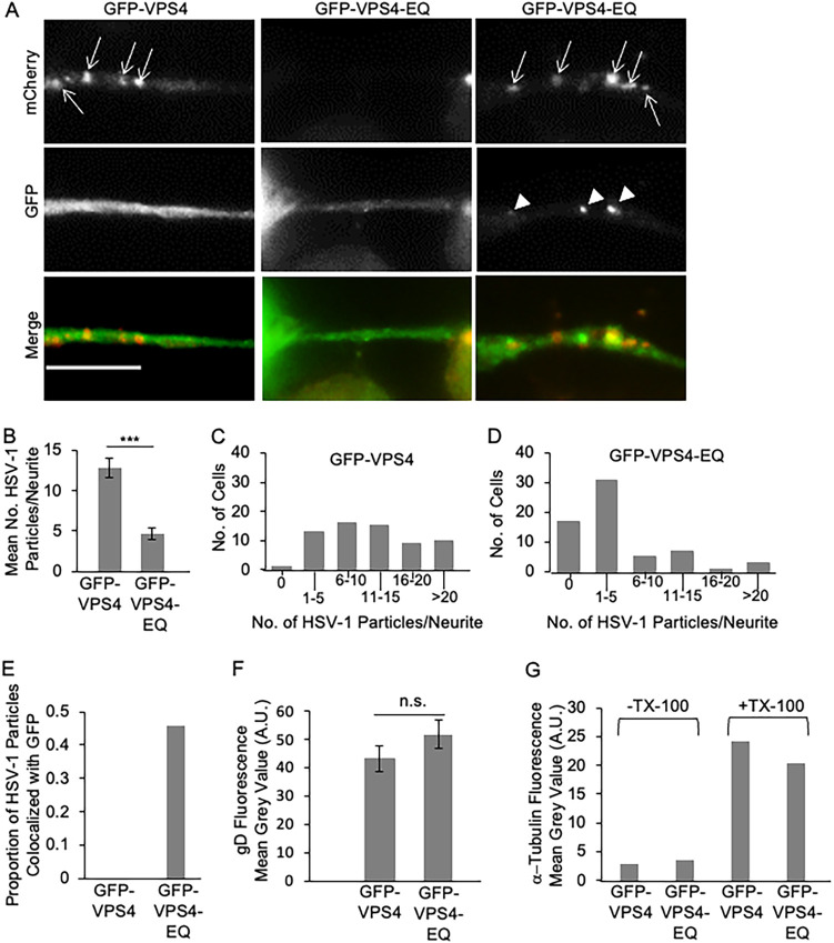
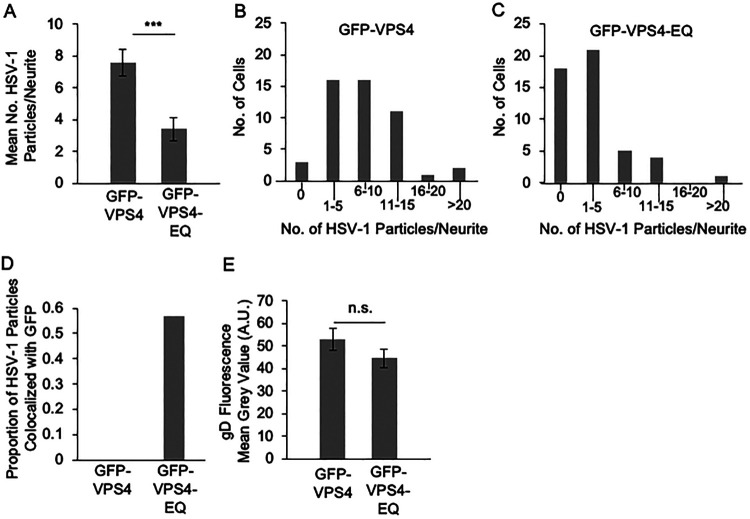
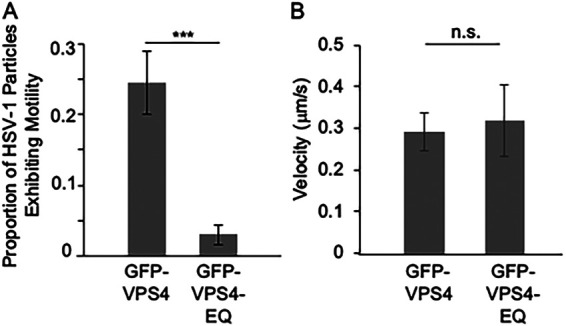
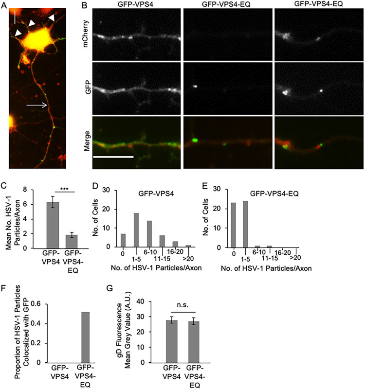
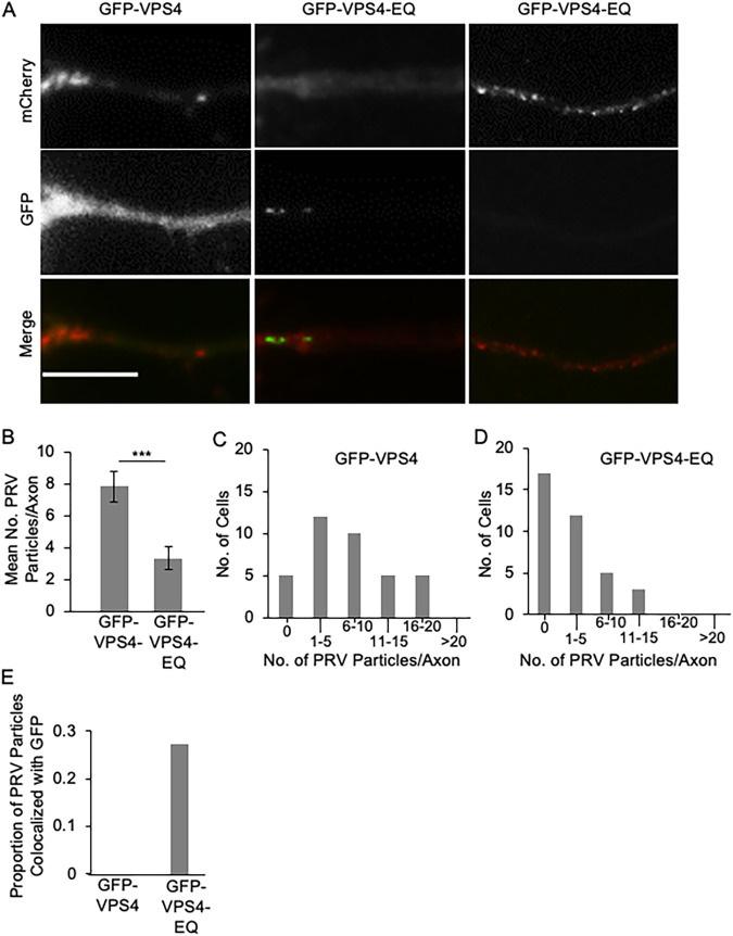
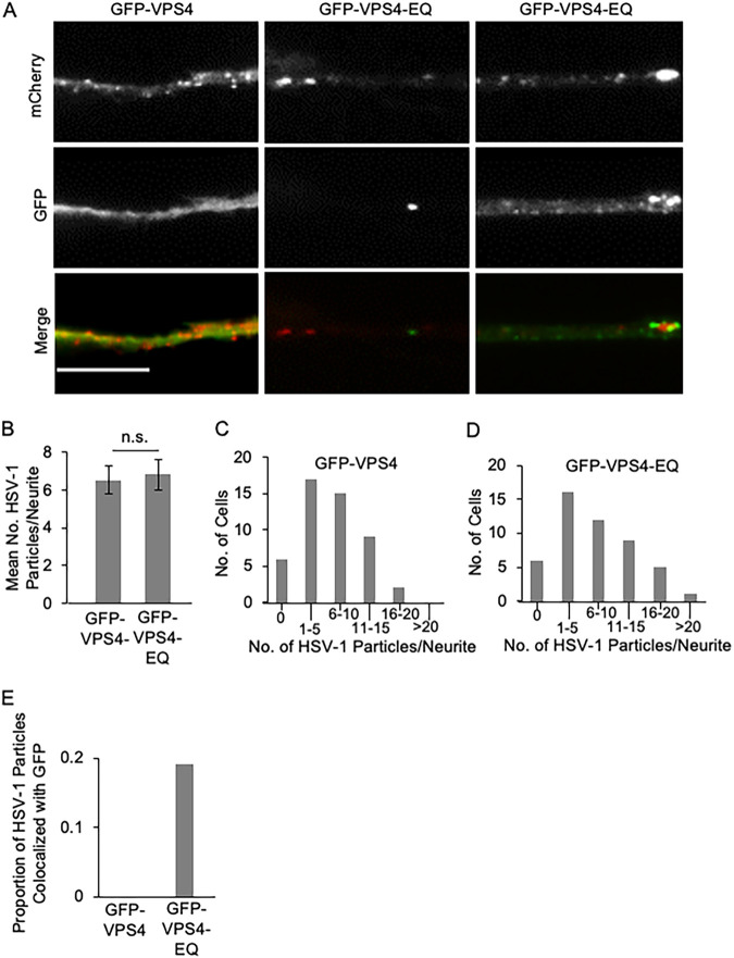
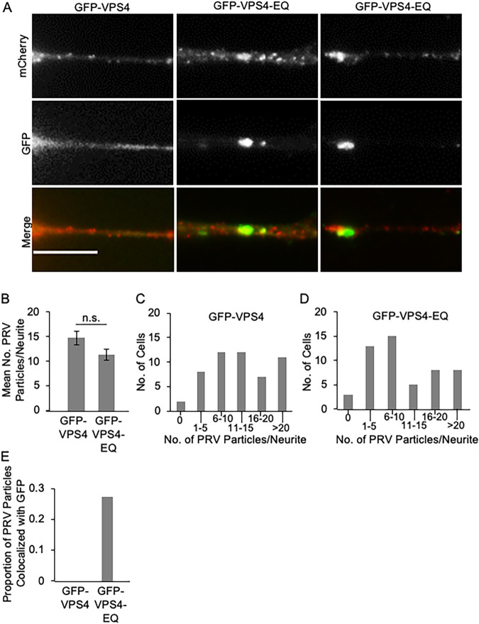
The assembly and egress of alphaherpesviruses, including herpes simplex virus 1 (HSV-1) and pseudorabies virus (PRV), within neurons is poorly understood. A key unresolved question is the structure of the viral particle that moves by anterograde transport along the axon, and two alternative mechanisms have been described. In the "married" model, capsids acquire their envelopes in the cell body and then traffic along axons as enveloped virions within a bounding organelle. In the "separate" model, nonenveloped capsids travel from the cell body into and along the axon, eventually encountering their envelopment organelles at a distal site, such as the nerve cell terminal. Here, we describe an "envelopment trap" to test these models using the dominant negative terminal endosomal sorting complex required for transport (ESCRT) component VPS4-EQ. Green fluorescent protein (GFP)-tagged VPS4-EQ was used to arrest HSV-1 or PRV capsid envelopment, inhibit downstream trafficking, and GFP-label envelopment intermediates. We found that GFP-VPS4-EQ inhibited trafficking of HSV-1 capsids into and along the neurites and axons of mouse CAD cells and rat embryonic primary cortical neurons, consistent with egress via the married pathway. In contrast, transport of HSV-1 capsids was unaffected in the neurites of human SK-N-SH neuroblastoma cells, consistent with the separate mechanism. Unexpectedly, PRV (generally thought to utilize the married pathway) also appeared to employ the separate mechanism in SK-N-SH cells. We propose that apparent differences in the methods of HSV-1 and PRV egress are more likely a reflection of the host neuron in which transport is studied rather than true biological differences between the viruses themselves. IMPORTANCE Alphaherpesviruses, including herpes simplex virus 1 (HSV-1) and pseudorabies virus (PRV), are pathogens of the nervous system. They replicate in the nerve cell body and then travel great distances along axons to reach nerve termini and spread to adjacent epithelial cells; however, key aspects of how these viruses travel along axons remain controversial. Here, we test two alternative mechanisms for transport, the married and separate models, by blocking envelope assembly, a critical step in viral egress. When we arrest formation of the viral envelope using a mutated component of the cellular ESCRT apparatus, we find that entry of viral particles into axons is blocked in some types of neurons but not others. This approach allows us to determine whether envelope assembly occurs prior to entry of viruses into axons or afterwards and, thus, to distinguish between the alternative models for viral transport.

Keywords: ESCRT; HSV-1; PRV; anterograde transport; envelopment; neuronal egress.


Conflict of interest statement

The authors declare no conflict of interest.

Figures

FIG 1
FIG 1
Transport of HSV-1 particles in the neurites of CAD cells transfected to express GFP-VPS4 or GFP-VPS4-EQ. Undifferentiated CAD cells were transfected with plasmids constructed to express GFP-VPS4 or GFP-VPS4-EQ under the control of a tetracycline-inducible promoter. They were then differentiated, infected with HSV-1, and GFP-VPS4 or GFP-VPS4-EQ expression induced by addition of doxycycline. After 20 h, cells were fixed, incubated with primary and fluorescent secondary antibodies as appropriate, and imaged by fluorescence microscopy. (A) Gallery of neurites from GFP-VPS4-expressing (left column) or GFP-VPS4-EQ-expressing (middle and right columns) CAD cells. Neurites were imaged in the red channel to detect puncta corresponding to mCherry-tagged HSV-1 capsids (top row, white arrows) or the green channel to detect GFP-tagged VPS4 alleles (middle row, white arrowheads indicate puncta enriched in GFP fluorescence). Bottom row shows merged images. Scale bar, 10 μm. (B) Number of mCherry-fluorescent HSV-1 capsid-associated puncta per neurite. Plotted values are the mean and standard deviation from the mean for neurites from 63 CAD cells expressing GFP-VPS4 (total of 822 puncta) or 63 CAD cells expressing GFP-VPS4-EQ (total of 297 puncta). (C, D) Frequency (y axes) at which neurites of infected CAD cells expressing GFP-VPS4 or GFP-VPS4-EQ (respectively) contain numbers of HSV-1 puncta in the ranges indicated (x axes). (E) Proportion of mCherry-fluorescent puncta, similar to those scored in panel B, exhibiting colocalization with GFP-VPS4 (total of 293 puncta) or GFP-VPS4-EQ fluorescence (total of 401 puncta) at levels enriched above that of background (see the text for more details). (F) Cell surface anti-gD immunofluorescence intensity plotted in arbitrary units (A.U.) for fixed, nonpermeabilized HSV-1-infected CAD cells expressing GFP-VPS4 or GFP-VPS4-EQ. (G) Similar experiment to panel F but plotting the intensity of anti-α-tubulin immunofluorescence displayed by fixed, Triton X-100 permeabilized (+TX-100) or nonpermeabilized (−TX-100) HSV-1-infected CAD cells expressing GFP-VPS4 or GFP-VPS4-EQ. For panels in this and all figures, no significant difference (n.s.) corresponds to P > 0.05; ***, P < 0.001 (Student's t test).
FIG 2
FIG 2
Transport of HSV-1 particles in the axons of CAD cells infected by baculoviruses expressing GFP-VPS4 or GFP-VPS4-EQ. CAD cells were differentiated and then infected with HSV-1 and subsequently with baculoviruses expressing GFP-VPS4 or GFP-VPS4-EQ. After 20 h, cells were fixed, incubated with primary and fluorescent secondary antibodies as appropriate, and imaged by fluorescence microscopy. (A) Number of mCherry-fluorescent HSV-1 capsid-associated puncta per neurite. Plotted values are the mean and standard deviation from the mean for neurites from 49 CAD cells expressing GFP-VPS4 (total of 374 puncta) or 49 CAD cells expressing GFP-VPS4-EQ (total of 169 puncta). (B, C) Frequency (y axes) at which neurites of infected CAD cells expressing GFP-VPS4 or GFP-VPS4-EQ (respectively) contain numbers of HSV-1 puncta in the ranges indicated (x axes). (D) Proportion of mCherry-fluorescent puncta, similar to those scored in panel A, exhibiting colocalization with GFP-VPS4 (total of 77 puncta) or GFP-VPS4-EQ fluorescence (total of 97 puncta) at levels enriched above that of background. (E) Cell surface anti-gD immunofluorescence intensity for fixed, nonpermeabilized HSV-1-infected CAD cells expressing GFP-VPS4 or GFP-VPS4-EQ.
FIG 3
FIG 3
Motility of HSV-1 viral particles in the neurites of living CAD cells in the presence of GFP-VPS4 or GFP-VPS4-EQ. Differentiated, HSV-1-, and baculovirus-infected CAD cells similar to those in Fig. 2 were prepared and HSV-1-associated puncta imaged in living neurites. (A) Proportion of total HSV-1 puncta per neurite that moved a distance at least three times their own diameter during the 60-s period of image analysis. Plotted value is mean and standard deviation from the mean for neurites from 33 GFP-VPS4-expressing or 33 GFP-VPS4-EQ-expressing CAD cells containing a total of 470 and 273 puncta, respectively. (B) Mean velocity and standard deviation from the mean of a subset of the motile HSV-1 particles scored in panel A. The velocity of 10 motile puncta was measured in the neurites of GFP-VPS4-expressing CAD cells and eight in the neurites of GFP-VPS4-EQ-expressing CAD cells.
FIG 4
FIG 4
Transport of HSV-1 particles in the axons of primary rat cortical neurons infected by baculoviruses expressing GFP-VPS4 or GFP-VPS4-EQ. PRCN were isolated and infected with HSV-1 and baculoviruses expressing GFP-VPS4 or GFP-VPS4-EQ. After 20 h, cells were fixed, incubated with primary and fluorescent secondary antibodies as appropriate, and puncta in the single long axon imaged by fluorescence microscopy. (A) Representative image of an HSV-1-infected PRCN showing the single, long axon-like process (white arrow) within which were scored viral puncta, and the cell body with short dendrite-like processes (white arrowheads). The image is a merge of the red channel (mCherry-tagged HSV-1 capsids) and green channel (GFP-VPS4). (B) Gallery of axons from GFP-VPS4-expressing (left column) or GFP-VPS4-EQ-expressing (middle and right columns) PRCN. Axons are imaged in the red channel to detect mCherry-tagged HSV-1 capsids (top row) or the green channel to detect GFP-tagged VPS4 alleles (middle row). Bottom row shows merged images. Scale bar, 10 μm. (C) Number of mCherry-fluorescent HSV-1 capsid-associated puncta per axon. Plotted values are the mean and standard deviation from the mean for 49 PRCN expressing GFP-VPS4 (total of 316 puncta) or 49 PRCN expressing GFP-VPS4-EQ (total of 89 puncta). (D, E) Frequency (y axes) at which axons of infected PRCN, expressing GFP-VPS4 or GFP-VPS4-EQ, respectively, contain numbers of HSV-1 puncta in the ranges indicated (x axes). (F) Proportion of mCherry-fluorescent puncta, similar to those scored in panel C, exhibiting colocalization with GFP-VPS4 (total of 47 puncta) or GFP-VPS4-EQ fluorescence (total of 33 puncta) at levels enriched above that of background. (G) Cell surface anti-gD immunofluorescence intensity for fixed, nonpermeabilized HSV-1-infected PRCN expressing GFP-VPS4 or GFP-VPS4-EQ.
FIG 5
FIG 5
Transport of PRV particles in the axons of primary rat cortical neurons infected by baculoviruses expressing GFP-VPS4 or GFP-VPS4-EQ. Similar study to that in Fig. 4 but examining the numbers of viral puncta in PRCN axons during a PRV infection. (A) Gallery of axons similar to that depicted in Fig. 4B, except that the red channel depicts mCherry-tagged PRV capsids. Scale bar, 10 μm. (B to D) Mean number of PRV puncta per axon and frequency distribution in the presence of GFP-VPS4 or GFP-VPS4-EQ. Axons were scored from PRV-infected PRCN-expressing GFP-VPS4 or GFP-VPS4-EQ (37 cells in each case, containing 290 and 120 axonal PRV puncta, respectively). (E) Proportion of mCherry-fluorescent puncta similar to those scored in panel B exhibiting colocalization with GFP-VPS4 (total of 74 puncta) or GFP-VPS4-EQ fluorescence (total of 77 puncta) at levels enriched above that of background.
FIG 6
FIG 6
Transport of HSV-1 particles in the neurites of SK-N-SH cells infected by baculoviruses expressing GFP-VPS4 or GFP-VPS4-EQ. Differentiated SK-N-SH cells were infected with HSV-1 and subsequently with baculoviruses expressing GFP-VPS4 or GFP-VPS4-EQ. After 20 h, cells were fixed and puncta in the neurite imaged by fluorescence microscopy. (A) Gallery of neurites from GFP-VPS4-expressing (left column) or GFP-VPS4-EQ-expressing (middle and right columns) SK-N-SH cells. Neurites are imaged in the red channel to detect mCherry-tagged HSV-1 capsids (top row) or the green channel to detect GFP-tagged VPS4 alleles (middle row). Bottom row shows merged images. Scale bar, 10 μm. (B) Number of mCherry-fluorescent HSV-1 capsid-associated puncta per neurite. Plotted values are the mean and standard deviation from the mean for 49 SK-N-SH cells expressing GFP-VPS4 (total of 335 puncta) or 49 SK-N-SH cells expressing GFP-VPS4-EQ (total of 347 puncta). (C, D) Frequency (y axes) at which infected SK-N-SH cells expressing GFP-VPS4 or GFP-VPS4-EQ, respectively, contain numbers of HSV-1 puncta in their neurites in the ranges indicated (x axes). (E) Proportion of mCherry-fluorescent puncta, similar to those scored in panel B, exhibiting colocalization with GFP-VPS4 (total of 207 puncta) or GFP-VPS4-EQ fluorescence (total of 214 puncta) at levels enriched above that of background.
FIG 7
FIG 7
Transport of PRV particles in the neurites of SK-N-SH cells infected by baculoviruses expressing GFP-VPS4 or GFP-VPS4-EQ. Similar study to that in Fig. 6 but examining the numbers of viral puncta in SK-N-SH neurites during a PRV infection. (A) Gallery of neurites similar to that depicted in Fig. 6A, except that the red channel depicts mCherry-tagged PRV capsids. Scale bar, 10 μm. (B to D) Mean number of PRV puncta per neurite, and frequency distribution, in the presence of GFP-VPS4 or GFP-VPS4-EQ. PRV-infected, GFP-VPS4-expressing or GFP-VPS4-EQ-expressing, SK-N-SH cells were scored (52 cells in each case, containing 709 and 572 neurite-associated PRV puncta, respectively). (E) Proportion of mCherry-fluorescent puncta similar to those scored in panel B exhibiting colocalization with GFP-VPS4 (total of 135 puncta) or GFP-VPS4-EQ fluorescence (total of 209 puncta) at levels enriched above that of background.

Similar articles

References

    1. Heming JD, Conway JF, Homa FL. 2017. Herpesvirus capsid assembly and DNA packaging. Adv Anat Embryol Cell Biol 223:119–142. 10.1007/978-3-319-53168-7_6. - DOI - PMC - PubMed
    1. Dai X, Zhou ZH. 2018. Structure of the herpes simplex virus 1 capsid with associated tegument protein complexes. Science 360:eaao7298. 10.1126/science.aao7298. - DOI - PMC - PubMed
    1. El Bilali N, Khadivjam B, Bonneil E, Thibault P, Lippe R. 2021. Proteomics of herpes simplex virus type 1 nuclear capsids. J Virol 95:e01842-19. 10.1128/JVI.01842-19. - DOI - PMC - PubMed
    1. Ahmad I, Wilson DW. 2020. HSV-1 cytoplasmic envelopment and egress. Int J Mol Sci 21:5969. 10.3390/ijms21175969. - DOI - PMC - PubMed
    1. Draganova EB, Thorsen MK, Heldwein EE. 2021. Nuclear egress. Curr Issues Mol Biol 41:125–170. 10.21775/cimb.041.125. - DOI - PMC - PubMed

Publication types

Substances

LinkOut - more resources