. 2022 Jan 18;119(3):e2109506119.
doi: 10.1073/pnas.2109506119.

Celebrating 20 years of live single-actin-filament studies with five golden rules

Affiliations

Celebrating 20 years of live single-actin-filament studies with five golden rules

Hugo Wioland et al. Proc Natl Acad Sci U S A. .

Abstract

The precise assembly and disassembly of actin filaments is required for several cellular processes, and their regulation has been scrutinized for decades. Twenty years ago, a handful of studies marked the advent of a new type of experiment to study actin dynamics: using optical microscopy to look at individual events, taking place on individual filaments in real time. Here, we summarize the main characteristics of this approach and how it has changed our ability to understand actin assembly dynamics. We also highlight some of its caveats and reflect on what we have learned over the past 20 years, leading us to propose a set of guidelines, which we hope will contribute to a better exploitation of this powerful tool.

Keywords: TIRF; biochemistry; biophysics; cytoskeleton; microscopy.


Conflict of interest statement

The authors declare no competing interest.

Figures

Fig. 1.
Fig. 1.
Main single-filament techniques used to study actin dynamics. (A) In TIRF microscopy, a shallow region of the sample, a few hundred nanometers above the coverslip, is illuminated by the evanescent wave resulting from the total reflection of the incoming light. Note that if the concentration of labeled proteins is low, TIRF may not be necessary, and epifluorescence can be used. (BD) Actin filaments can be maintained close to the surface by specific anchors (A), by a crowding agent present in solution (B), or by a microfluidic flow while they are anchored by one end only (here, via a seed; C). (B, Right; C, Right; and D, Right) Actin filaments (10% labeled with AlexaFluor-488 and 1% biotinylated actin subunits on surface lysines) anchored to a streptavidin functionalized coverslip (A), actin filaments (10% labeled with AlexaFluor-568) with 0.3% methylcellulose (4,000 cP at 2%; B), and actin filaments (10% labeled with AlexaFluor-488) polymerized from spectrin-actin seeds inside a microfluidic chamber (C). All filaments are made with rabbit alpha-skeletal actin, imaged in the same buffer (5 mM Tris⋅HCl, pH 7.4, 50 mM KCl, 1 mM MgCl2, 0.2 mM EGTA, 0.2 mM ATP, 10 mM DTT, 1 mM DABCO). EGTA, ethylene glycol tetraacetic acid; DTT, dithiothreitol; DABCO, diazabicyclooctane.
Fig. 2.
Fig. 2.
A selection of key live single-actin-filament assays and discoveries. (A) Sketches of typical live single-filament experiments. For clarity, filaments (F-actin) are depicted straight, with time passing from left to right. (B) A time line presenting some of the key scientific achievements of single-actin-filament studies and important technical milestones that led to these discoveries. ADF, actin depolymerizing factor; CAP, cyclase-associated protein; CP, capping protein; DIP, dia-interacting protein; SPIN90, SH3 protein interacting with Nck; VASP, vasodilator-stimulated phosphoprotein; VCA, verprolin homology, central and acidic domains. The timeline contains additional references (–115).
Fig. 3.
Fig. 3.
Surface anchoring and fluorescent labeling are common sources of artifacts. (A) Five actin subunits in a filament based on the cryo-EM structure of ADP-actin filaments from ref. (Protein Data Bank ID code 6DJO) shown from different angles using ChimeraX (104). One actin subunit is colored in blue, and the amino acids commonly targeted for labeling with synthetic dyes are highlighted. Actin can also be fused to a fluorescent protein at its C or N terminus (for cell studies mostly). (B) Fragmentation, induced by illumination, of actin filaments (10% labeled with AlexaFluor-568 on surface lysines). Images are acquired with the power of the lamp (Xcite, Lumen Dynamics) set at 40% of its maximum. The curves were obtained by observing the severing of ≥10 filaments per experiment. Lines and shaded surfaces represent the average and SD over three independent experiments. Note the lag, which indicates that photo-induced severing is not a simple first-order reaction. (C) Depolymerization of an actin filament (10% labeled with AlexaFluor-488 and 1% biotinylated actin subunits on surface lysines) anchored to a streptavidin-functionalized coverslip, at 25 °C. Filament length was measured using the JFilament ImageJ plugin (105). A pause occurs when the depolymerizing barbed end reaches an anchoring point, which can be identified as a static and brighter point along the filament. In B and C, filaments are made with rabbit alpha-skeletal actin and imaged in the same buffer (5 mM Tris⋅HCl, pH 7.4, 50 mM KCl, 1 mM MgCl2, 0.2 mM EGTA, 0.2 mM ATP, 10 mM DTT, 1 mM DABCO) supplemented with 0.3% (B) and 0.5% (C) methylcellulose (4,000 cP at 2%). Data from B and C can be found in Dataset S1. EGTA, ethylene glycol tetraacetic acid; DTT, dithiothreitol; DABCO, diazabicyclooctane.
Fig. 4.
Fig. 4.
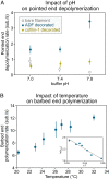
Impact of pH and temperature on reaction rates. (A) Depolymerization of actin filament pointed ends (15% labeled with AlexaFluor-568 on surface lysines) at different buffer pH (5 mM Tris⋅HCl, 50 mM KCl, 1 mM MgCl2, 0.4 mM CaCl2, 0.2 mM EGTA, 0.2 mM ATP, 10 mM DTT, 1 mM DABCO) supplemented with 1 to 2 µM ADF or cofilin-1 as indicated. Filaments are attached to the coverslip at their barbed end by gelsolin in a microfluidics chamber. Depolymerization of bare and ADF/cofilin-decorated filaments was recorded for up to 30 min at one frame every 30 to 60 s. Each data point represents the mean and SD over 14 to 36 filaments (from one experiment). Data adapted from ref. with permission from the American Chemical Society (ACS), further permission should be directed to the ACS. (B) Elongation of the actin filament barbed end from anchored spectrin-actin seeds in a microfluidics flow, at different temperatures, exposed to 1 µM Mg-ATP-actin (10% AlexaFluor-488 labeled on surface lysines) in buffer (5 mM Tris⋅HCl, pH 7.4, 50 mM KCl, 1 mM MgCl2, 0.2 mM EGTA, 0.2 mM ATP, 10 mM DTT, 1 mM DABCO). Elongation was recorded for 5 min at one frame every 10 s, and n > 30 filaments were analyzed for each condition. (Inset) The slope of the linear relation of log(elongation rate) as a function of the inverse of the temperature (Arrhenius plot) allows us to estimate the free energy associated with actin monomer addition at the barbed end to be 37.8 kJ/mol (9.03 kcal/mol), similar to the value reported by Drenckhahn and Pollard (100) using EM. Data can be found in Dataset S1. ADF, actin depolymerizing factor; EGTA, ethylene glycol tetraacetic acid; DTT, dithiothreitol; DABCO, diazabicyclooctane.

Similar articles

Cited by

References

    1. Bugyi B., Kellermayer M., The discovery of actin: “To see what everyone else has seen, and to think what nobody has thought.” J. Muscle Res. Cell Motil. 41, 3–9 (2020). - PMC - PubMed
    1. Depue R. H. Jr., Rice R. V., F-actin is a right-handed helix. J. Mol. Biol. 12, 302–303 (1965). - PubMed
    1. Pollard T. D., Mooseker M. S., Direct measurement of actin polymerization rate constants by electron microscopy of actin filaments nucleated by isolated microvillus cores. J. Cell Biol. 88, 654–659 (1981). - PMC - PubMed
    1. Pollard T. D., Rate constants for the reactions of ATP- and ADP-actin with the ends of actin filaments. J. Cell Biol. 103, 2747–2754 (1986). - PMC - PubMed
    1. Kouyama T., Mihashi K., Fluorimetry study of N-(1-pyrenyl)iodoacetamide-labelled F-actin. Local structural change of actin protomer both on polymerization and on binding of heavy meromyosin. Eur. J. Biochem. 114, 33–38 (1981). - PubMed

Publication types

LinkOut - more resources