. 2021 Oct 19;4(1):60-75.
doi: 10.1096/fba.2021-00005. eCollection 2022 Jan.

Differential ER stress as a driver of cell fate following ricin toxin exposure

Affiliations

Differential ER stress as a driver of cell fate following ricin toxin exposure

Claire Peterson-Reynolds et al. FASEB Bioadv. .

Abstract

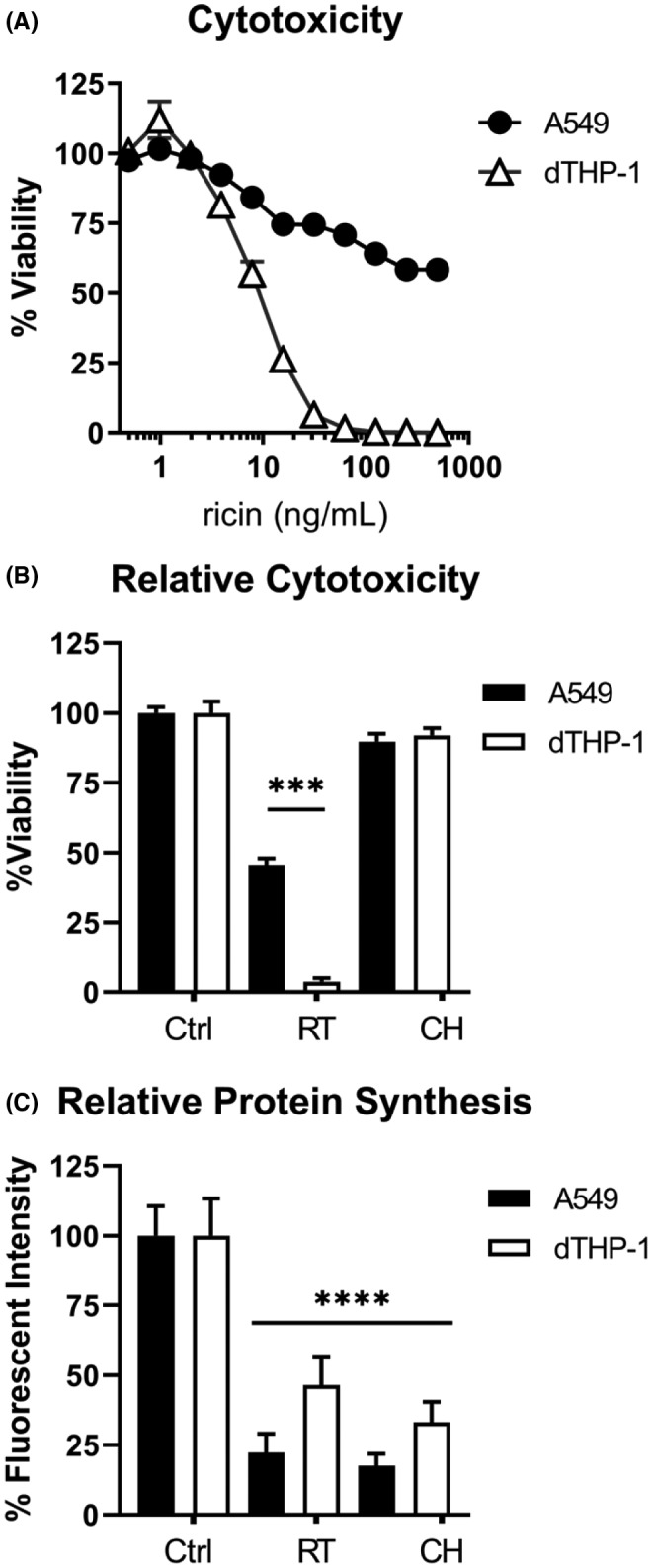
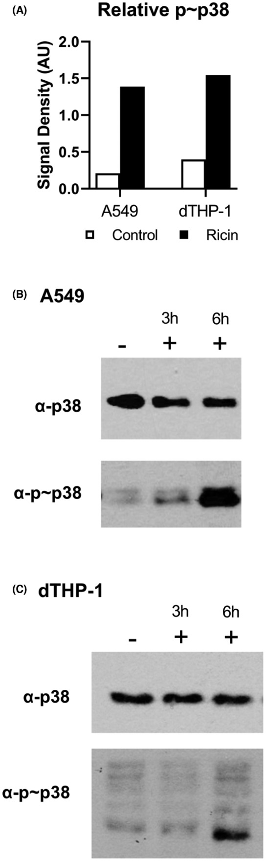
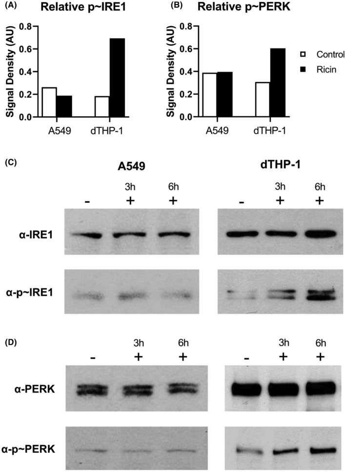
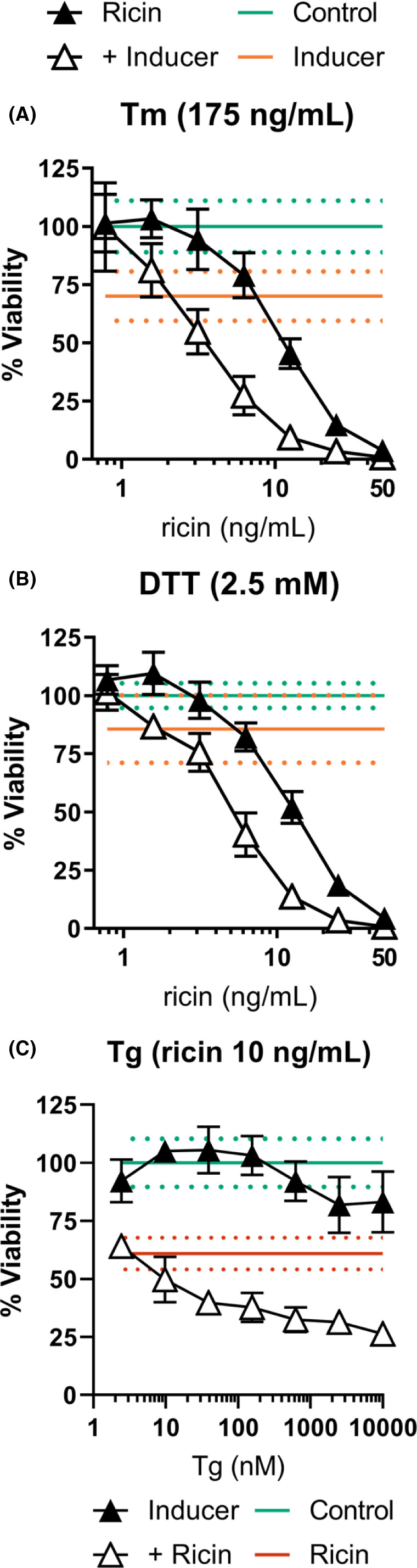
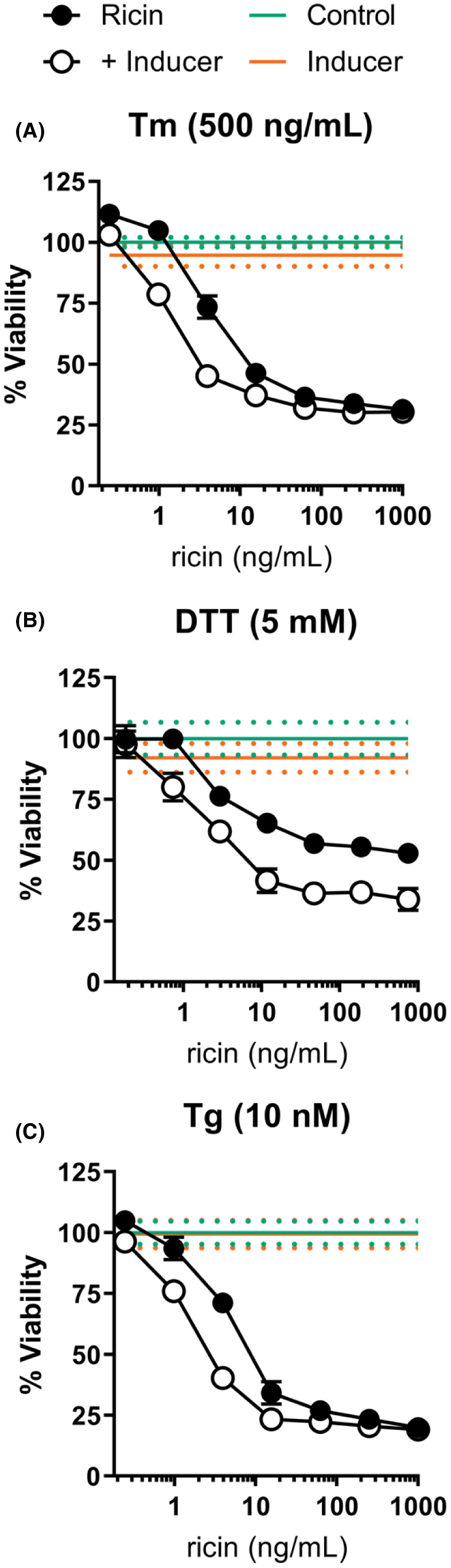
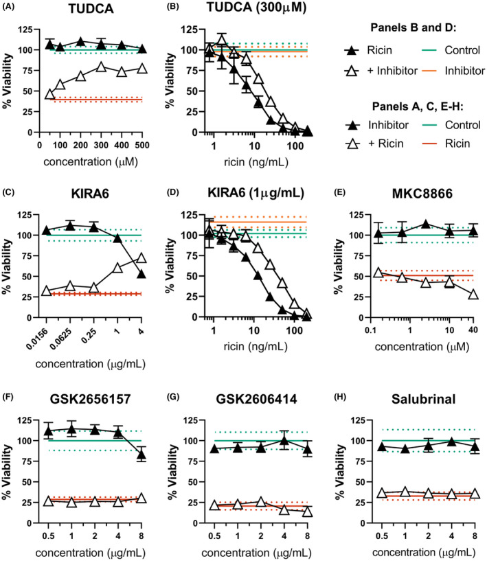
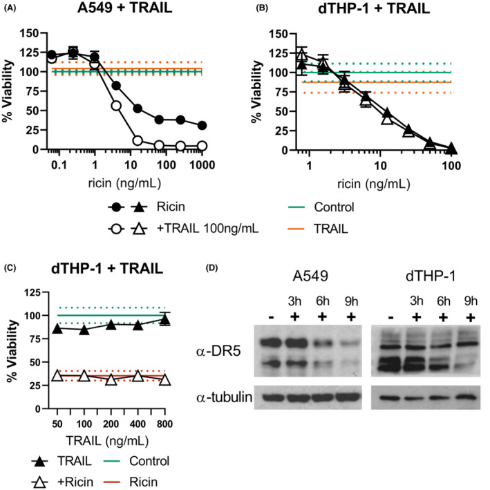
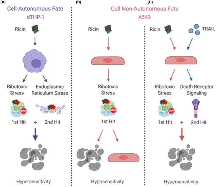
Inhalation of trace amounts of ricin toxin, a plant-derived ribosome-inactivating protein, results in ablation of alveolar macrophages, widespread epithelial damage, and the onset of acute respiratory distress syndrome (ARDS). While ricin's receptors are ubiquitous, certain cell types are more sensitive to ricin-induced cell death than others for reasons that remain unclear. For example, we demonstrate in side-by-side studies that macrophage-like differentiated THP-1 (dTHP-1) cells are hyper-sensitive to ricin, while lung epithelium-derived A549 cells are relatively insensitive, even though both cell types experience similar degrees of translational inhibition and p38 MAPK activation in response to ricin. Using a variety of small molecule inhibitors, we provide evidence that ER stress contributes to ricin-mediated cytotoxicity of dTHP-1 cells, but not A549 cells. On the other hand, the insensitivity of A549 cells to ricin was overcome by the addition of (TNF)-related apoptosis-inducing ligand (TRAIL; CD253), a known stimulator of extrinsic programmed cell death. These results have implications for understanding the complex pathophysiology of ricin-induced ARDS in that they demonstrate that intrinsic (e.g., ER stress) and extrinsic (e.g., TRAIL) factors may ultimately determine the fate of specific cell types following ricin intoxication.

Keywords: apoptosis; epithelium; inflammation; lung; macrophage; stress; toxin.


Conflict of interest statement

The authors have no financial or other competing interests to declare.

Figures

FIGURE 1
FIGURE 1
Comparative sensitivity of A549 and dTHP‐1 cells to ricin toxin. (A) Side‐by‐side 96‐well plate cytotoxicity assays were used to compare ricin sensitivity across a range of doses between A549 and dTHP‐1 cells. Cells were exposed to ricin for 4 h, washed, and allowed to recover for 24 h prior to assay development. Data presented are the mean of eight replicate wells with the 95% confidence interval. Viability as expressed by AUC ±95% CI (area under the curve with 95% confidence interval) was significantly different, with a value of 1428 ±93 for THP‐1 cells and 31210 ±1100 for A549 cells. (B) Cytotoxicity and (C) protein synthesis inhibition were compared side‐by‐side in both cell types following 4 h exposure to 20 ng/ml ricin (RT) or 50 µg/ml cycloheximide (CH). Circles represent A549 cells, triangles represent dTHP‐1 cells. In panel (B), viability was significantly reduced in both cell types following ricin treatment (p < 0.0001). When comparing ricin‐treated dTHP‐1 and A549 cells, dTHP‐1 cells exhibited a significantly greater reduction in viability (p < 0.0001). In panel (C), both RT and CH treatment significantly reduced protein synthesis compared to the untreated controls (p < 0.0001). Within each cell type, RT and CH treatment were equally effective at inhibiting protein synthesis. Although ricin treatment had a significantly greater suppressive effect on protein synthesis in A549 cells (p = 0.0065), its lethality was significantly lower in this cell type. (a) N = 7–8, (b) N = 14–16, (c) N = 8–10. The significance of “*** and ****” correspond to <0.001 and <0.0001, respectively
FIGURE 2
FIGURE 2
Ribotoxic stress is evident in both A549 and dTHP‐1 cells. (A) The relative abundance of phosphorylated p38 in response to 6 h ricin treatment (black bars, 20 ng/ml) was dramatically increased over the baseline level observed in untreated cells (white bars) in both A549 and dTHP‐1 cells. Bars represent semi‐quantitative densitometric analysis of the phosphorylated form of each protein relative to the total pool of that protein. (C, D) Digitized film images of western blots. Membrane swatches bearing electrophoresed whole cell lysate from A549 cells (left) and dTHP‐1 cells (right) were probed with anti‐phospho‐p38 monoclonal antibody (Cell Signaling Technology Cat# 4511, RRID:AB_2139682) and then stripped and re‐probed with anti‐p38 polyclonal antibody (Cell Signaling Technology Cat# 8690, RRID:AB_10999090). Representative blot results are shown from among three to four replicate experiments. −, indicates the absence of ricin; +, indicates the presence of ricin. Lysates were collected after 3 or 6 h of ricin exposure. Results from 3 h lysates were variable between experiments and were thus excluded from the bar graph. Refer to Figures S1 and S2 to observe the original films from which these images were derived
FIGURE 3
FIGURE 3
Evidence for ER stress in dTHP‐1 but not A549 cells. The relative abundance of phosphorylated IRE1 (A, C) and phosphorylated PERK (B, D) in response to 6 h ricin treatment (black bars; 20 ng/ml) or controls (white bars) in dTHP‐1 cells. Bars represent the semi‐quantitative signal density of the phosphorylated form of each protein relative to the total pool of that protein probed sequentially on the same membrane swatch. Digitized films of representative western blots selected from among three to four replicate experiments. Membrane swatches were probed with anti‐phospho‐PERK (Cell Signaling Technology Cat# 3179, RRID:AB_2095853) or anti‐IRE1 (Cell Signaling Technology Cat# 3294, RRID:AB_823545), then stripped and re‐probed with anti‐PERK (Cell Signaling Technology Cat# 3192, RRID:AB_2095847) or anti‐phospho‐IRE (Thermo Fisher Scientific Cat# PA1‐16927, RRID:AB_2262241) antibodies. Results at 3 h treatments were variable and thus were not included in the semi‐quantitative graphs in (A) and (B). Refer to Figures S3–S6 to observe the original films from which these images were derived
FIGURE 4
FIGURE 4
External induction of ER stress further sensitizes dTHP‐1 cells to ricin‐induced death. (A) dTHP‐1 cells treated with Tm (175 ng/ml) for 2 h followed by 2 h Tm +ricin resulted in significantly enhanced ricin‐induced cell death (AUC of 554.5 ±76.5 vs. 1504 ±103 of ricin‐only). (B) Treatment with DTT (2.5 mM) for 2 h followed by a 2 h ricin exposure significantly enhanced ricin‐induced cell death (AUC of 709.1 ±70.1 vs. 1677 ±73 of ricin‐only). (C) Treatment with varying concentrations of Tg for 2 h followed by a 2 h co‐treatment with ricin and Tg. Tg significantly increased ricin‐induced cell death at all concentrations but the two lowest concentrations tested (p ≤ 0.0014) without independent toxicity. Tm and DTT treatment exhibited significant independent toxicity, which was corrected for in the dual‐treatment curves. Closed symbols, ricin‐only; open symbols, dual treatment, error bars represent the 95% confidence interval (CI). Green line and dots represent the mean and 95% CI of control; orange line and dots represent the mean and 95% CI of the indicated ER stress‐inducing compound; red line and dots represent the mean and 95% CI of ricin treatment. Experiments were independently repeated in triplicate with eight replicate wells per treatment condition, with controls for independent effects included in each trial. Representative survival curves are shown. Graphical representation of the unnormalized data and results of pairwise statistical testing for treatment interactions are available in Figure S7
FIGURE 5
FIGURE 5
Treatment with ER stress inducers enhanced ricin toxicity in A549 cells. (A) A549 cells treated with Tm (500 ng/ml) for 2 h followed by a 4 h Tm +ricin co‐treatment significantly enhanced ricin‐induced cell death (AUC of 30,830 ±1609 vs. 33,962 ±1236 of ricin‐only). (B) Treatment with DTT (5 mM) for 2 h prior to a 4 h ricin exposure significantly enhanced ricin‐induced cell death (AUC of 27,082 ±3935 vs. 41,420 ±2200 of ricin‐only) (C) Treatment with Tg (10 nM) for 2 h followed by a 4 h Tg +ricin co‐treatment significantly enhanced ricin‐induced cell death (AUC of 20,541 ±1344 vs. 23,326 ±769 of ricin‐only). Closed symbols, ricin‐only; open symbols, dual treatment, error bars represent the 95% CI. The green line and dots represent the mean and 95% CI of control cell viability; orange line and dots represent the mean and 95% CI of cell viability for the indicated ER stress‐inducing compound. Tm and DTT treatment exhibited independent toxicity, which was corrected for in the combinatorial treatment curves. Experiments were independently repeated in triplicate with six to eight replicate wells per treatment condition with controls for independent effects included in each trial. Representative survival curves are shown. Graphical representation of the unnormalized data and results of pairwise statistical testing for treatment interactions are available in Figure S8
FIGURE 6
FIGURE 6
ER stress contributes to dTHP‐1 ricin sensitivity through IRE1 activity. dTHP‐1 cells were pre‐treated with the indicated ERS suppressing compound for 18 h prior to 2 h co‐treatment with ricin (A & C, 10 ng/ml; E–H,12.5 ng/ml). (A) Pre‐treatment with TUDCA was able to partially rescue ricin sensitivity in dTHP‐1 cells. Rescue was statistically significant from 200 µM (p ≤ 0.03). (B) Pre‐treatment with 300 µM TUDCA significantly reduced cytotoxicity of ricin exposure (AUC 3302 ±207 vs. 1604 ±174 of ricin‐only). (C) Specific inhibition of IRE1 with the small molecule KIRA6 partially rescued ricin‐induced dTHP‐1 cell death, with significant independent toxicity at doses exceeding 1 μg/ml. Rescue was statistically significant in all but the lowest doses (p < 0.0001). (D) Pre‐treatment with 1 µg/ml KIRA6 significantly reduced cytotoxicity of ricin exposure (AUC of 5879 ±386 vs. 2165 ±142 of ricin‐only) (E) Pre‐treatment with IRE1 RNase inhibitor MKC8866 enhanced ricin toxicity at 40 µM (p < 0.001) with no effect seen at lower doses. (F and G) Specific inhibition of PERK using the small molecule inhibitors GSK2656157 and GSK2606414 had no effect on ricin‐induced cell death. GSK2656157 exhibited significant independent toxicity above 8 μg/ml, while GSK2606414 was well tolerated. (H) Treatment with Salubrinal had no independent toxicity and no effect on ricin‐induced cell death. Green lines and dots and red lines and dots represent the mean and 95% CI of control‐ and ricin‐treated cells, respectively, while orange lines and dots similarly represent the independent effect of KIRA6 or TUDCA. Closed symbols, ricin‐only; open symbols, dual treatment; error bars represent the 95% CI. Data presented are the mean of at least six replicate wells. For (A), (C), and (E, F), points in the dual‐treatment group have been individually corrected for significant independent effects on viability by the inhibitor treatment as appropriate. Graphical representation of the unnormalized data (panels B, C, and D) and results of pairwise statistical testing for treatment interactions (panels B and D) are available in Figure S9
FIGURE 7
FIGURE 7
Impact of TRAIL on A549 and dTHP‐1 sensitivities to ricin. (A) Ricin +TRAIL co‐treatment sensitizes A549 cells to ricin‐induced cell death (AUC of 5278 ±321 vs. 36,190 ±4027 for ricin‐only treated), but (B) TRAIL has no effect on ricin cytotoxicity in dTHP‐1 cells (AUC of 1924 ±223 vs. 2161 ±167 for ricin‐only treated). (C) TRAIL co‐treatment failed to influence ricin‐induced dTHP‐1 cell death even at much higher doses (12.5 ng/ml ricin). (D) Western blots analysis of A549 and dTHP‐1 cells demonstrate DR5/TRAILR2 expression in both cell types, the primary death receptor for TRAIL (Cell Signaling Technology Cat# 8074, RRID:AB_10950817). α‐tubulin served as the loading control (Cell Signaling Technology Cat# 5346, RRID:AB_1950376). Closed symbols, ricin‐only; open symbols, dual treatment, error bars represent the 95% CI. Green lines and dots and red lines and dots represent the mean and 95% CI of control‐ and ricin‐treated cells, respectively. Orange lines and dots represent the independent effect of TRAIL. Data presented are the mean of eight replicate wells with the 95% confidence interval. Graphical representation of the unnormalized data and results of pairwise statistical testing for treatment interactions are available in Figure S10. Refer to Figure S11 to observe the original films from which DR5 blot images were derived
FIGURE 8
FIGURE 8
Two two‐hit models to explain cell type‐specific ricin sensitivity. Under normal conditions, dTHP‐1 cells exhibit ricin hypersensitivity (A), while A549 cells exhibit ricin hyposensitivity (B) based on the percent of cultured cells remaining alive after ricin challenge. We have identified ER stress and resulting activation of the unfolded protein response as a contributing factor to the unique sensitivity of dTHP‐1 cells, which are efficiently killed by cell‐autonomous ricin‐induced pro‐apoptotic signaling. Supplementation with TRAIL, a source of extrinsic pro‐apoptotic activation, is sufficient to induce cell non‐autonomous ricin hypersensitivity in A549 cells (C). Addition of TRAIL to dTHP‐1 cells had no effect on their response to ricin, further supporting their fully cell‐autonomous cell fate determination

Similar articles

Cited by

References

    1. Gal Y, Mazor O, Falach R, Sapoznikov A, Kronman C, Sabo T. Treatments for pulmonary ricin intoxication: current aspects and future prospects. Toxins (Basel). 2017:9:311. - PMC - PubMed
    1. Sapoznikov A, Gal Y, Falach R, et al. Early disruption of the alveolar‐capillary barrier in a ricin‐induced ARDS mouse model: neutrophil‐dependent and ‐independent impairment of junction proteins. Am J Physiol Lung Cell Mol Physiol. 2019;316:L255‐L268. - PubMed
    1. Wong J, Magun BE, Wood LJ. Lung inflammation caused by inhaled toxicants: a review. Int J Chron Obstruct Pulmon Dis. 2016;11:1391‐1401. - PMC - PubMed
    1. Rapak A, Falnes PO, Olsnes S. Retrograde transport of mutant ricin to the endoplasmic reticulum with subsequent translocation to cytosol. Proc Natl Acad Sci USA. 1997;94:3783‐3788. - PMC - PubMed
    1. Sowa‐Rogozińska N, Sominka H, Nowakowska‐Gołacka J, Sandvig K, Słomińska‐Wojewódzka M Intracellular transport and cytotoxicity of the protein toxin ricin. Toxins. 2019;11(6):350. - PMC - PubMed

LinkOut - more resources