. 2022 Mar 9;96(5):e0182721.
doi: 10.1128/JVI.01827-21. Epub 2022 Jan 12.

Human Cytomegalovirus Hijacks WD Repeat Domain 11 for Virion Assembly Compartment Formation and Virion Morphogenesis

Affiliations

Human Cytomegalovirus Hijacks WD Repeat Domain 11 for Virion Assembly Compartment Formation and Virion Morphogenesis

Bo Yang et al. J Virol. .

Abstract

Human cytomegalovirus (HCMV) has a large (∼235 kb) genome with more than 200 predicted open reading frames that exploits numerous cellular factors to facilitate its replication. A key feature of HCMV-infected cells is the emergence of a distinctive membranous cytoplasmic compartment termed the virion assembly compartment (vAC). Here, we report that host protein WD repeat domain 11 (WDR11) plays a key role in vAC formation and virion morphogenesis. We found that WDR11 was upregulated at both mRNA and protein levels during HCMV infection. At the late stage of HCMV replication, WDR11 relocated to the vAC and colocalized with markers of the trans-Golgi network (TGN) and vAC. Depletion of WDR11 hindered HCMV-induced membrane reorganization of the Golgi and TGN, altered vAC formation, and impaired HCMV secondary envelopment and virion morphogenesis. Further, motifs critical for the localization of WDR11 in TGN were identified by alanine-scanning mutagenesis. Mutation of these motifs led to WDR11 mislocation outside the TGN and loss of vAC formation. Taken together, these data indicate that host protein WDR11 is required for efficient viral replication at the stage of virion assembly, possibly by facilitating the remodeling of the endomembrane system for vAC formation and virion morphogenesis. IMPORTANCE During the late phase of human cytomegalovirus (HCMV) infection, the endomembrane system is dramatically reorganized, resulting in the formation of a unique structure termed the virion assembly compartment (vAC), which is critical for the assembly of infectious virions. The mechanism of HCMV-induced vAC formation is still not fully understood. In this report, we identified a host factor, WDR11, that plays an important role in vAC formation. Our findings argue that WDR11 contributes to the relocation of the Golgi and trans-Golgi network to the vAC, a membrane reorganization process that appears to be required for efficient virion maturation. The present work provides new insights into the vAC formation and HCMV virion morphogenesis and a potential novel target for antiviral treatment.

Keywords: WDR11; human cytomegalovirus; virion assembly compartment; virion morphogenesis; virion structure.


Conflict of interest statement

The authors declare no conflict of interest.

Figures

FIG 1
FIG 1
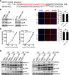
Human cytomegalovirus (HCMV) infection induces WDR11. HFFs were mock-infected (M) or infected with HCMV Towne strain (V) at a multiplicity of infection (MOI) of 3 and then harvested at the times indicated. (A) WDR11 mRNA levels were determined by quantitative reverse transcriptase PCR (qRT-PCR) and normalized to levels in mock-infected cells at 6 hours postinfection (hpi). (B) Infected cell lysates were assayed for the indicated proteins by immunoblotting (IB). Protein levels determined by densitometry and normalized to mock-infected cells at 24 h are graphed below. (C) HFFs were mock-infected (M), HCMV-infected (V), or infected with UV-inactivated HCMV (UV) at an MOI of 3 in the presence of vehicle (dimethyl sulfoxide [DMSO]), ganciclovir (GCV), or phosphonoacetic acid (PAA). At 72 hpi, samples were collected and analyzed by IB for the indicated proteins. Protein levels determined by densitometry and normalized to mock-infected cells are graphed below. Glyceraldehyde-3-phosphate dehydrogenase (GAPDH) serves as a loading control. The data were collected from three independent experiments and analyzed by one-way analysis of variance (ANOVA); the results are presented as averages ± standard deviation (SD). **, P < 0.01; ***, P < 0.001; NS, not significant; IE, immediate-early; gB, glycoprotein B.
FIG 2
FIG 2
Knockdown of WDR11 results in reduced virion yield. (A) Efficiency of WDR11 knockdown. Human foreskin fibroblasts (HFFs) were transduced with lentiviruses expressing scrambled shRNA (shScram) or WDR11-specific shRNAs (shW1 and shW2), and WDR11 levels were examined by IB. The WDR11 protein levels relative to shScram are listed below each blot. (B) Cell viability. Cell viability was determined by 3-(4,5-dimethylthiazol-2-yl)-2,5-diphenyltetrazolium bromide (MTT) assay. Data were collected at the indicated times from three independent experiments and analyzed by one-way ANOVA. The results are presented as means ± SD. NS, not significant. (C, D) Growth kinetics of virus. Control cells (shScram) and WDR11 knockdown cells (shW1 and shW2) were infected with HCMV at MOIs of 3 or 0.05, and supernatants collected at the indicated times postinfection were titrated by plaque assay. The results were obtained from three independent experiments, each conducted in triplicate and analyzed by one-way ANOVA. **, P < 0.01; ***, P < 0.001.
FIG 3
FIG 3
Effect of WDR11 overexpression (OE) and knockout (KO) on viral entry, genome replication, and viral gene expression. (A) Sequence of genomic target site of WDR11 knockout clone. DNA was extracted from CTL and WDR11-KO cells, PCR-amplified with WDR11-specific primers, and sequenced. The PAM motif is underlined, and the gRNA sequence is in red. (B) HELFs were transduced with lentiviral vectors that overexpress WDR11 (WDR11-OE) or knock out WDR11 (WDR11-KO) or relative empty vector (Vector and CTL), then exposed to HCMV at an MOI of 5 for 1 h at 4°C to allow virus attach, and then transferred to 37°C incubation for 1 h to allow virus entry. Finally, cells were washed by citric acid (pH 3) to remove noninternalized virus and harvested by trypsinization. WDR11 and cell-associated pp65 levels were determined by IB. The numbers below each blot indicate WDR11 or pp65 levels, determined by densitometry, normalized to GAPDH and to controls. (C) Viral entry determined by pp65 staining. Cells on coverslips were infected with HCMV at an MOI of 1 and fixed 6 hpi. HCMV pp65 protein (red) was detected by immunofluorescence assay (IFA), and the nuclei were counterstained with 4´,6-diamidino-2-phenylindole (DAPI) (blue). Scale bar, 50 μm. Total cells and nuclear pp65-positive cells were counted in 20 random fields selected from each experiment. The data were analyzed by one-way ANOVA. Representative images (left panel) and quantifications (right panel) are shown. The results are medians ± SD. NS, not significant. (D) Cells were infected with HCMV at an MOI of 1 and HCMV genome copy numbers were determined by quantitative PCR (qPCR) and analyzed by one-way ANOVA. The results shown are means ± SD. (E) Viral protein level. Cells were infected with HCMV at an MOI of 1, and lysates were analyzed by IB to detect the indicated proteins. The results shown are representative of three independent experiments.
FIG 4
FIG 4
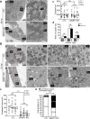
Analysis of infected cells by transmission electron microscopy (TEM). Cells infected with HCMV at an MOI of 1 were analyzed by TEM. (A) Sections prepared 96 hpi were imaged at 1,700× (panels a and b), and nuclear compartments containing capsids (indicated by white dashed boxes) were enlarged to 7,800× (panels a1 and b1). Total numbers of capsids located within 10 nuclei from each culture were quantitated (panel c). The numbers of A, B, or C capsids in 10 nuclei from each culture were determined based on capsid morphology, and the percentage of each capsid type was calculated (panel d). (B) Sections prepared 120 hpi were imaged at 1,700× (panels a and b), and cytoplasmic compartments containing capsids (indicated by white-dashed boxes) were enlarged to 7,800× (panels a1 to a3 and b1 to b3). The magnified images of the matured virions (panel a3, indicated with arrowhead) and unenveloped virions (panel b3, indicated with arrow) are presented in panels a4 and b4, respectively. Typical (panel a) and abnormal vAC (panel b) are delineated by dotted red lines. Total numbers of cytoplasmic capsids in 10 cells from each culture were quantitated (panel c). Medians for three independent experiments are indicated to the right of each plot, and the means of the three experiments are labeled above each group. The data were analyzed by Kruskal-Wallis test. NS, not significant; ***, P < 0.001. Total numbers of enveloped or nonenveloped capsids in 10 cells from each culture were quantitated percentages of each were calculated (d). The data were analyzed by chi-square test. n, total numbers of capsids analyzed; ***, P < 0.001.
FIG 5
FIG 5
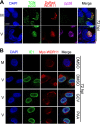
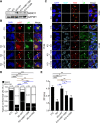
Subcellular localization of WDR11 redistribution to vAC. (A) Human telomerase (hTERT)-immortalized HEL cells (HELFs) were transfected with a plasmid expressing a DsRed-WDR11 fusion protein then mock-infected (M) or HCMV-infected (V) at an MOI of 1. At 72 hpi, the cells were fixed and costained with antibodies to viral protein pp28 (purple) and trans-Golgi network (TGN) marker p230 (green). Nuclei were counterstained with DAPI (blue). Scale bar, 10 μm. (B) HELFs were transfected with a plasmid expressing a Myc-WDR11 fusion protein and infected as in panel A in the presence of dimethyl sulfoxide (DMSO), ganciclovir (GCV) (100 μg/ml), or phosphonoacetic acid (PAA) (100 μg/ml). At 72 hpi, the cells were fixed and costained with antibodies to Myc (red) and the HCMV IE1 protein (green). The nuclei were counterstained with DAPI (blue). Scale bar, 10 μm.
FIG 6
FIG 6
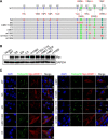
Alteration of WDR11 subcellular localization by mutagenesis of sorting motifs. (A) Illustration of WDR11 protein sequence showing putative sorting signals that were targeted by alanine substitutions in mutants indicated below. [DE]XXXL[LI] motifs are shown in green, tyrosine-based motifs (YXXØ) are blue, and amino acids substituted with alanines are red. (B) HEK293T cells were transfected with plasmids expressing wild-type WDR11 (WT) or the indicated WDR11 mutants and analyzed by immunoblotting (IB) 48 h posttransfection. (C) HELF cells were transfected as in panel B and costained by IF for TGN (green) and Myc (red). The nuclei were counterstained with DAPI (blue). Scale bar, 10 μm.
FIG 7
FIG 7
WDR11 mutants with altered localization lower virus yields. (A) WDR11-KO cells (KO) were transduced with lentiviruses encoded wild-type WDR11 (WT) or mutants m967–1006 and m1069 and selected in G418 sulfate (500 μg/ml; catalog no. A600958, Sangon Biotech) for 1 week and generated KO+WT, KO+m967–1006, and KO+m1069 cells. Cell lysates were prepared and analyzed by IB to detect WDR11. (B and C) CTL, KO, KO+WT, KO+m967–1006, and KO+m1069 cells were infected with HCMV at an MOI = 1. At 96 hpi, cells were fixed and costained by IF for pp65 (red) and gM (green) in panel B or TGN (red), MCP (cyan), and gB (green) in panel C. The nuclei were counterstained with DAPI (blue). White arrowheads indicate typical vACs; white arrows indicate abnormal vACs. Scale bar, 10 μm. (D) Based on morphology of gB staining shown in panels B and C, vACs were classified as typical or abnormal, quantitated, and analyzed by chi-square test. n, number of infected cells used for quantitation; ***, P < 0.001. (E) Culture supernatants from CTL or WDR11-KO cells that were transfected and infected as in panels B and C were collected at 96 hpi and titrated by plaque assay. The results shown were from three independent experiments each conducted with triplicates. Significance was analyzed by one-way ANOVA. *, P < 0.05; **, P < 0.01; ***, P < 0.001.
FIG 8
FIG 8
Deletion of WDR11 alters Golgi recruitment to vAC. CTL and WDR11-KO cells were infected by mock (A, B) or by HCMV at an MOI of 1 (C, D). The cells were harvested at 96 hpi and fractionated as described under Material and Methods. Fractions were analyzed by IB for markers for early endosome (EE, early endosome antigen 1 [EEA1]), multivesicular body (MVB, hepatocyte growth factor-regulated tyrosine kinase substrate [HGS]), endoplasmic reticulum (ER, calnexin [CANX]), or Golgi (GM130), and viral vAC markers gB and pp28. In each fraction, protein levels were quantified by densitometry, and viral genome copies were determined by qPCR. All were expressed as percentages of the totals and plotted together versus fraction number.
FIG 9
FIG 9
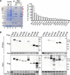
Identification of HCMV proteins that interact with WDR11. (A) HFFs were transduced with a Myc-WDR11-expressing lentivirus and mock-infected (M) or HCMV-infected (V) with an MOI of 1. At 72 hpi, cell lysates were immunoprecipitated (IP) with anti-Myc antibody followed by SDS-PAGE and Coomassie brilliant blue staining (left). Infected-cell-specific bands (arrows) in lane 6 were excised and analyzed by LC-MS/MS. The viral proteins identified and their corresponding spectral counts are shown (right panel). (B) HEK293T cells were cotransfected with plasmids expressing Myc-WDR11 and Flag-tagged viral proteins or empty vector. At 48 h, posttransfection lysates were immunoprecipitated using anti-Myc antibody, and both input and immunoprecipitated samples were immunoblotted and probed with antibodies to Myc, Flag, or GAPDH.

Similar articles

Cited by

References

    1. Cobbs CS, Harkins L, Samanta M, Gillespie GY, Bharara S, King PH, Nabors LB, Cobbs CG, Britt WJ. 2002. Human cytomegalovirus infection and expression in human malignant glioma. Cancer Res 62:3347–3350. - PubMed
    1. Wen L, Qiu Y, Cheng S, Jiang X, Ma YP, Fang W, Wang W, Cui J, Ruan Q, Zhao F, Hu F, Luo MH. 2018. Serologic and viral genome prevalence of HSV, EBV, and HCMV among healthy adults in Wuhan, China. J Med Virol 90:571–581. 10.1002/jmv.24989. - DOI - PubMed
    1. Lazzarotto T, Guerra B, Gabrielli L, Lanari M, Landini MP. 2011. Update on the prevention, diagnosis and management of cytomegalovirus infection during pregnancy. Clin Microbiol Infect 17:1285–1293. 10.1111/j.1469-0691.2011.03564.x. - DOI - PubMed
    1. Jean Beltran PM, Cristea IM. 2014. The life cycle and pathogenesis of human cytomegalovirus infection: lessons from proteomics. Expert Rev Proteomics 11:697–711. 10.1586/14789450.2014.971116. - DOI - PMC - PubMed
    1. Tandon R, Mocarski ES. 2012. Viral and host control of cytomegalovirus maturation. Trends Microbiol 20:392–401. 10.1016/j.tim.2012.04.008. - DOI - PMC - PubMed

Publication types

MeSH terms