. 2021 Dec 30;14(1):173.
doi: 10.3390/cancers14010173.

The Tumour Suppressor CYLD Is Required for Clathrin-Mediated Endocytosis of EGFR and Cetuximab-Induced Apoptosis in Head and Neck Squamous Cell Carcinoma

Affiliations

The Tumour Suppressor CYLD Is Required for Clathrin-Mediated Endocytosis of EGFR and Cetuximab-Induced Apoptosis in Head and Neck Squamous Cell Carcinoma

Rin Liu et al. Cancers (Basel). .

Abstract

Epidermal growth factor receptor (EGFR) is frequently overexpressed in head and neck squamous cell carcinoma (HNSCC) and is a target for the therapeutic antibody cetuximab (CTX). However, because only some patients have a significant clinical response to CTX, identification of its predictive biomarkers and potentiation of CTX-based therapies are important. We have recently reported a frequent downregulation of cylindromatosis (CYLD) in primary HNSCC, which led to increased cell invasion and cisplatin resistance. Here, we show that CYLD located mainly in lipid rafts was required for clathrin-mediated endocytosis (CME) and degradation of the EGFR induced by EGF and CTX in HNSCC cells. The N-terminus containing the first cytoskeleton-associated protein-glycine domain of CYLD was responsible for this regulation. Loss of CYLD restricted EGFR to lipid rafts, which suppressed CTX-induced apoptosis without impeding CTX's inhibitory activity against downstream signalling pathways. Disruption of the lipid rafts with cholesterol-removing agents overcame this resistance by restoring CME and the degradation of EGFR. Regulation of EGFR trafficking by CYLD is thus critical for the antitumour activity of CTX. Our findings suggest the usefulness of a combination of cholesterol-lowering drugs with anti-EGFR antibody therapy in HNSCC.

Keywords: CYLD; EGFR; cetuximab; clathrin-mediated endocytosis; head and neck squamous cell carcinoma.


Conflict of interest statement

The authors declare no conflict of interest.

Figures
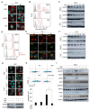
Figure 1
Figure 1
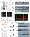
CME of EGFR is essential for CTX-induced apoptosis. (A,B) Localization of EGFR after EGF stimulation. HSC3, Ca9-22, and TSU cells were stimulated with 100 ng/mL EGF for 60 min. (A) EGFR localization was analysed via immunofluorescence staining. Scale bars, 10 μm. (B) Cell-surface EGFR was analysed using flow cytometry. (C) Amount of total EGFR after EGF stimulation. Cells were stimulated with 100 ng/mL EGF for the indicated times and total EGFR expression was analysed by using Western blotting. (D) Cell-surface EGFR expression after CTX treatment. Cells were treated with 100 μg/mL CTX for 60 min, and then cell-surface EGFR was stained with an anti-EGFR antibody (clone LA1). (E) Co-localization of EGFR with endosomes and lysosomes after CTX treatment. The localizations of EGFR and the endosome and lysosome markers were analysed via immunofluorescence staining after 100 μg/mL CTX treatment for 30 min (Rab5 and Rab7) or 60 min (LAMP1). Scale bars, 10 μm. (F) Amount of total EGFR expression after EGF stimulation. Cells were stimulated with 100 μg/mL CTX for the indicated times and total EGFR expression was analysed by using Western blotting. (G) Effects of CPZ on EGFR internalization and degradation after EGF or CTX treatment. HSC3 cells were pretreated with 5 μM CPZ for 30 min and were then stimulated with 100 ng/mL of EGF or 100 μg/mL of CTX for 60 min. CHX was added before adding CPZ. The localization of EGFR was analysed via immunofluorescence staining (upper panels). Total EGFR expression was analysed via Western blotting (lower panels). Scale bars, 10 μm. (H) Apoptosis after CTX given with CPZ. HSC3 cells were cultured in the presence of 5 μM CPZ for 30 min, followed by incubation with 100 μg/mL of CTX for 12 h in serum-free medium. Cells were harvested and stained with Annexin V-APC and 7-AAD. NT, no treatment. Bars indicate the percentage of apoptotic cells. * p < 0.05. (I) Phosphorylation of EGFR and major downstream molecules after CTX given with CPZ. Cells were pretreated with 5 μM CPZ for 30 min, after which they were treated with 100 μg/mL of CTX for the indicated times.
Figure 2
Figure 2
CYLD downregulation inhibits EGF- and CTX-induced CME of EGFR. (A) CYLD and EGFR expression after CYLD knockdown. HSC3, Ca9-22, and TSU cells were transfected with siRNA and then incubated for 48 h. The expression of total CYLD and EGFR was analysed by using Western blotting (left panels). Cell-surface EGFR expression was analysed by means of flow cytometry (right panels). (B) Internalization of EGFR after EGF or CTX treatment in CYLD-downregulated cells. HSC3 cells were treated with 100 ng/mL of EGF or 100 μg/mL of CTX for 60 min. EGFR localization was analysed via immunofluorescence staining (upper panels). Cell-surface EGFR expression was analysed via flow cytometry (lower panels). Scale bars, 10 μm. (C) Total EGFR expression levels after treatment with EGF or CTX in CYLD-downregulated cells. HSC3, Ca9-22, and TSU cells were transfected with siRNA and were then stimulated with 100 ng/mL of EGF or 100 μg/mL of CTX for the indicated times before harvesting. The cell lysate was immunoblotted with antibodies against the indicated proteins. (D) EGFR and CTX localization in CYLD-downregulated cells. HSC3, Ca9-22, and TSU cells were transfected with siRNA and were then treated with 100 μg/mL of CTX for 60 min. Immunofluorescence staining was used to analyse the localization of EGFR and CTX. Scale bars, 20 μm. (E) Cell viability after CTX treatment in CYLD-downregulated cells. HSC3, Ca9-22, and TSU cells were transfected with siRNA for 48 h before treatment with 100 μg/mL of CTX for 72 h in serum-free medium. * p < 0.05; † p < 0.01; § p < 0.005 (siCtrl vs. siCYLD or siCYLD-UTR). (F) Apoptosis after CTX treatment in CYLD-downregulated cells. HSC3, Ca9-22, and TSU cells were transfected with siRNA and incubated for 48 h. The medium was changed to serum-free medium and then 100 μg/mL of CTX was added. After a 12 h incubation with CTX, cells were harvested and analysed by means of Annexin V-APC and 7-AAD. * p < 0.01; † p < 0.001; § p < 0.005. (G) Phosphorylation changes in EGFR and the downstream signalling molecules after treatment with CTX in CYLD-downregulated cells. HSC3 cells were transfected with siRNA and incubated for 48 h. The medium was changed to serum-free medium, and cells were then treated with 100 μg/mL of CTX for the indicated times. Cell lysates were immunoblotted with antibodies against the indicated proteins.
Figure 3
Figure 3
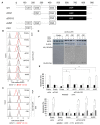
Impact of CYLD domain deficiency on CTX’s efficacy. (A) Domain structure of human CYLD protein and constructs encoding deletion mutants of CYLD. Three CG domains (CG1–3) and the USP domain are shown as white and black boxes, respectively. (B) The effects of CYLD mutants on cell-surface EGFR expression after EGF stimulation. HSC3 cells were co-transfected with CYLD deletion mutants and siRNA. After a 48 h incubation, cells were stimulated with 100 ng/mL of EGF for 15 min. Flow cytometry was used to analyse cell-surface EGFR expression. (C) The effects of WT-CYLD and CG1 constructs on cell-surface EGFR expression after EGF stimulation in combination with CPZ. HSC3 cells were co-transfected with WT-CYLD or CYLD mutants and siRNA. After a 48 h incubation, cells were stimulated with 100 ng/mL of EGF for 15 min. CPZ (5 μM) was added 30 min before adding the EGF. Flow cytometry was used to analyse cell-surface EGFR expression. (D) The effects of CYLD mutants on total EGFR expression. HSC3 cells were co-transfected with WT-CYLD or CG1 constructs and siRNA. After incubation for 48 h, the medium was changed to a serum-free medium and then the incubation continued for 12 h. Cells were stimulated with 100 ng/mL of EGF or 100 μg/mL of CTX for 60 min and were analysed by immunoblotting. (E,F) The effects of CYLD mutants on apoptosis induced by CTX. HSC3 cells were co-transfected with WT-CYLD or CYLD deletion constructs and siRNA. After incubation for 12 h with 100 μg/mL of CTX in serum-free medium, cells were harvested and analysed with Annexin V-APC and 7-AAD. * p < 0.001 (E). CPZ (5 μM) was added 30 min before adding CTX. * p < 0.05; † p < 0.01; § p < 0.0001 (F).
Figure 4
Figure 4
CYLD expression and subcellular EGFR localization in human HNSCC tissues. (A) Relationship between CYLD expression score and membrane EGFR score in primary HNSCC tissues. (B) Percentage of specimens with low or high CYLD expression scores compared with membrane EGFR scores. The membrane EGFR score was determined based on the percentage of tumour cells showing dominant EGFR localization in the cell membranes (see Materials and Methods for scoring details). * p < 0.01 (Pearson’s χ2 test). (C) Examples of high CYLD expression for low membrane EGFR scores and low CYLD expression for high membrane EGFR scores. The images on the right provide enlargements of the boxed areas in the middle images. Scale bars, 100 μm.
Figure 5
Figure 5
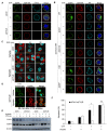
Effect of cholesterol depletion on CYLD-downregulated cells. (A,B) Localization of EGFR, lipid rafts, and CYLD (A) or anti-hemagglutinin (HA) (B) as analysed by immunofluorescence staining. HSC3 cells were transfected with the indicated siRNA and plasmids expressing deletion constructs of CYLD. After incubation for 48 h, cells were starved for 12 h in a serum-free medium. Cells were then stained with the appropriate antibodies and observed under fluorescent microscopy. Scale bars, 10 µm. (C,D) Effects of nystatin on EGF- or cetuximab (CTX)-induced EGFR endocytosis. HSC3 cells were transfected with siRNA and incubated for 48 h, then, after 12 h of incubation in a serum-free medium, cells were pretreated with 25 µg/mL of nystatin for 30 min before stimulation with 100 ng/mL of EGF for the indicated times (C) or 100 µg/mL of CTX for 60 min (D). EGFR localization was analysed via immunofluorescence staining. Scale bars, 20 µm. (E) Effects of nystatin on total EGFR expression after CTX treatment in CYLD-downregulated cells. HSC3 cells were transfected with siRNA and were then incubated for 48 h. Cells were pretreated with 25 µg/mL of nystatin for 30 min before treatment with 100 µg/mL of CTX for the indicated times. Total EGFR protein expression was analysed via Western blotting. CHX was added 1 h before nystatin treatment. (F) Effects of nystatin on CTX-induced apoptosis. HSC3 cells were transfected with siRNA and then incubated for 48 h. Cells were pretreated with 25 µg/mL of nystatin for 30 min before treatment with 100 µg/mL of CTX in a serum-free medium for 12 h. Apoptosis was analysed using Annexin V-APC and 7-AAD. * p < 0.001; n.s., not significant.

Similar articles

Cited by

References

    1. Joseph A.W., D’Souza G. Epidemiology of human papillomavirus-related head and neck cancer. Otolaryngol. Clin. N. Am. 2012;45:739–764. doi: 10.1016/j.otc.2012.04.003. - DOI - PubMed
    1. van der Heijden M., Essers P., de Jong M.C., de Roest R.H., Sanduleanu S., Verhagen C.V., Vens C. Biological determinants of chemo-radiotherapy response in HPV-negative head and neck cancer: A multicentric external validation. Front. Oncol. 2020;9:1470. doi: 10.3389/fonc.2019.01470. - DOI - PMC - PubMed
    1. Machiels J.P., Leemans C.R., Golusinski W., Grau C., Licitra L., Gregoire V. Squamous cell carcinoma of the oral cavity, larynx, oropharynx and hypopharynx: EHNS-ESMO-ESTRO Clinical Practice Guidelines for diagnosis, treatment and follow-up. Ann. Oncol. 2020;31:1462–1475. doi: 10.1016/j.annonc.2020.07.011. - DOI - PubMed
    1. Lerch S., Berthold S., Ziemann F., Dreffke K., Subtil F.S., Senger Y., Jensen A., Engenhart-Cabillic R., Dikomey E., Wittig A., et al. HPV-positive HNSCC cell lines show strongly enhanced radiosensitivity after photon but not after carbon ion irradiation. Radiother. Oncol. 2020;151:134–140. doi: 10.1016/j.radonc.2020.07.032. - DOI - PubMed
    1. Meccariello G., Maniaci A., Bianchi G., Cammaroto G., Iannella G., Catalano A., Vicini C. Neck dissection and trans oral robotic surgery for oropharyngeal squamous cell carcinoma. Auris Nasus Larynx. 2021;S0385-8146:00163-2. doi: 10.1016/j.anl.2021.05.007. - DOI - PubMed