. 2022 Jan:197:105232.
doi: 10.1016/j.antiviral.2021.105232. Epub 2021 Dec 29.

S-adenosylmethionine-dependent methyltransferase inhibitor DZNep blocks transcription and translation of SARS-CoV-2 genome with a low tendency to select for drug-resistant viral variants

Affiliations

S-adenosylmethionine-dependent methyltransferase inhibitor DZNep blocks transcription and translation of SARS-CoV-2 genome with a low tendency to select for drug-resistant viral variants

Ram Kumar et al. Antiviral Res. 2022 Jan.

Abstract

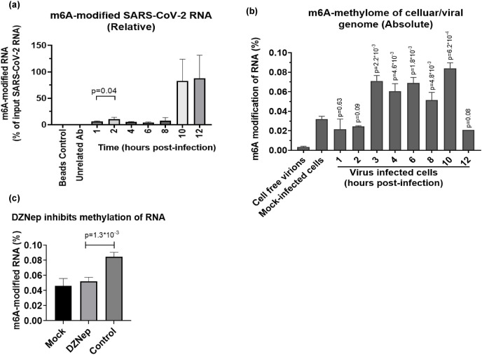
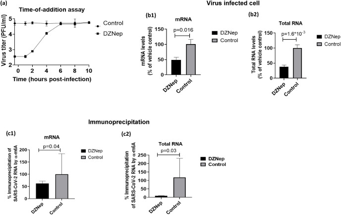
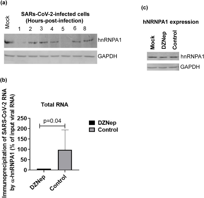
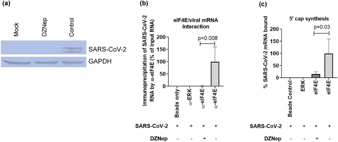
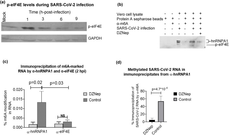
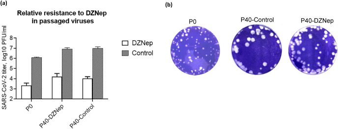
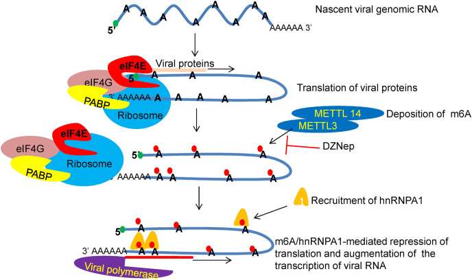
We report the in vitro antiviral activity of DZNep (3-Deazaneplanocin A; an inhibitor of S-adenosylmethionine-dependent methyltransferase) against SARS-CoV-2, besides demonstrating its protective efficacy against lethal infection of infectious bronchitis virus (IBV, a member of the Coronaviridae family). DZNep treatment resulted in reduced synthesis of SARS-CoV-2 RNA and proteins without affecting other steps of viral life cycle. We demonstrated that deposition of N6-methyl adenosine (m6A) in SARS-CoV-2 RNA in the infected cells recruits heterogeneous nuclear ribonucleoprotein A1 (hnRNPA1), an RNA binding protein which serves as a m6A reader. DZNep inhibited the recruitment of hnRNPA1 at m6A-modified SARS-CoV-2 RNA which eventually suppressed the synthesis of the viral genome. In addition, m6A-marked RNA and hnRNPA1 interaction was also shown to regulate early translation to replication switch of SARS-CoV-2 genome. Furthermore, abrogation of methylation by DZNep also resulted in defective synthesis of the 5' cap of viral RNA, thereby resulting in its failure to interact with eIF4E (a cap-binding protein), eventually leading to a decreased synthesis of viral proteins. Most importantly, DZNep-resistant mutants could not be observed upon long-term sequential passage of SARS-CoV-2 in cell culture. In summary, we report the novel role of methylation in the life cycle of SARS-CoV-2 and propose that targeting the methylome using DZNep could be of significant therapeutic value against SARS-CoV-2 infection.

Keywords: DZNep; Drug resistance; Epitranscriptomic; SARS-CoV-2; Virus replication.


Conflict of interest statement

The authors declare that they have no known competing financial interests or personal relationships that could have appeared to influence the work reported in this paper.

Figures

Fig. 1
Fig. 1
m6A modifications positively regulate SARS-CoV-2 replication. (a) Determination of the Cytotoxicity of DZNep. Indicated concentrations of DZNep or equivalent volumes of vehicle control (DMSO) were incubated with cultured Vero cells for 96 h and % cell viability was measured by MTT assay. (b).DZNep inhibits SARS-CoV-2 replication. Vero cells, in triplicates, were infected with SARS-CoV-2 at MOI of 0.1 in the presence of indicated concentrations of DZNep or vehicle-control. The virus particles released in the infected cell culture supernatants at 48 hpi were quantified by plaque assay. (c-e) siRNA knockdown. Vero cells, in triplicates, were transfected with indicated siRNAs along with negative control, followed by SARS-CoV-2 infection at MOI of 1. The virus yields in the infected cell culture supernatants at 24 hpi were quantified by plaque assay. The virus yield in METTL3 (c), hnRNPA1 (d) and MAT2A (e) knockdown cells is shown. (f) Antiviral efficacy of DZNep against Delta variant of SARS-CoV-2.1 μg/ml of DZNep was used to assess its antiviral efficacy against Delta variant of SARS-CoV-2. (g) Antiviral efficacy of DZNep in BHK 21 cells. Antiviral efficacy of DZNep at a non-cytotoxic concentration (1 μg/ml) against SARS-CoV-2 (MOI = 0.1) carried out in BHK-21 cells is shown. Values are means ± SD and representative of the result of at least 3 independent experiments. p value indicates the level of statistically significant difference.
Fig. 2
Fig. 2
SARS-CoV-2 infection leads to reprogramming of m6A methylome (a) Kinetics of the m6A modification in SARS-CoV-2 genome. Vero cells, in triplicates were infected with SARS-CoV-2 at MOI of 5 followed by washing with PBS and addition of fresh MEM. Cell lysates were prepared at the indicated time points and subjected to CHIP assay. The cell lysates were incubated with α-m6A to immunoprecipitate the m6A-modified RNA. The levels of SARS-CoV-2 RNA (“N” gene) at different time points were quantified by qRT-PCR and expressed as % of the input viral RNA. (b) Quantitation of m6A methylome. Five hundred millilitre of virus (SARS-CoV-2 infected cell culture supernatant) was filtered using 0.45 μm syringe filter, treated with RNase A and DNAse-I to eliminate the uncapsidated cellular RNA/DNA and then ultracentrifuged at 30,000 rpm for 1 h. The resulting pellet was resuspended in 1 ml PBS. Purified virus particles and cell lysates from mock- or SARS-CoV-2-infected cells at indicated time points were subjected to RNA isolation. Equal amount of RNA was evaluated for the determination of the absolute level of m6A modified RNA by EpiQuikTM m6A RNA Methylation Quantification Kit (Colorimetric). The OD values were normalized with negative controls and absolute amount of m6A modified RNA (%) was calculated by comparing it with the positive control. (c) DZNep inhibits methylation of RNA. Vero cells, in triplicates were infected with SARS-CoV-2 at MOI of 5 followed by washing with PBS and addition of DZNep (1 μg/ml) or 0.05% DMSO. Cell lysates were prepared at 10 hpi. Equal amount of total RNA was evaluated for the determination of the absolute level of m6A modified RNA by EpiQuikTM m6A RNA Methylation Quantification Kit as described above. Values are means ± SD and representative of the result of at least 3 independent experiments. p value indicates the level of statistically significant difference.
Fig. 3
Fig. 3
m6A modifications facilitate synthesis of SARS-CoV-2 genome. (a) Time-of-addition-addition assay. Confluent monolayers of Vero cells, in triplicate, were infected, with SARS-CoV-2 at MOI of 5 for 1 h, washed 6 times with PBS and fresh medium with either DZNep (1 μg/ml) or 0.05% DMSO was added at indicated times. Supernatant was collected at 12 hpi and quantified by plaque assay. (b) Effect of DZNep on synthesis of viral RNA. Confluent monolayers of Vero cells, in triplicates, were infected with SARS-CoV-2 for 1 h at MOI of 5. DZNep (1 μg/ml) was added at 3 hpi and cells were harvested at 10 hpi to determine levels of SARS-CoV-2 RNA by qRT-PCR. Oligo dT and random hexamer primers were used to detect mRNA and total RNA respectively. Threshold cycle (Ct) values were analyzed to determine relative fold-change in copy numbers of mRNA (b1) and total RNA (b2). (c) m6A modifications are essential for synthesis of SARS-CoV-2 genome. Vero cells, in triplicates were infected with SARS-CoV-2 at MOI of 5 followed by washing with PBS and addition of fresh MEM. DZNep (1 μg/ml) or equivalent volume of vehicle control were applied at 3 hpi and cell lysates were prepared at 8 hpi to isolate the RNA. Equal amount of SARS-CoV-2 RNA (normalized by qRT-PCR) was incubated with α-m6A to immunoprecipitate the m6A-modified RNA. The amount of SARS-CoV-2 RNA in the immunoprecipitate was quantified by qRT-PCR. Threshold cycle (Ct) values were analyzed to determine relative fold-change (% of vehicle control) in copy numbers of mRNA (c1) and total RNA (c2). Values are means ± SD and representative of the result of at least 3 independent experiments. p value indicates the level of statistically significant difference.
Fig. 4
Fig. 4
m6A modifications facilitate binding of hNRNPA1 with viral RNA to optimally synthesize viral genome.(a) Kinetics of hnRNPA1 expression in SARS-CoV-2-infected Vero cells. Vero cells were infected with SARS-CoV-2 at MOI of 5 and the cell lysates were prepared at the indicated time points to determine the levels of hnRNPA1 and GAPDH in a Western blot analysis. (b) hnRNPA1 interacts with m6A-modified SARs-CoV-2 RNA and this interaction is essential for optimal synthesis of the viral genome. Vero cells, in triplicates were infected with SARS-CoV-2 at MOI of 5 followed by washing with PBS and addition of fresh medium. DZNep (1 μg/ml) or equivalent volume of vehicle control were applied at 3 hpi and cell lysates were prepared at 10 hpi, Cell lysates were incubated with α-hnRNPA1 to immunoprecipitate the RNA associated with it. The relative levels of SARS-CoV-2 RNA (N gene) in the immunoprecipitate were determined by qRT-PCR and expressed as % of the input viral RNA. (c) Effect of DZNep on hnRNPA1 expression. Vero cells were infected with SARS-CoV-2 at MOI of 5 and the cell lysates were prepared at 3 hpi to determine the levels of hnRNPA1 and GAPDH in a Western blot analysis. Values are means ± SD and are representative of the result of at least 3 independent experiments. p value indicates the level of statistically significant difference.
Fig. 5
Fig. 5
Methylation of the cap-adjacent nucleotides in the 5′ cap of SARS-CoV-2 mRNA is essential for eIF4E-mediated translation of viral proteins.(a) Effect of DZNep on synthesis of SARS-CoV-2 protein. Vero cells were infected with SARS-CoV-2 at MOI of 5. DZNep (1 μg/ml) or equivalent volume of vehicle control were added at 3 hpi. Cell lysates were prepared at 8 hpi to detect the levels of viral proteins by Western blot analysis by using serum from a COVID-19-infected patient. The levels of viral proteins (upper panel), along with housekeeping GAPDH protein (lower panels) are shown. (b) Cap-adjacent epitranscriptomic modifications in the 5′ cap of SARS-CoV-2 mRNA djacent epitranscriptomic modifications in the 5′ cap of SARS-CoV-2 mRNA are essential for interaction with eIF4E. Vero cells, in triplicates were infected with SARS-CoV-2 at MOI of 5 followed by washing with PBS and addition of fresh MEM. DZNep (1 μg/ml) or equivalent volume of vehicle control were applied at 4 hpi and cell lysates were prepared at 8 hpi, Cell lysates were incubated with α-eIF4E to immunoprecipitate the RNA. The amount of SARS-CoV-2 RNA in the immunoprecipitate was quantified by qRT-PCR and expressed as % of input viral RNA. (c) DZNep leads to defective synthesis of the 5′ cap of viral mRNA. The p-eIF4E was purified from uninfected Vero cells as described in the material and method section. Next, Vero cells, in triplicates were infected with SARS-CoV-2 at MOI of 5 followed by washing with PBS and addition of fresh MEM. DZNep (1 μg/ml) or equivalent volume of vehicle control(s) were applied at 3 hpi. At 8 hpi, cells were subjected to RNA extraction using TRI reagent. Equal amount of viral RNA from DZNep and vehicle control-treated cells (RNA levels were normalized by qRT-PCR) were incubated with purified p-eIF4E (described above) for 30 min. The immunoprecipitate was subjected to RNA extraction, cDNA preparation (using oligo dT) and quantitation of SARS-CoV-2 mRNA (N gene) by qRT-PCR. Values are means ± the result of at least 3 independent experiments. **=p < 0.01, *=p < 0.05. p value indicates the level of statistically significant difference
Fig. 6
Fig. 6
m6A modifications of viral RNA act as a molecular switch from translation to replication of SARS-CoV-2 RNA. (a) Kinetics of eIF4E activation in SARS-CoV-2-infected cells. Vero cells were infected with SARS-CoV-2 at MOI of 5 and the cell lysates were subjected to determination of the levels of p-eIF4E and GAPDH in a Western blot analysis at indicated time points. (b) Levels of α-hnRNPA1 and α-eIF4E in the cell lysate immunoprecipitated by α-m6A. Confluent monolayers of Vero cells were infected with SARS-CoV-2 at MOI of 5 for 1 h, followed by washing with PBS and the addition of fresh MEM having DZNep (1 μg/ml) or equivalent volume of DMSO. At 2 hpi cells were subjected to covalently cross-link proteins and nucleic acid for 10 min. The cells lysates and cytosolic fractions were prepared as described in materials and methods under CLIP assay. The cytosolic fraction was subjected to immunoprecipitation by α-m6A. Proteins (hnRNPA1 and eIF4E) interacting with m6A-marked-RNA were probed from the immunoprecipiate (protein-RNA complex) by Western blot analysis. (c) Levels of m6A-modifed RNA in the cell lysates (at 2 hpi) immunoprecipitated with α-hnRNPA1 and α-eIF4E. Confluent monolayers of Vero cells were infected with SARS-CoV-2 at MOI of 5 for 1h in the presence of DZNep (1 μg/ml) or equivalent volume of DMSO, followed by washing with PBS and the addition of fresh MEM having either DZNep or vehicle control. At 2 hpi, cells were subjected to covalent cross-linking. The cells lysates were then incubated with α-hnRNPA1 or α-eIF4E and the immunoprecipitates were subjected to determination of m6A methylome by EpiQuikTM m6A RNA Methylation Quantification Kit (Colorimetric). (d) Levels of m6A-modifed SARS-CoV-2 RNA. DZNep-treated or vehicle control-treated cells (at 2 hpi) were first immunprecipitated by α-hnRNPA1. The hnRNPA1-bound RNA (immunoprecipitate) was purified (Triazol) and again subjected to immunoprecipitation using α-m6A. The relative levels of m6A-modified SARS-CoV-2 RNA in the immunoprecipitate were quantified by qRT-PCR and expressed as % of input (RNA immunoprecipitated by α-hnRNPA1) SARS-CoV-2 RNA. Values are means ± SD and representative of the result of at least 3 independent experiments. p value indicates the level of statistically significant difference. NS indicates nonsignificant diffference.
Fig. 7
Fig. 7
Selection of DZNep-resistant SARS-CoV-2 mutants. Vero cells were infected with SARS-CoV-2 at MOI of 0.01 and grown in the presence of either 0.5 μg/ml of DZNep or vehicle control (0.05% DMSO). The progeny virus particles released in the supernatant was harvested either at 48–72 hpi or when ∼75% cells exhibited CPE. Forty (40) such sequential passages were made. Thereafter, Vero cells, in triplicate, were infected with P0, P40-DZNep or P40-Control passaged viruses (SARS-CoV-2) at MOI of 0.1 in the presence of either 1 μg/ml of DZNep or 0.05% DMSO and the progeny virus particles released in the supernatant at 24 hpi were quantified by plaque assay (a).Values are means ± SD and representative of the result of at least 3 independent experiments. Plaque morphology of P0, P40-Control and P40-DZNep viruses is also shown (b).
Fig. 8
Fig. 8
In ovo antiviral efficacy of DZNep against IBV: SPF embryonated chicken eggs, in triplicates, were infected with IBV at EID50 of 100 via allantoic route in the presence of indicated concentrations of DZNep or equivalent volume of DMSO and observed daily for mortality of the embryos. LD50 was determined by the Reed-Muench method (a). Duration of the survival of chicken embryos following IBV challenge as determined by Kaplan-Meier (survival) curve is shown (b). Statistical analysis in survival curves was made using Log-rank (Mantel-Cox) Test using GraphPad Prism 8.0 software. Morphological changes in the chicken embryos at different drug regimens following IBV challenge is also shown (c). * = P < 0.05, ** = P < 0.01.
Fig. 9
Fig. 9
Role of epitranscriptomic machinery in SARS-CoV-2 replication. Immediately following infection (∼1h), the nascent positive sense SARS-CoV-2 RNA interacts with cap-dependent translational initiation machinery to directly translate the viral polyprotein which is further cleaved to produce 16 NSPs. After sometime (∼2h), viral RNA is subjected to m6A modifications (eight m6A sites in SARs-CoV-2 genome) via cellular writers such as METTL3 and METTL14. m6A deposition facilitates recruitment of hnRNPA1 (three hnRNPA1 binding sites-two at 3′ end and one in “S” gene) which eventually repress translation and facilitate transcription-switch of viral RNA from translation to transcription. DZNep treatment inhibits deposition of m6A mark on SARS-CoV-2 RNA which eventually inhibits recruitment of hnRNPA1 and hence reduces synthesis of the viral RNA.

Similar articles

Cited by

References

    1. Anreiter I., Mir Q., Simpson J.T., Janga S.C., Soller M. New twists in detecting mRNA modification dynamics. Trends Biotechnol. 2021;39:72–89. - PMC - PubMed
    1. Beig Parikhani A., Bazaz M., Bamehr H., Fereshteh S., Amiri S., Salehi-Vaziri M., Arashkia A., Azadmanesh K. The inclusive review on SARS-CoV-2 biology, epidemiology, diagnosis, and potential management options. Curr. Microbiol. 2021;78:1099–1114. - PMC - PubMed
    1. Boccaletto P., Baginski B. MODOMICS: an operational guide to the use of the RNA modification pathways database. Methods Mol. Biol. 2021;2284:481–505. - PubMed
    1. Brocard M., Ruggieri A., Locker N. m6A RNA methylation, a new hallmark in virus-host interactions. J. Gen. Virol. 2017;98:2207–2214. - PubMed
    1. Burgess H.M., Depledge D.P., Thompson L., Srinivas K.P., Grande R.C., Vink E.I., Abebe J.S., Blackaby W.P., Hendrick A., Albertella M.R., Kouzarides T., Stapleford K.A., Wilson A.C., Mohr I. Targeting the m(6)A RNA modification pathway blocks SARS-CoV-2 and HCoV-OC43 replication. Genes Dev. 2021;35:1005–1019. - PMC - PubMed

Publication types

MeSH terms

Substances