. 2021 Dec 9:12:796260.
doi: 10.3389/fimmu.2021.796260. eCollection 2021.

Lymphatic Reconstruction in Kidney Allograft Aggravates Chronic Rejection by Promoting Alloantigen Presentation

Affiliations

Lymphatic Reconstruction in Kidney Allograft Aggravates Chronic Rejection by Promoting Alloantigen Presentation

Jinwen Lin et al. Front Immunol. .

Erratum in

Abstract

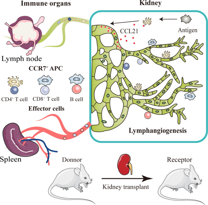
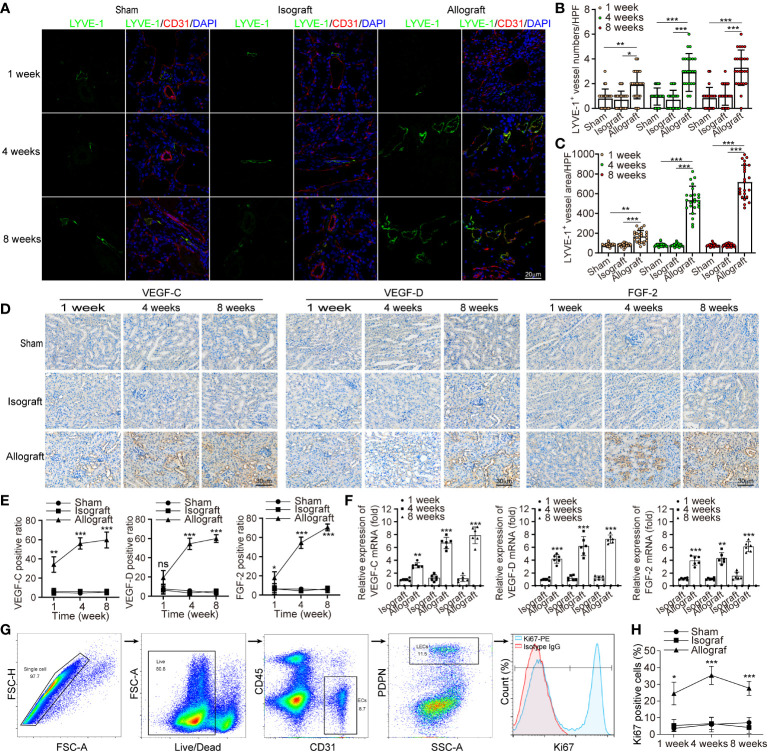
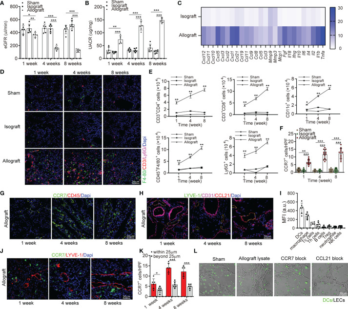
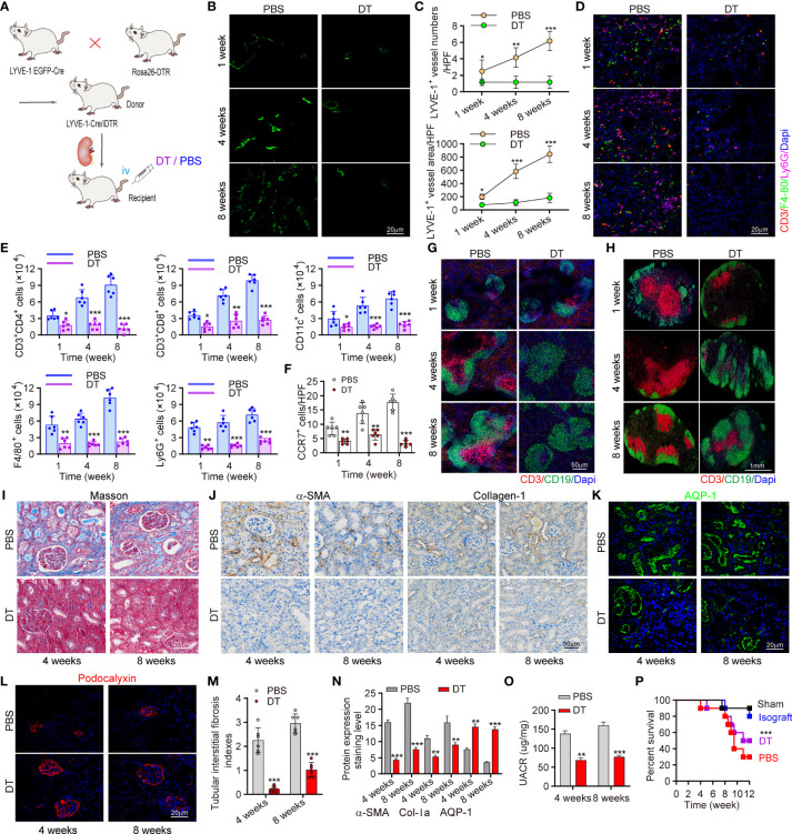
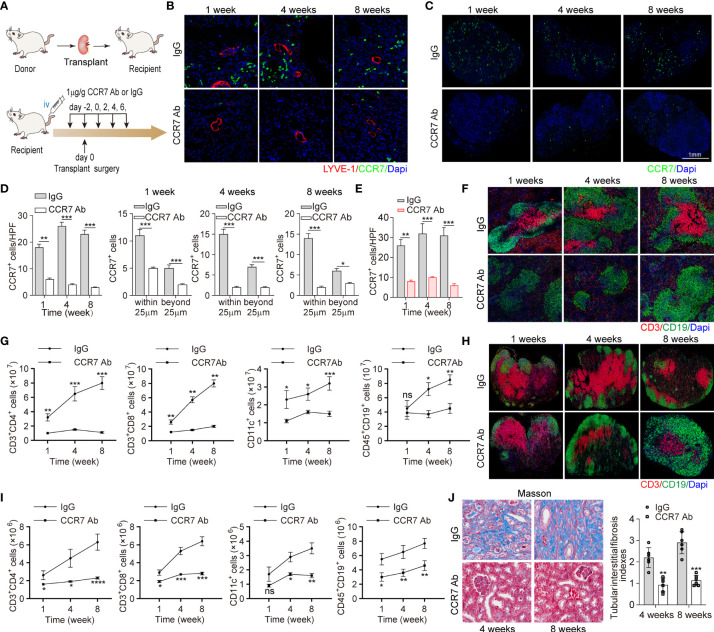
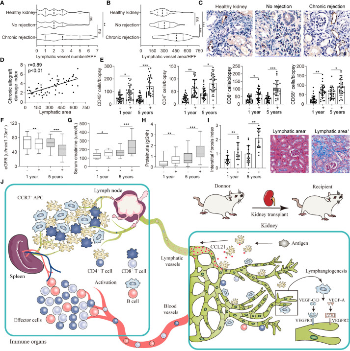
Chronic rejection of the renal allograft remains a major cause of graft loss. Here, we demonstrated that the remodeling of lymphatic vessels (LVs) after their broken during transplantation contributes to the antigen presenting and lymph nodes activating. Our studies observed a rebuilt of interrupted lymph draining one week after mouse kidney transplantation, involving preexisting lymphatic endothelial cells (LECs) from both the donor and recipient. These expanding LVs also release C-C chemokine ligand 21 (CCL21) and recruit CCR7+ cells, mainly dendritic cells (DCs), toward lymph nodes and spleen, evoking the adaptive response. This rejection could be relieved by LYVE-1 specific LVs knockout or CCR7 migration inhibition in mouse model. Moreover, in retrospective analysis, posttransplant patients exhibiting higher area density of LVs presented with lower eGFR, severe serum creatinine and proteinuria, and greater interstitial fibrosis. These results reveal a rebuilt pathway for alloantigen trafficking and lymphocytes activation, providing strategies to alleviate chronic transplantation rejection.

Keywords: allograft; chronic rejection; inflammation; lymphangiogenesis; renal transplantation.


Conflict of interest statement

The authors declare that the research was conducted in the absence of any commercial or financial relationships that could be construed as a potential conflict of interest.

Figures

Graphical Abstract
Graphical Abstract
After kidney transplantation, a triad crosstalk of CCR7-expressing antigen presenting cells (APCs), CCL21-expressing lymphatic vessels, and effector lymphocytes was demonstrated, which promote alloimmune response. Injured lymphatic endothelial cells release CCL21, guiding the migration of CCR7+ APCs to adjacent lymph nodes through broken lymphatic vessels. This migration then activates effector lymphocytes and aggravate lymphangiogenesis, leading to more severer organ rejection.
Figure 1
Figure 1
Chronic rejection is associated with lymphangiogenesis in renal allograft. (a) Representative immunofluorescence images of LYVE-1, CD31, and DAPI within sham (n=6 mice), isograft (n=6 mice) and allograft (n=6 mice) kidneys at 1, 4 and 8 weeks respectively. (b and c) Numbers and area counting of LYVE-1+ vessels in high-power field (HPF) at 1, 4 and 8 weeks respectively. (d) Immunohistochemistry of VEGF-C, VEGF-D and FGF-2 expression within sham, isograft and allograft kidneys at 1, 4 and 8 weeks respectively. (e) Positive ratio of VEGF-C, VEGF-D and FGF-2 within sham, isograft and allograft kidneys at 1, 4 and 8 weeks respectively. (f) Relative mRNA expression of VEGF-C, VEGF-D and FGF-2 by qRT-PCR within isograft and allograft kidneys at 1, 4 and 8 weeks respectively, using sham as a reference. (g) Live single CD45- PDPN+ CD31+ LECs isolating from renal allograft by gating technology via flow cytometry, and the population of Ki67+ cells. (h) The ratio of Ki67+ cells in PDPN+CD31+ LECs within sham, isograft and allograft kidneys at 1, 4 and 8 weeks respectively. *P < 0.05, **P < 0.01, ***P < 0.001. Values are mean ± SEM.
Figure 2
Figure 2
Lymphangiogenesis in renal allograft accompanied with degenerated renal function and inflammatory cell infiltration mediated by CCL21 expression in lymphatic endothelial cells. (A) eGFR level in sham (n=6 mice), isograft (n=6 mice), and allograft (n=6 mice) groups at 1, 4 and 8 weeks respectively. (B) UACR level in sham, isograft, and allograft groups at 1, 4 and 8 weeks respectively. (C) Messenger RNA expression of pro-inflammatory cytokines by multiplex reverse transcription-polymerase chain reaction assay. The heat map displays the relative expression level in isografts and allografts compared to that in the sham control. (D) Representative immunofluorescence images showing F4-80+, CD3+, Ly6G+ cells in sham, isograft, and allograft groups at 1, 4 and 8 weeks respectively. (E) Numbers of Th cells, macrophages, dendritic cells, B cells, and neutrophile granulocytes in sham, isograft, and allograft groups determined by flow cytometry at 1, 4 and 8 weeks respectively. (F) CCR7+ cell counting in sham, isograft, and allograft groups at 1, 4 and 8 weeks respectively. (G) Immunofluorescence of CCR7+, and CD45+ cells in allografts at 1, 4 and 8 weeks respectively. (H) Representative immunofluorescence images showing LYVE-1+, CD31+ and CCL21+ cells in allografts at 1, 4 and 8 weeks respectively. (I) FACS analysis of the different CCR7+ cell types in kidney allograft infiltrated CD45+ immune cells: DCs (CD45+ and CD11c+), macrophage (CD11b+ and F4/80+), cytotoxic T cells (CD8+), T helper cells (CD4+), B cells (CD19+), Treg cells (CD4+, CD25+ and FOXP3+), neutrophils (CD11b+ and Ly6G+) and NK cells (NK1.1+). (J) Representative immunofluorescence images showing the distribution of CCR7+ cells and LYVE-1+ cells in allografts to confirm the chemotaxis at 1, 4 and 8 weeks respectively. (K) Cell counting of CCR7+ cells that have different distances (within or beyond 25 μm) from lymphatic endothelial cells. (L) Representative images of dendritic cells migrating to lymphatic capillary-like structures. All scale bars represent 20 μm. *P < 0.05, **P < 0.01, ***P < 0.001. Values are mean ± SEM.
Figure 3
Figure 3
Determination of the reconnection and origin of LVs (A) Staining of renal draining lymph nodes (yellow arrows) after the injection of Evans Blue under renal capsule in sham (n=6 mice), isograft (n=6 mice), and allograft (n=6 mice) groups at 1, 4 and 8 weeks respectively. (B) Representative immunofluorescence images showing the dendritic cells separated from peripheral blood of Itgax-Cre-GFP mice in renal draining lymph nodes after injection of these cells under the renal capsule at the 1st, 4th, and 8th week after surgery. Scale bar represents 1 mm. (C) Co-localization of LYVE-1+ and EGFP+ cells to explore the donor or recipient origin of lymphatic vessels at 1, 4 and 8 weeks respectively. Scale bar represents 20 μm.
Figure 4
Figure 4
Conditional knockout of LVs attenuated CCR7+ cells expansion and ameliorates allograft function. (A) Scheme showing the establishment of conditional LVs KO mouse model and allograft model. Intervention controls: DT (n=6 mice) or PBS (n=6 mice) injection. (B) Immunofluorescence of LYVE-1 labeling intrarenal LVs. (C) Numbers and area of intrarenal LYVE-1+ vessel counted in HPF. (D) Representative immunofluorescence images of intrarenal CD3+, F4/80+ and Ly6G+ cells. (E) Inflammatory cells infiltration in kidneys analyzed by flow cytometry. (F) Visual counting of CCR7+ cells with confocal microscopy. (G, H) Immunofluorescence of B cells and T cells in spleen (left) and RDLN (right). (I) Masson staining of renal tissues. (J) The expression of α-SMA and collagen-1 assessed by immunohistochemistry. (K, L) Immunofluorescence of AQP-1 and Podocalyxin. (M) The assessment of tubular interstitial fibrosis indexes. (N) Quantitative analysis of fibrosis–related protein expression. (O) Changes of UARC in kidney transplant mice. (P) Precent survival of recipient mice in different groups. *P < 0.05, **P < 0.01, ***P < 0.001. Values are mean ± SEM. iv, intravenously; DT, diphtheria toxin; RDLN, renal draining lymph node; AQP, aquaporin-1.
Figure 5
Figure 5
CCR7 blocking antibody attenuated lymphocyte recruitment and allograft function losing. (A) Scheme showing recombinant anti-CCR7 antibody intervention in C57BL/6 (H-2b) mouse model (n=6 mice). Control group (n=6 mice): rabbit anti-mouse IgG. (B) LYVE-1 and CCR7 staining of kidney tissues following IgG or CCR7 antibody treatment. (C) Representative images of CCR7 in RDLN. (D) Cell counting of intrarenal CCR7+ cells at the 1, 4, and 8 weeks after surgery, including the exact cell counting within or beyond 25 μm respectively. (E) Cell counting of CCR7+ cells in RDLN. (F, H) Immunofluorescence staining of CD3+ and CD19+ in spleen (upper) and RDLN (bottom). (G, I) The numbers of CD3+CD4+ and CD3+CD8+ T cells, CD11c+DCs and CD45+CD19+ B cells in spleen (upper) and RDLN (bottom) determined by flow cytometry. (J) Masson staining of renal tissues (left), and tubular interstitial fibrosis indexes (right) in recipient mice at the 4 and 8 weeks after surgery. *P < 0.05, **P < 0.01, ***P < 0.001. Values are mean ± SEM.
Figure 6
Figure 6
Intrarenal lymphangiogenesis negatively correlated with allograft function in transplant patients. (A, B) Lymphatic vessels number and area counted in high-power field (HPF) respectively in healthy (n=11), no rejection (n=80) and chronic rejection kidneys (n=54) from patients. (C) Representative images of immunohistochemical staining of LYVE-1 in healthy, no rejection and chronic rejection kidneys from patients to identify the situation of lymphangiogenesis. (D) Linear regression graph of chronic allograft damage index and lymphatic area. (E) Cell counting of different immune cells, include CD45+ (myeloid cells), CD4+ (Th cells), CD8+ (CTL cells), CD68+ (macrophage), in lymphatic area+ (defined as lymphatic vessels area more than 458.74 μm2) and lymphatic area- (defined as lymphatic vessels area less than 458.74 μm2) group from chronic rejection kidneys at 1 year and 5 years determined by flow cytometry. (F) Quantitative data of eGFR (μL/min/1.73m2) in lymphatic area+ (n=36) and lymphatic area- (n=18) group from chronic rejection kidneys at 1 year and 5 years. (G) Quantitative data of serum creatinine (μmol/L) in lymphatic area+ and lymphatic area- group from chronic rejection kidneys at 1 year and 5 years. (H) Quantitative data of proteinuria (g/24h) in lymphatic area+ and lymphatic area- group from chronic rejection kidneys at 1 year and 5 years. (I) Interstitial fibrosis index in lymphatic area+ and lymphatic area- group from chronic rejection kidneys at 1 year and 5 years. Representative image of Masson staining showing the situation of interstitial fibrosis within lymphatic area+ and lymphatic area- groups. (J) Schematic diagram concluding the crosstalk between kidney and immune organs in allograft rejection lymphangiogenesis. *P < 0.05, **P < 0.01, ***P < 0.001. Values are mean ± SEM.

Similar articles

Cited by

References

    1. Lai X, Zheng X, Mathew JM, Gallon L, Leventhal JR, Zhang ZJ. Tackling Chronic Kidney Transplant Rejection: Challenges and Promises. Front Immunol (2021) 12:661643. doi: 10.3389/fimmu.2021.661643 - DOI - PMC - PubMed
    1. Carrasco YR, Batista FD. B Cells Acquire Particulate Antigen in a Macrophage-Rich Area at the Boundary Between the Follicle and the Subcapsular Sinus of the Lymph Node. Immunity (2007) 27(1):160–71. doi: 10.1016/j.immuni.2007.06.007 - DOI - PubMed
    1. Bergtold A, Desai DD, Gavhane A, Clynes R. Cell Surface Recycling of Internalized Antigen Permits Dendritic Cell Priming of B Cells. Immunity (2005) 23(5):503–14. doi: 10.1016/j.immuni.2005.09.013 - DOI - PubMed
    1. Suzuki K, Grigorova I, Phan TG, Kelly LM, Cyster JG. Visualizing B Cell Capture of Cognate Antigen From Follicular Dendritic Cells. J Exp Med (2009) 206(7):1485–93. doi: 10.1084/jem.20090209 - DOI - PMC - PubMed
    1. Marino J, Babiker-Mohamed MH, Crosby-Bertorini P, Paster JT, LeGuern C, Germana S, et al. . Donor Exosomes Rather Than Passenger Leukocytes Initiate Alloreactive T Cell Responses After Transplantation. Sci Immunol (2016) 1(1):8759–69. doi: 10.1126/sciimmunol.aaf8759 - DOI - PMC - PubMed

Publication types