. 2022 Jan 25;50(2):1128-1146.
doi: 10.1093/nar/gkab1267.

CryoEM of RUVBL1-RUVBL2-ZNHIT2, a complex that interacts with pre-mRNA-processing-splicing factor 8

Affiliations

CryoEM of RUVBL1-RUVBL2-ZNHIT2, a complex that interacts with pre-mRNA-processing-splicing factor 8

Marina Serna et al. Nucleic Acids Res. .

Abstract

Biogenesis of the U5 small nuclear ribonucleoprotein (snRNP) is an essential and highly regulated process. In particular, PRPF8, one of U5 snRNP main components, requires HSP90 working in concert with R2TP, a cochaperone complex containing RUVBL1 and RUVBL2 AAA-ATPases, and additional factors that are still poorly characterized. Here, we use biochemistry, interaction mapping, mass spectrometry and cryoEM to study the role of ZNHIT2 in the regulation of the R2TP chaperone during the biogenesis of PRPF8. ZNHIT2 forms a complex with R2TP which depends exclusively on the direct interaction of ZNHIT2 with the RUVBL1-RUVBL2 ATPases. The cryoEM analysis of this complex reveals that ZNHIT2 alters the conformation and nucleotide state of RUVBL1-RUVBL2, affecting its ATPase activity. We characterized the interactions between R2TP, PRPF8, ZNHIT2, ECD and AAR2 proteins. Interestingly, PRPF8 makes a direct interaction with R2TP and this complex can incorporate ZNHIT2 and other proteins involved in the biogenesis of PRPF8 such as ECD and AAR2. Together, these results show that ZNHIT2 participates in the assembly of the U5 snRNP as part of a network of contacts between assembly factors required for PRPF8 biogenesis and the R2TP-HSP90 chaperone, while concomitantly regulating the structure and nucleotide state of R2TP.


Figures

Figure 1.
Figure 1.
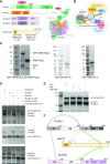
Purification and assembly of RUVBL1–RUVBL2–ZNHIT2ΔC. (A) Left top panels, domain architecture of the components of R2TP (TPRs, tetratricopeptide repeats; PBD, PIH1D1 binding domain; RBD, RUVBL2 binding domain; PIH, protein interacting with HSP90 domain; CS, CHORD-containig proteins and SGT1 domain). Domains involved in interactions within R2TP are indicated as links with dashed lines. Left bottom panel, constructs of human ZNHIT2 used in this work (H, HIT domain). Right panel, cartoon of the structure of the R2TP complex. (B) Diagram of the network of interactions described in this work for R2TP, ZNHIT2, ECD, AAR2 and PRPF8. (C) SDS-PAGE of purified ZNHIT2ΔC, GST-ZNHIT2ΔC, GST-ZNHIT2-C and GST-ZNHIT2-N. (D) GST-pull-down experiments to test the interaction of GST-ZNHIT2ΔC, GST-ZNHIT2-C and GST-ZNHIT2-N with RUVBL1, RUVBL2 and RUVBL1–RUVBL2 complexes (RUVBL1/2 for simplicity in figure legends hereafter). RUVBL1/2-ΔDII corresponds to mutants lacking part of the DII domain. RUVBL1/2-WB corresponds to mutants in the Walker B domains, able to bind ATP but affected in ATP hydrolysis. Control lanes for these experiments are shown in Supplementary Figure S1B, C. (E) GST-pull-down experiment to evaluate the effect of the presence or absence of the nucleotides ATP, ADP or AMP–PNP in the interaction between RUVBL1–RUVBL2 and ZNHIT2ΔC. Control lanes for these experiments are shown in Supplementary Figure S1D. (F) Cartoon showing the crosslinks found in RUVBL1–RUVBL2–ZNHIT2 complex detected by XL-MS.
Figure 2.
Figure 2.
CryoEM of the RUVBL1–RUVBL2-ZNHIT2ΔC complex. (A) SDS-PAGE of the purified RUVBL1–RUVBL2–ZNHIT2ΔC complex used for structural studies. (B) Representative 2D averages obtained from the cryoEM images of RUVBL1–RUVBL2- ZNHIT2ΔC. Grey arrowheads label some of the RUVBL1–RUVBL2 DII domains, while yellow arrowheads indicate the position of ZNHIT2ΔC. (C) Two views of the consensus cryoEM map of the RUVBL1–RUVBL2-ZNHIT2ΔC complex at 4.4 Å resolution. Density for ZNHIT2ΔC (yellow color) locates at the DII-face of the ring (grey color). (D) A view of one of the cryoEM maps obtained after classification using cryoDRGN (39), showing the DII-side of the RUVBL1–RUVBL2 ring. The density for ZNHIT2ΔC complex is divided in two lobules that correlate with the two domains predicted for ZNHIT2ΔC by AlphaFold (40). (E) Left panel, a tilted view of the cryoEM map in D showing the interaction of the large ZNHIT2ΔC domain. Both the atomic model of the RUVBL1–RUVBL2 complex and the ZNHIT2ΔC C-terminal domain are fitted within the map. Linked residues, identified by XL-MS, are shown as green dots. Each RUVBL subunit is labelled from 1 to 3 (subscript). Right panel, close-up view to highlight the proximity between the cross-linked residues in ZNHIT2 and the adjacent DII domains in the RUVBL1–RUVBL2 model.
Figure 3.
Figure 3.
ZNHIT2ΔC alters the conformation and ATPase activity of RUVBL2. (A) CryoEM density of the AAA-ring in RUVBL1–RUVBL2 after being processed independently of the flexible density for ZNHIT2ΔC and the DII domains. Density for nucleotides is only present in the RUVBL1 subunits (green density with ADP fitted inside). ADP molecules, without density accounting for them, are fitted in the described nucleotide binding sites of RUVBL2 only to highlight the lack of density in the region where nucleotide is present in human RUVBL1–RUVBL2 crystal structures (PDB ID 2XSZ). (B) Right panel, crystal structure of RUVBL1–RUVBL2 (PDB ID 2XSZ). Grey and pink arrowheads indicate the N-terminal regions of RUVBL1 and RUVBL2 respectively. These were absent in the atomic structure of RUVBL1–RUVBL2–ZNHIT2ΔC (indicated as pink empty arrowheads). Nucleotides are shown in green colour. (C) Close-up of nucleotide-binding sites for every RUVBL subunit after the interaction with ZNHIT2ΔC. Nucleotide molecules are show in green and nucleotide densities are displayed in semi-transparent green density. In RUVBL2, density for nucleotide is not observed, and this is indicated by placing ADP in the binding-site. (D) ATPase activity of RUVBL1–RUVBL2, wild type and mutants (RUVBL1WB and RUVBL2WB stand for the mutants whose Walker B motif has been altered to impair ATPase activity), in the presence and absence of ZNHIT2ΔC using the value of RUVBL1–RUVBL2 alone as 100%. Significance value of the paired Student's T-Test is shown for the RUVBL1–RUVBL2–ZNHIT2ΔC complex in comparison with the RUVBL1–RUVBL2 complex alone.
Figure 4.
Figure 4.
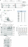
ZNHIT2 forms a complex with R2TP in vitro and in cells. (A) Consecutive pull-down experiments to analyze the formation of R2TP-ZNHIT2ΔC complexes. His-RUVBL1 was used in a first pulldown step, and then the eluted material was the input of a second pulldown experiment using the GST tag in ZNHIT2ΔC. (B) Volcano plot comparing interactors of RPAP3M626A/F630R versus RPAP3-WT is shown. Two-sample Student's t-test was performed. Only interactors with a P-value <0.05 and a log2 ratio ≤ 2 were considered downregulated in the mutant condition. The permutation-based FDR was estimated to be below 5%. (C) The 22 interactors significantly down-represented in RPAP3M626A/F630R versus RPAP3-WT are indicated in the table. Those linked with U5 snRNP or its maturation are in bold. (D) Immunoprecipitation (IP) of transiently transfected HEK293T cells with RPAP3-WT RPAP3M626A/F630R. Anti-FLAG-IPs, whole-cell extracts (WCE, 1%) and flow through (FT) were subjected to western analysis using the indicated antibodies. Blots are representative of 5 independent experiments. Data (mean ± SEM of five independent experiments) were normalized by total FLAG and expressed as fold-change of the different proteins indicated association with FLAG with respect to the RPAP3-WT condition. Statistical significance was analyzed using unpaired t-test. **P < 0.01, ***P < 0.001.
Figure 5.
Figure 5.
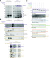
RUVBL1–RUVBL2 interacts with AAR2, ECD and ZNHIT2ΔC. (A) Pull-down experiments using the GST tag in ZNHIT2ΔC showing that ZNHIT2ΔC interacts with RUVBL1–RUVBL2 and ECD. ZNHIT2ΔC contaminants (*) coelute with AAR2 so the interaction between ZNHIT2ΔC and AAR2 could not be determined in this experiment. Electrophoretic protein mobility is shown with coloured arrowheads according to the protein labels. Main results are highlighted within rectangles. (B) Pull-down experiments using the Strep II tag in ECD confirmed its interaction with ZNHIT2ΔC and RUVBL1–RUVBL2. Electrophoretic protein mobility is shown with coloured arrowheads according to the protein labels. Main results are highlighted within rectangles. (C) Pull-downs using the His-tag in AAR2 confirmed its interaction with ECD and ZNHIT2ΔC. Albeit the His-tag been removed from RUVBL1–RUVBL2 some unspecific interaction was detected in the RUVBL1–RUVBL2 control lane (**). Electrophoretic protein mobility is shown with coloured arrowheads according to the protein labels. Main results are highlighted within rectangles. (D) Pull-down experiments using the Strep II tag in AAR2 probed that AAR2 interacts with RUVBL1–RUVBL2 both in the absence and in the presence of ZNHIT2ΔC. Main results are highlighted within rectangles.
Figure 6.
Figure 6.
Interactions between PRPF8 and RUVBL1–RUVBL2, RPAP3-PIH1D1, AAR2, ECD and ZNHIT2 (A) Flag-pull-down experiments showed a direct interaction between PRPF8 (bait) and RUVBL1–RUVBL2 (prey), which is not affected by the presence of ATP. Asterisks indicate contaminants that co-eluted with PRPF8. (B) Upper panel, pull-down experiments using Flag-PRPF8 (bait) and RPAP3-PIH1D1 in the presence or absence of RUVBL1–RUVBL2 were analyzed by western blot. Stained SDS-PAGE gels can be found in Supplementary Figure S8B. Lower panel, pull-down experiment using the GST tag in RPAP3 within the RPAP3-PIH1D1 complex, analyzed by western blot. MW, molecular weight markers. (C) In the presence of ECD, ZNHIT2ΔC, RUVBL1–RUVBL2, RPAP3-PIH1D1 and AAR2 (preys), PRPF8 (bait) interacted with RUVBL1–RUVBL2, RPAP3-PIH1D1, AAR2 and ECD as show in the Flag-pull-down assay. Control experiments using a cell extract lacking the overexpressed PRPF8, but overexpressing the Flag-tag were run in parallel SDS-PAGE for the experiments and they are shown in Supplementary Figure S8. Stained SDS-PAGE gels can be found in Supplementary Figure S8C.The presence of PRPF8, RUVBL1–RUVBL2, ECD, AAR2, RPAP3, PIH1D1 and ZNHIT2 in the Flag-pull-down assays was analyzed by western blot.

Similar articles

Cited by

References

    1. Galej W.P., Oubridge C., Newman A.J., Nagai K.. Crystal structure of Prp8 reveals active site cavity of the spliceosome. Nature. 2013; 493:638–643. - PMC - PubMed
    1. Nguyen T.H., Li J., Galej W.P., Oshikane H., Newman A.J., Nagai K.. Structural basis of Brr2-Prp8 interactions and implications for U5 snRNP biogenesis and the spliceosome active site. Structure. 2013; 21:910–919. - PMC - PubMed
    1. Malinova A., Cvackova Z., Mateju D., Horejsi Z., Abeza C., Vandermoere F., Bertrand E., Stanek D., Verheggen C.. Assembly of the U5 snRNP component PRPF8 is controlled by the HSP90/R2TP chaperones. J. Cell Biol. 2017; 216:1579–1596. - PMC - PubMed
    1. Cloutier P., Poitras C., Durand M., Hekmat O., Fiola-Masson E., Bouchard A., Faubert D., Chabot B., Coulombe B.. R2TP/Prefoldin-like component RUVBL1/RUVBL2 directly interacts with ZNHIT2 to regulate assembly of U5 small nuclear ribonucleoprotein. Nat. Commun. 2017; 8:15615. - PMC - PubMed
    1. Pearl L.H. Review: The HSP90 molecular chaperone-an enigmatic ATPase. Biopolymers. 2016; 105:594–607. - PMC - PubMed

Publication types