. 2021 Nov 30;11(12):1801.
doi: 10.3390/biom11121801.

Andrographolide Attenuates Established Pulmonary Hypertension via Rescue of Vascular Remodeling

Affiliations

Andrographolide Attenuates Established Pulmonary Hypertension via Rescue of Vascular Remodeling

Xiaowei Nie et al. Biomolecules. .

Abstract

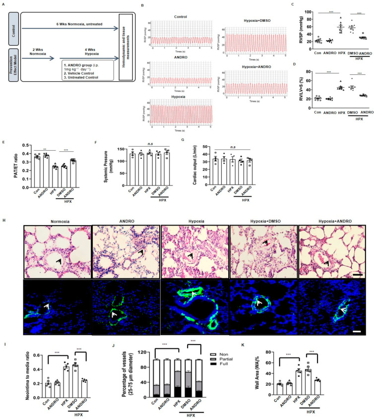
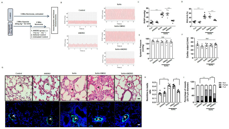
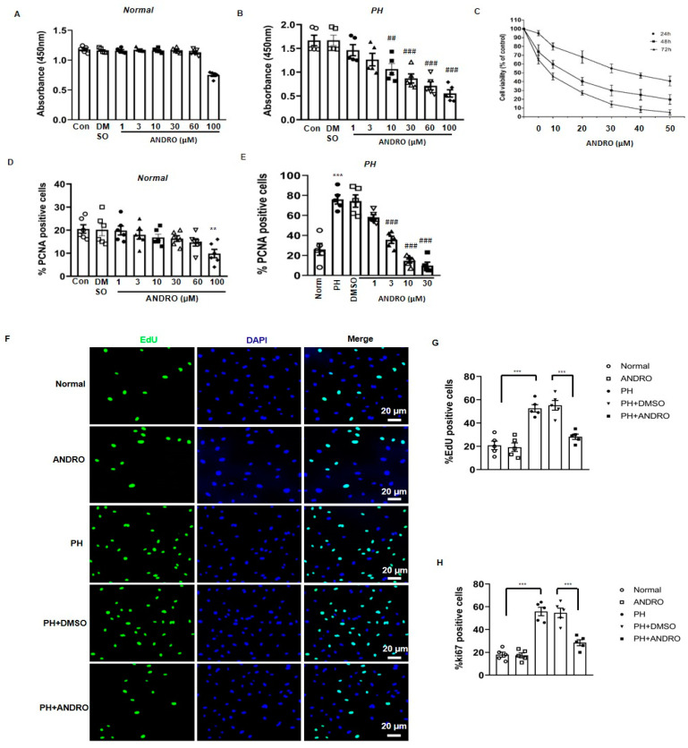
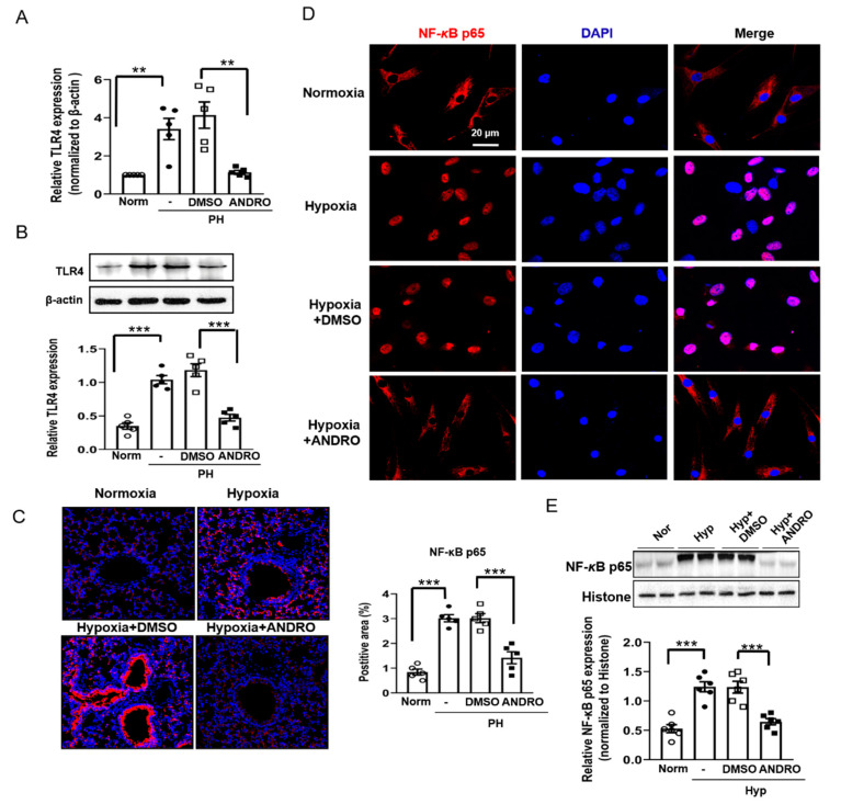
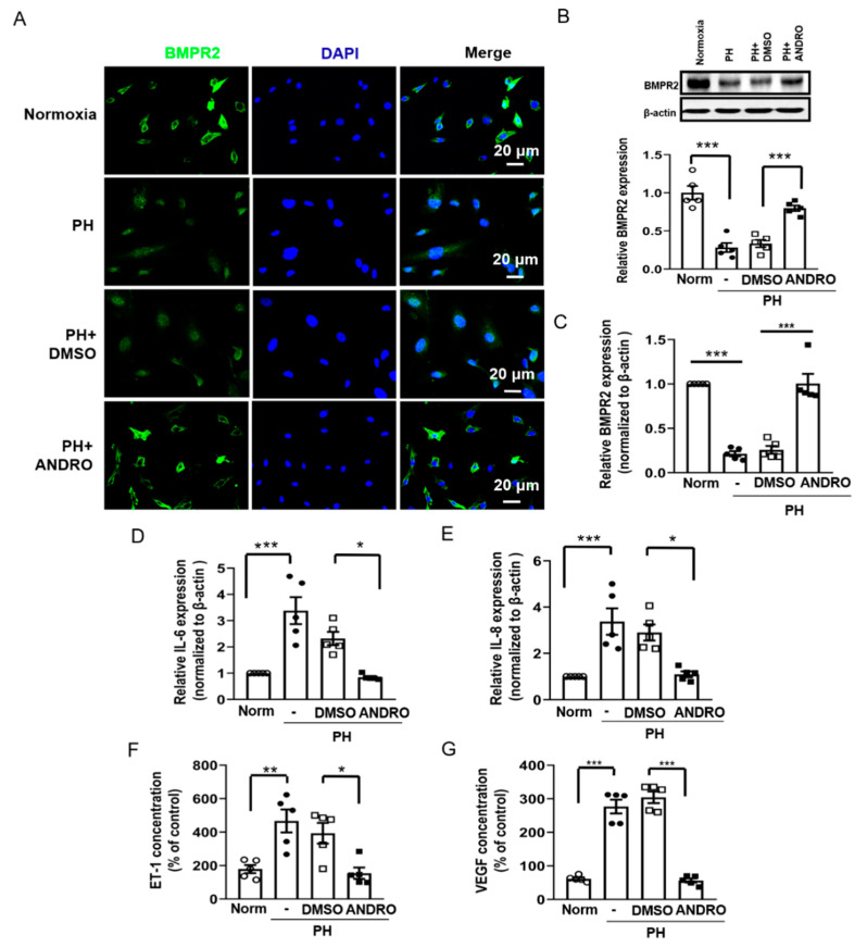
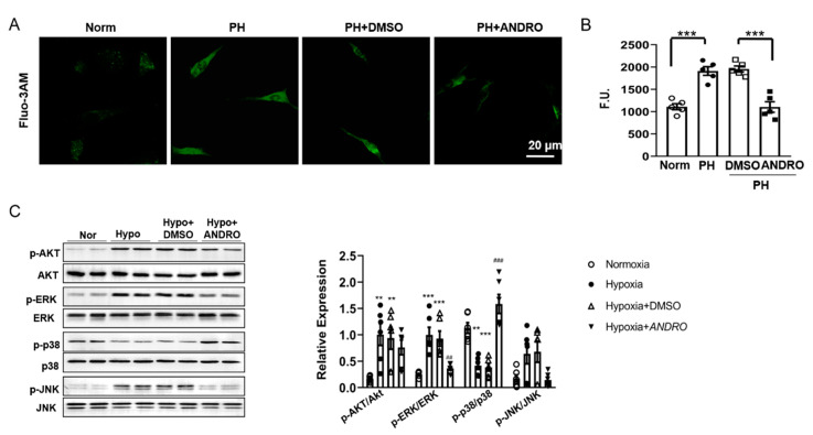
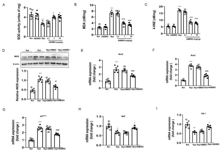
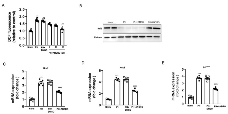
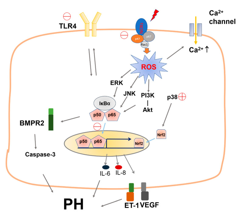
Pulmonary hypertension (PH) is characterized by vascular remodeling caused by marked proliferation of pulmonary artery smooth muscle cells (PASMCs). Andrographolide (ANDRO) is a potent anti-inflammatory agent which possesses antioxidant, and has anticarcinogenic activity. The present study examined potential therapeutic effects of ANDRO on PH in both chronic hypoxia and Sugen5416/hypoxia mouse PH models. Effects of ANDRO were also studied in cultured human PASMCs isolated from either healthy donors or PH patients. In vivo, ANDRO decreased distal pulmonary arteries (PAs) remodeling, mean PA pressure and right ventricular hypertrophy in chronic hypoxia- and Sugen/hypoxia-induced PH in mice. ANDRO reduced cell viability, proliferation and migration, but increased cell apoptosis in the PASMCs isolated from PH patients. ANDRO also reversed the dysfunctional bone morphogenetic protein receptor type-2 (BMPR2) signaling, suppressed [Ca2+]i elevation, reactive oxygen species (ROS) generation, and the upregulated expression of IL-6 and IL-8, ET-1 and VEGF in PASMCs from PH patients. Moreover, ANDRO significantly attenuated the activation of TLR4/NF-κB, ERK- and JNK-MAPK signaling pathways and reversed the inhibition of p38-MAPK in PASMCs of PH patients. Further, ANDRO blocked hypoxia-triggered ROS generation by suppressing NADPH oxidase (NOX) activation and augmenting nuclear factor erythroid 2-related factor 2 (Nrf2) expression both in vitro and in vivo. Conventional pulmonary vasodilators have limited efficacy for the treatment of severe PH. We demonstrated that ANDRO may reverse pulmonary vascular remodeling through modulation of NOX/Nrf2-mediated oxidative stress and NF-κB-mediated inflammation. Our findings suggest that ANDRO may have therapeutic value in the treatment of PH.

Keywords: andrographolide; pulmonary artery smooth muscle cells; pulmonary hypertension; vascular remodeling.


Conflict of interest statement

The authors declare no conflict of interest.

Figures

Figure 1
Figure 1
Andrographolide (ANDRO) prevented mice from hypoxia-induced pulmonary hypertension (PH). (A) Protocol of animal experiments. For hypoxia-induced PH model, mice were intraperitoneally injected with ANDRO (1 mg·kg−1·day−1) or a vehicle for two weeks followed by hypoxia for four weeks. Control mice were subjected to six weeks normoxia only. (B) Representative tracings showing the effect of ANDRO on pulmonary artery systolic pressure in hypoxia (HPX)-induced PH mouse model. n = 8. (C) ANDRO treatment significantly attenuated the elevated right ventricular systolic pressure (RVSP) in HPX-induced PH mice. Scale bars are 100 μm. n = 8. (D) ANDRO reversed right ventricular hypertrophy in HPX-induced PH in mice assessed invasively by the right ventricular hypertrophy index (RV/LV+S). n = 8. (E) Echocardiography analysis showing the decreased PAT/ET in hypoxia-induced PH mice were reversed by ANDRO. n = 6. (F,G) ANDRO failed to affect systemic pressure (F) and cardiac output (G). n = 8. (H) Representative H&E staining (upper panel) and α-SMA immunofluorescence staining (lower panel) images showing the vascular remodeling in various groups. (I) Quantitative analyses showing that ANDRO significantly reduced the ratio of neointima/media. n = 5. (J) Quantitative assessment of pulmonary arterial muscularization. Non-, partially and fully muscularized arteries as a percentage of total alveolar wall and duct arteries were scored (n = 5, one-way ANOVA for fully muscularized vessels). (K) ANDRO treatment significantly reduced the relative area of wall area (WA)%. n = 5. Data represent mean ± SEM. ** p < 0.01, *** p < 0.001. DMSO, Dimethyl sulfoxide; n.s.: no significant difference. Scale bars are 100 μm.
Figure 2
Figure 2
Therapeutic effects of ANDRO in a SU5416-hypoxia (SuHx)-induced mouse PH model. (A) Protocol of animal experiments. For SuHx-induced PH group, mice were subjected to three weeks hypoxia + Su5416 injection. ANDRO (1 mg/kg/day, i.p.) or DMSO (vehicle) were given for two weeks after three weeks of hypoxia + Su5416 injection. Control mice were subjected to five weeks normoxia only. n = 8. (B) Representative tracings showing the effect of ANDRO on PA systolic pressure in SuHx-induced PH mouse model. n = 8. (C) ANDRO treatment significantly attenuated the elevated RVSP in SuHx-induced PH mice. n = 8. (D) ANDRO reversed RV hypertrophy in SuHx-induced PH mice assessed invasively by the right ventricular hypertrophy index. (E,F) ANDRO failed to affect systemic pressure or cardiac output. n = 8. (G) Representative H&E staining (upper panel) and α-SMA immunofluorescence staining (lower panel) images showing the vascular remodeling in various groups. (H) Quantitative analyses showing that ANDRO significantly reduced the ratio of neointima/media. (I) Quantification of non-, partially and fully muscularized arteries as a percentage of total alveolar wall and duct arteries (n = 5, one-way ANOVA for fully muscularized vessels). (JO) Representative immunohistochemical photomicrographs of serial lung sections and group data showing that ANDRO significantly reduced the increased expression of α-SMA (WA%, K), ratio of Ki67 positive cells (L) and ratio of PCNA positive cells (M) in SuHx-induced PH mice. ANDRO also significantly increased apoptosis, as reflected by cleaved-caspase-3 (N). This is further supported by Western blotting analysis (O). n = 5. Results are expressed in percent (%) calculated as follows: (number of positive cells divided by the total number of hematoxylin-stained nuclei) ×100, from six arteries per group. Scale bars, 100 μm. Data represent mean ± SEM. ** p < 0.01; *** p < 0.001; n.s.: no significant difference.
Figure 2
Figure 2
Therapeutic effects of ANDRO in a SU5416-hypoxia (SuHx)-induced mouse PH model. (A) Protocol of animal experiments. For SuHx-induced PH group, mice were subjected to three weeks hypoxia + Su5416 injection. ANDRO (1 mg/kg/day, i.p.) or DMSO (vehicle) were given for two weeks after three weeks of hypoxia + Su5416 injection. Control mice were subjected to five weeks normoxia only. n = 8. (B) Representative tracings showing the effect of ANDRO on PA systolic pressure in SuHx-induced PH mouse model. n = 8. (C) ANDRO treatment significantly attenuated the elevated RVSP in SuHx-induced PH mice. n = 8. (D) ANDRO reversed RV hypertrophy in SuHx-induced PH mice assessed invasively by the right ventricular hypertrophy index. (E,F) ANDRO failed to affect systemic pressure or cardiac output. n = 8. (G) Representative H&E staining (upper panel) and α-SMA immunofluorescence staining (lower panel) images showing the vascular remodeling in various groups. (H) Quantitative analyses showing that ANDRO significantly reduced the ratio of neointima/media. (I) Quantification of non-, partially and fully muscularized arteries as a percentage of total alveolar wall and duct arteries (n = 5, one-way ANOVA for fully muscularized vessels). (JO) Representative immunohistochemical photomicrographs of serial lung sections and group data showing that ANDRO significantly reduced the increased expression of α-SMA (WA%, K), ratio of Ki67 positive cells (L) and ratio of PCNA positive cells (M) in SuHx-induced PH mice. ANDRO also significantly increased apoptosis, as reflected by cleaved-caspase-3 (N). This is further supported by Western blotting analysis (O). n = 5. Results are expressed in percent (%) calculated as follows: (number of positive cells divided by the total number of hematoxylin-stained nuclei) ×100, from six arteries per group. Scale bars, 100 μm. Data represent mean ± SEM. ** p < 0.01; *** p < 0.001; n.s.: no significant difference.
Figure 3
Figure 3
ANDRO reduced cell viability and proliferation of pulmonary artery smooth muscle cells from PH patients (PH-PASMCs). (A,B) Treatment with ANDRO for 24 h only significantly reduced cell viability of normal PASMCs at 100 μM, but caused a significant reduction in PAH-PASMCs at the concentration range of 1–100 μM. (C) The dose- and time-dependent effects of ANDRO on cell viability of PH-PASMCs. (D,E) Treatment with ANDRO for 24 h only significantly reduced cell proliferation of normal PASMCs at 100 μM, but produced obvious effect in PH-PASMCs starting from 1 to 100 μM. (F,G) Representative EdU staining (F) and group data (G) showing that ANDRO treatment (30 μM, 24 h) reversed PH-PASMCs proliferation. (H) Ki67 staining assay showing that ANDRO (30 μM, 24 h) reversed PH-PASMCs proliferation. Mean ± SEM, n = 5. ** p < 0.01, *** p < 0.001 vs. normal; ## p < 0.01, ### p < 0.001 vs. PAH-DMSO.
Figure 4
Figure 4
ANDRO reversed the resistance of cells to apoptosis and decreased migration of PH-PASMCs. (A,B) Representative TdT-mediated dUTP nick end labeling (TUNEL) assay image (A) and group data (B) showing that ANDRO reversed apoptosis resistance of PH-PASMCs when compared to the healthy control. (C) The dose-dependent effect of ANDRO (1–30 μM) on cell apoptosis in PH-PASMCs. (D,E) ANDRO only increased cell apoptosis at higher concentrations (60 μM) in healthy PASMCs, both under normal (10% FBS) (D) or serum deprivation (0.1% FBS) (E)conditions. Values are expressed as mean ± SEM, n = 5. ** p < 0.01, *** p < 0.001 vs. normal; ### p < 0.001 vs. PH-DMSO. (F,G) Representative images of wound-healing assay (F) and group data (G) showing that ANDRO (30 μM, 24 h) significantly reversed cell migration in PH-PASMCs. n = 5. Data are shown as mean ± SEM. *** p < 0.001.
Figure 5
Figure 5
ANDRO suppressed the activation of NF-κB in human PH-PASMCs or PAs of PH mice. (A,B) qPCR (A) and Western blotting (B) analysis showing that ANDRO (30 μM, 24 h) suppressed the mRNA and protein expression of TLR4. (C) Representative immunohistochemical staining and group data showing that ANDRO inhibited the expression of phosphorylated-NF-κB p65 (Ser276) in the lung tissues of hypoxia-induced PH mice. Scale bars are 50 μm. (D,E) immunofluorescence (D) and Western blotting (E) analysis showing that ANDRO (30 μM, 24 h) reversed hypoxia-induced translocation of p65 to nuclei. Data represent mean ± SEM. n = 5, ** p < 0.01, *** p < 0.001.
Figure 6
Figure 6
Effect of ANDRO on NF-κB-related signaling cascade in PH-PASMCs. (AC) Immunofluorescence staining (A), Western blots (B) and qRT-PCR (C) showing the effect of ANDRO on the expression of BMPR2 in PH-PASMCs. (D,E) QRT-PCR analysis showing that ANDRO significantly decreased IL-6 (D) and IL-8 (E) expression in PH-PASMCs. (F,G) ELISA assay showing that ANDRO significantly decreased ET-1 (F) and VEGF (G) expression in PH-PASMC. Data represent mean ± SEM. n = 5. * p < 0.05, ** p < 0.01, *** p < 0.001.
Figure 7
Figure 7
Effect of ANDRO on [Ca2+]i, and related signaling pathways. (A,B) Representative image (A) and group data (B) showing that ANDRO significantly reduced [Ca2+]i elevation in PH-PASMCs compared to healthy PASMC (n = 18 from five independent experiments). Scale bars are 20μm. (C) Western blot analysis showing the effects of ANDRO on phosphorylation of AKT, ERK, p38 and JNK. n = 6. Data represent mean ± SEM. ** p < 0.01, *** p < 0.001 vs. control; ## p < 0.01, ### p < 0.001 vs. PH-DMSO.
Figure 8
Figure 8
ANDRO treatment inhibited hypoxia-induced pulmonary oxidative/nitrative stress. (AC) Effect of ANDRO on the levels of SOD (A), MDA (B), and 4-HNE (C) in homogenized fresh lung tissues. (D) Western blot analysis showing that ANDRO reversed the increased iNOS level in hypoxia-treated mice. (EG) Real-time qPCR analysis showing that ANDRO treatment reduced hypoxia-upregulated mRNA expression of the ROS-generating NADPH oxidases (NOX2, NOX4, and p47 phox) in the lung tissues. (H, I) Real-time qPCR analysis showing that the down-regulated mRNA expressions of Nrf2 and HO-1 in the lung tissues were attenuated by ANDRO treatment. ** p < 0.01, *** p < 0.001, **** p < 0.0001, compared to normoxia control; # p < 0.05, ## p < 0.01, #### p < 0.001, compared to Hyp-DMSO.
Figure 9
Figure 9
ANDRO treatment blocked ROS generation via regulating NOX/Nrf2 redox imbalance in human PH-PASMCs. (A) Intracellular ROS was measured with DCFH-DA probe using confocal microscopy. ANDRO at 30 μM attenuated ROS production in human PH-PASMCs. (B) Representative Western blot analysis showing that ANDRO treatment attenuated the reduced Nrf2 expression during PH. (CE) Real-time PCR analysis showing that ANDRO treatment significantly attenuated the upregulated mRNA levels of NADPH oxidases (NOX2, NOX4, and p47 phox) in PH-PASMCs. *** p < 0.001, **** p < 0.0001, compared to healthy control; ## p < 0.01, #### p < 0.0001, compared to PH-DMSO.
Figure 10
Figure 10
Schematic illustration of the mechanisms for the beneficial effects of ANDRO on PH. ANDRO inhibits ROS generation by decreasing NOX2, NOX4 and p47phox expression and suppresses the TLR4/NF-κB pathway by inhibiting the protein expression of TLR4 and the translocation of NF-kb. ANDRO upregulates BMPR2 expression and inhibits cytokines like IL-6 and IL-8 expression, and ET-1 and VEGF protein expression. ANDRO also inhibits ROS generation and [Ca2+]i elevation and the activation of JNK-/ERK-MAPKs. Interestingly, ANDRO stimulates p38-MAPK which is suppressed during PH. All these events contribute to the therapeutic effects of ANDRO.

Similar articles

Cited by

References

    1. Austin E.D., Loyd J.E. The genetics of pulmonary arterial hypertension. Circ. Res. 2014;115:189–202. doi: 10.1161/CIRCRESAHA.115.303404. - DOI - PMC - PubMed
    1. Grant J.S., White K., MacLean M.R., Baker A.H. MicroRNAs in pulmonary arterial remodeling. Cell. Mol. Life Sci. 2013;70:4479–4494. doi: 10.1007/s00018-013-1382-5. - DOI - PMC - PubMed
    1. Guignabert C., Tu L., Le Hiress M., Ricard N., Sattler C., Seferian A., Huertas A., Humbert M., Montani D. Pathogenesis of pulmonary arterial hypertension: Lessons from cancer. Eur. Respir. Rev. Off. J. Eur. Respir. Soc. 2013;22:543–551. doi: 10.1183/09059180.00007513. - DOI - PMC - PubMed
    1. Gomberg-Maitland M., Maitland M.L., Barst R.J., Sugeng L., Coslet S., Perrino T.J., Bond L., Lacouture M.E., Archer S.L., Ratain M.J. A dosing/cross-development study of the multikinase inhibitor sorafenib in patients with pulmonary arterial hypertension. Clin. Pharmacol. Ther. 2010;87:303–310. doi: 10.1038/clpt.2009.217. - DOI - PMC - PubMed
    1. Ma B.L., Ma Y.M. Pharmacokinetic herb-drug interactions with traditional Chinese medicine: Progress, causes of conflicting results and suggestions for future research. Drug Metab. Rev. 2016;48:1–26. doi: 10.3109/03602532.2015.1124888. - DOI - PubMed

Publication types

MeSH terms