Review
. 2021 Nov 26;10(12):3328.
doi: 10.3390/cells10123328.

Lipotoxicity and β-Cell Failure in Type 2 Diabetes: Oxidative Stress Linked to NADPH Oxidase and ER Stress

Affiliations
Review

Lipotoxicity and β-Cell Failure in Type 2 Diabetes: Oxidative Stress Linked to NADPH Oxidase and ER Stress

Eloisa Aparecida Vilas-Boas et al. Cells. .

Abstract

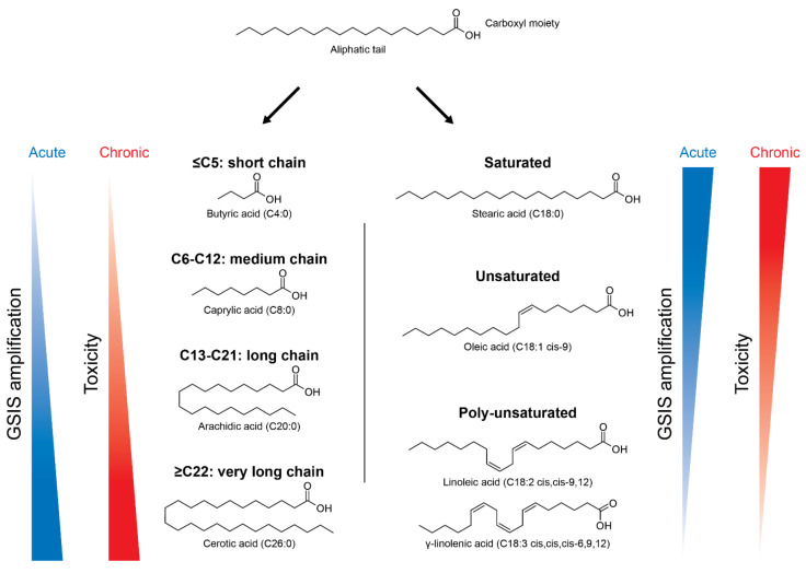
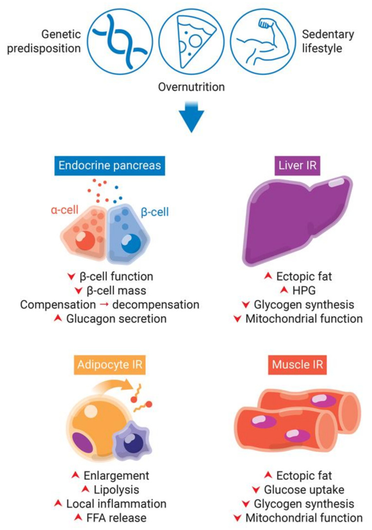
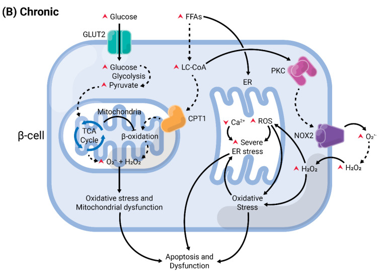
A high caloric intake, rich in saturated fats, greatly contributes to the development of obesity, which is the leading risk factor for type 2 diabetes (T2D). A persistent caloric surplus increases plasma levels of fatty acids (FAs), especially saturated ones, which were shown to negatively impact pancreatic β-cell function and survival in a process called lipotoxicity. Lipotoxicity in β-cells activates different stress pathways, culminating in β-cells dysfunction and death. Among all stresses, endoplasmic reticulum (ER) stress and oxidative stress have been shown to be strongly correlated. One main source of oxidative stress in pancreatic β-cells appears to be the reactive oxygen species producer NADPH oxidase (NOX) enzyme, which has a role in the glucose-stimulated insulin secretion and in the β-cell demise during both T1 and T2D. In this review, we focus on the acute and chronic effects of FAs and the lipotoxicity-induced β-cell failure during T2D development, with special emphasis on the oxidative stress induced by NOX, the ER stress, and the crosstalk between NOX and ER stress.

Keywords: ER stress; NADPH oxidase; lipotoxicity; oxidative stress; pancreatic β-cell; type 2 diabetes.


Conflict of interest statement

The authors declare no conflict of interest.

Figures

Figure 1
Figure 1
Nomenclature of different types of fatty acids (FAs) and their effects in pancreatic β-cells according to chain length and degree of saturation. FAs are composed by a carboxyl moiety bonded to an aliphatic tail, with variable number of carbons. Depending on the number of carbons, FAs can be divided in short-chain (C ≤ 5), medium-chain (C6-C12), long-chain (C13-C21) and very long-chain (C ≥ 22). They can also be: (i) saturated, with no double bonds, as stearic acid (C18:0); (ii) unsaturated, with one double bond, as oleic acid (C18:1); and (iii) poly-unsaturated, with more than two double bonds, as linoleic acid (C18:2) and γ-linolenic acid (C18:3). FAs are known to increase the glucose-stimulated insulin secretion (GSIS), but can also be toxic in the long-term. The potency of GSIS amplification is positively influenced by the carbon chain length and negatively influenced by the degree of unsaturation.
Figure 2
Figure 2
Obesity-induced pancreatic β-cells dysfunction and insulin resistance (IR) in peripheral tissues. Genetic predisposition, overnutrition, and a sedentary lifestyle are involved in obesity and type 2 diabetes development. The lipotoxicity involved in chronic obesity and hyperglycemia leads to an increase of insulin demand due to IR in peripheral tissues. While in a first moment, β-cells try to compensate for IR by increasing insulin synthesis, they start to decompensate in late stages, with decrease of β-cell function and mass. IR in adipocytes eliminate insulin-induced repression of lipolysis. Impaired adipogenesis and dysfunctional hypertrophy of adipocytes increase plasma levels of free fatty acids (FFAs). Additionally, dysfunctional adipose tissue releases increased levels of insulin resistance-inducing pro-inflammatory adipokines, leading to local inflammation. The increased FFAs flux to the liver and skeletal muscle leads to increase of fat deposition, decrease of glycogen synthesis and mitochondrial dysfunction. Additionally, the hallmark of insulin resistance in the liver is increased hepatic glucose production (HPG).
Figure 3
Figure 3
NADPH oxidase (NOX) family: NOX1, NOX2, NOX3, NOX4, NOX5, DUOX1, and DUOX2. NOX1-4 are stabilized by p22phox subunit. NOX1 is activated by NOXO1/NOXA1 and Rac subunits, and NOX2 by p47phox/p67phox/p40phox and Rac. NOX3 has constitutive activity, but binding to NOXO1/NOXA1 increases reactive oxygen species (ROS) production. NOX4 is constitutively activated. NOX4, NOX5, and DUOX1-2 do not interact with regulatory subunits. NOX5 and DUOX1-2 do not depend on p22phox for stabilization and are activated by Ca2+. NOX1, NOX2, NOX3, and NOX5 produce superoxide (O2•−), and NOX4 and DUOX1-2 produce hydrogen peroxide (H2O2).
Figure 4
Figure 4
Summary of acute versus chronic effects of fatty acids (FAs) in pancreatic β-cells. Glucose enters the β-cell through specific transporters located at the plasma membrane, GLUT2 (in rodents). After its phosphorylation by glucokinase, it undergoes various modifications by enzymes from the glycolysis, until the generation of pyruvate. This enters the mitochondria and is oxidized in the tricarboxylic acid (TCA) cycle. The electrons are transferred to the electron transport chain, resulting in the generation of reactive oxygen species (ROS), as byproducts, and ATP. (A) In acute conditions, FAs enter the cells and are converted into long-chain fatty-acyl CoA (LC-CoA), which is translocated to the mitochondria via carnitine palmitoyltransferase 1 (CPT1) to be oxidized in the β-oxidation, generating ATP and ROS. The increase of ATP/ADP leads to the closure of K+ channels sensitive to ATP (KATP) at the plasma membrane. The consequent membrane depolarization leads to the opening of L-type voltage-gated Ca2+ channels (L-VGCC). The rapid Ca2+ influx mobilizes insulin granules, which are released. Long-chain saturated or unsaturated FAs can also bind to Gαq-protein coupled receptor GPR40 at the plasma membrane. This activates the phospholipase C (PLC)/diacylglycerol (DAG) pathway, which respectively activates PKC and mobilizes Ca2+ from the endoplasmic reticulum (ER), potentiating GSIS. Activation of PKC may also activate NADPH oxidase 2 (NOX2) at the plasma to produce ROS, which are second messengers for GSIS. (B) Chronic exposure to FAs leads to depletion of ER Ca2+ and activation of ER stress, and potentiates ROS formation in all compartments (cytosol, mitochondria, and ER). Prolonged and unresolved oxidative stress, ER stress, and mitochondrial dysfunction culminate in apoptosis and dysfunction.
Figure 4
Figure 4
Summary of acute versus chronic effects of fatty acids (FAs) in pancreatic β-cells. Glucose enters the β-cell through specific transporters located at the plasma membrane, GLUT2 (in rodents). After its phosphorylation by glucokinase, it undergoes various modifications by enzymes from the glycolysis, until the generation of pyruvate. This enters the mitochondria and is oxidized in the tricarboxylic acid (TCA) cycle. The electrons are transferred to the electron transport chain, resulting in the generation of reactive oxygen species (ROS), as byproducts, and ATP. (A) In acute conditions, FAs enter the cells and are converted into long-chain fatty-acyl CoA (LC-CoA), which is translocated to the mitochondria via carnitine palmitoyltransferase 1 (CPT1) to be oxidized in the β-oxidation, generating ATP and ROS. The increase of ATP/ADP leads to the closure of K+ channels sensitive to ATP (KATP) at the plasma membrane. The consequent membrane depolarization leads to the opening of L-type voltage-gated Ca2+ channels (L-VGCC). The rapid Ca2+ influx mobilizes insulin granules, which are released. Long-chain saturated or unsaturated FAs can also bind to Gαq-protein coupled receptor GPR40 at the plasma membrane. This activates the phospholipase C (PLC)/diacylglycerol (DAG) pathway, which respectively activates PKC and mobilizes Ca2+ from the endoplasmic reticulum (ER), potentiating GSIS. Activation of PKC may also activate NADPH oxidase 2 (NOX2) at the plasma to produce ROS, which are second messengers for GSIS. (B) Chronic exposure to FAs leads to depletion of ER Ca2+ and activation of ER stress, and potentiates ROS formation in all compartments (cytosol, mitochondria, and ER). Prolonged and unresolved oxidative stress, ER stress, and mitochondrial dysfunction culminate in apoptosis and dysfunction.

Similar articles

Cited by

References

    1. DeFronzo R.A., Ferrannini E., Groop L., Henry R.R., Herman W.H., Holst J.J., Hu F.B., Kahn C.R., Raz I., Shulman G.I., et al. Type 2 diabetes mellitus. Nat. Rev. Dis. Primers. 2015;1:15019. doi: 10.1038/nrdp.2015.19. - DOI - PubMed
    1. Defronzo R.A. Banting Lecture. From the triumvirate to the ominous octet: A new paradigm for the treatment of type 2 diabetes mellitus. Diabetes. 2009;58:773–795. doi: 10.2337/db09-9028. - DOI - PMC - PubMed
    1. Fuchsberger C., Flannick J., Teslovich T.M., Mahajan A., Agarwala V., Gaulton K.J., Ma C., Fontanillas P., Moutsianas L., McCarthy D.J., et al. The genetic architecture of type 2 diabetes. Nature. 2016;536:41–47. doi: 10.1038/nature18642. - DOI - PMC - PubMed
    1. Grarup N., Sandholt C.H., Hansen T., Pedersen O. Genetic susceptibility to type 2 diabetes and obesity: From genome-wide association studies to rare variants and beyond. Diabetologia. 2014;57:1528–1541. doi: 10.1007/s00125-014-3270-4. - DOI - PubMed
    1. Meigs J.B., Cupples L.A., Wilson P.W. Parental transmission of type 2 diabetes: The Framingham Offspring Study. Diabetes. 2000;49:2201–2207. doi: 10.2337/diabetes.49.12.2201. - DOI - PubMed

Publication types

MeSH terms