. 2022 Feb;602(7897):475-480.
doi: 10.1038/s41586-021-04326-0. Epub 2021 Dec 20.

VLDLR and ApoER2 are receptors for multiple alphaviruses

Affiliations

VLDLR and ApoER2 are receptors for multiple alphaviruses

Lars E Clark et al. Nature. 2022 Feb.

Abstract

Alphaviruses, like many other arthropod-borne viruses, infect vertebrate species and insect vectors separated by hundreds of millions of years of evolutionary history. Entry into evolutionarily divergent host cells can be accomplished by recognition of different cellular receptors in different species, or by binding to receptors that are highly conserved across species. Although multiple alphavirus receptors have been described1-3, most are not shared among vertebrate and invertebrate hosts. Here we identify the very low-density lipoprotein receptor (VLDLR) as a receptor for the prototypic alphavirus Semliki forest virus. We show that the E2 and E1 glycoproteins (E2-E1) of Semliki forest virus, eastern equine encephalitis virus and Sindbis virus interact with the ligand-binding domains (LBDs) of VLDLR and apolipoprotein E receptor 2 (ApoER2), two closely related receptors. Ectopic expression of either protein facilitates cellular attachment, and internalization of virus-like particles, a VLDLR LBD-Fc fusion protein or a ligand-binding antagonist block Semliki forest virus E2-E1-mediated infection of human and mouse neurons in culture. The administration of a VLDLR LBD-Fc fusion protein has protective activity against rapidly fatal Semliki forest virus infection in mouse neonates. We further show that invertebrate receptor orthologues from mosquitoes and worms can serve as functional alphavirus receptors. We propose that the ability of some alphaviruses to infect a wide range of hosts is a result of their engagement of evolutionarily conserved lipoprotein receptors and contributes to their pathogenesis.


Conflict of interest statement

The authors declare no competing interests.

Figures

Fig. 1
Fig. 1. A CRISPR–Cas9 screen identifies VLDLR as a host factor for SFV E2–E1-mediated infection.
a, Results of MAGeCK analysis for the screen performed with SFV RVPs in HEK 293T-Cas9 cells showing enriched genes on the basis of top robust rank aggregation (RRA) scores. b, Wild-type (WT) cells, VLDLR-knockout (KO) cells and VLDLR-knockout cells transiently transfected with cDNA encoding VLDLR with an N-terminal Flag tag (VLDLR–Flag) were infected with SFV single-cycle RVPs expressing GFP, and infection was measured by fluorescence-activated cell sorting (FACS). VLDLR cell surface expression was monitored by immunostaining (Extended Data Fig. 2b). c, Infection of HEK 293T cells with single-cycle SFV RVPs in the presence of an antibody against VLDLR or a control antibody against human leukocyte antigen (HLA), measured by FACS. d, Infection of Vero cells with SFV or CHIKV single-cycle RVPs expressing GFP in the presence of the indicated antibodies. Cells were imaged by fluorescence microscopy. Scale bar, 100 μm. The experiment was performed twice with representative images shown. e, Infection of Vero cells with replication-competent SINV chimeras expressing GFP and the structural proteins of SFV (SINV-SFV) or CHIKV (SINV-CHIKV) at a multiplicity of infection (MOI) of 1 in the presence of the indicated antibodies. GFP expression was measured by FACS 24 h after infection. f, Infection of HEK 293T cells with GFP-expressing single-cycle RVPs in the presence of receptor-associated protein (RAP) or transferrin (Tf) control, measured by FACS. Data are mean ± s.d. from two experiments (n = 6) (b, c, e, f). One-way ANOVA with Tukey’s multiple comparisons test, ****P < 0.0001 (b, e). Two-way ANOVA with Šídák’s multiple comparison test, ****P < 0.0001 (c, f). Source data
Fig. 2
Fig. 2. The VLDLR ligand-binding domain supports SFV E2–E1-mediated infection.
a, Infection of wild-type K562 cells or K562 cells expressing indicated proteins infected with SFV or CHIKV single-cycle RVPs expressing GFP. Cells were imaged by fluorescence microscopy. Scale bar, 100 μm. The experiment was performed twice independently with similar results and representative images are shown. b, Infection of wild-type or transduced K562 cells with GFP-expressing SFV or CHIKV single-cycle RVPs measured by FACS. NRP2 is a control membrane protein. c, VLDLR ectodomain and deletion constructs. LBD LA repeats are numbered. d, Infection of K562 cells transduced to express the constructs shown in c or NRP2 with GFP-expressing SFV single-cycle RVPs, measured by FACS. e, Infection of HEK 293T cells with SFV single-cycle RVPs after pre-incubation with VLDLRLBD–Fc or a control NRP2 a1 domain (NRP2a1)–Fc fusion protein, measured by FACS. Cell surface expression of NRP2–Flag, VLDLR–Flag and MXRA8 (a) and VLDLR–Flag variants (d) was confirmed with immunostaining (see Extended Data Fig. 3a). Data are mean ± s.d. from two experiments performed in triplicate (n = 6) (b) or three experiments performed in duplicate (d, e) (n = 6). One-way ANOVA with Tukey’s multiple comparisons test, ****P < 0.0001 (b, d). Two-way ANOVA with Šídák’s multiple comparison test, ****P < 0.0001 (e). Source data
Fig. 3
Fig. 3. Human VLDLR and ApoER2 support E2–E1-mediated entry of divergent alphaviruses.
a, Cell surface expression of VLDLRLBD–Fc or MXRA8ect–Fc in HEK 293T cells transfected with plasmids encoding alphavirus E3–E2–(6K/TF)–E1 proteins. PE, R-phycoerythrin. b, BLI-based binding analysis of VLPs to sensor tips coated with VLDLRLBD–Fc or MXRA8ect–Fc after pre-dipping into buffer or solution containing RAP or transferrin (Tf). The maximal response value is plotted. Sensorgrams are shown in Extended Data Fig. 4c. c, xy slice and 3D volume renderings of representative images of WGA (green)-stained transduced K562 cells incubated with fluorescently labelled VLPs (pink) imaged by live-cell confocal microscopy after co-incubation of cells and VLPs at the indicated temperatures (see Extended Data Fig. 6). Scale bars, 5 μm. d, Number of VLPs bound to individual cell membranes (membr.) or found in the cytoplasm (cyto.) of individual cells at the indicated temperatures (see Extended Data Fig. 8). Data are mean ± s.d. from two experiments performed in triplicate (n = 6); two-way ANOVA with Šídák’s multiple comparison test, ****P < 0.0001 (a). Mean of values obtained from two experiments; one-way ANOVA with Tukey’s multiple comparisons test, ****P < 0.0001; ***P = 0.0003 (d). Source data
Fig. 4
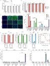
Fig. 4. Lipoprotein receptors mediate neuronal infection and divergent receptor orthologues support E2–E1 mediated entry.
a, Infection of mouse primary cortical neurons with GFP-expressing SFV single-cycle RVPs in the presence of the indicated proteins. Cells were imaged by fluorescence microscopy. Scale bars, 100 µm. Phase-contrast images are shown in Extended Data Fig. 9c. The experiment was performed twice, and representative images are shown. b, Quantification of infection in the experiment shown in a using a live cell imaging system (see Methods). c, d, Ten-day-old mice were administered a VLDLRLBD–Fc fusion protein or an isotype control antibody intraperitoneally 6 h before intraperitoneal inoculation with 100 PFU (c) or 1,000 PFU (d) of SFV A774. Survival of the mice was monitored daily. e, Infection of wild-type K562 or K562 cells transduced with VLDLR orthologues with single-cycle RVPs expressing GFP. f, Infection of wild-type K562 cells or K562 cells transduced with ApoER2 orthologues with single-cycle RVPs expressing GFP. Cell surface expression of constructs used in e and f, was confirmed by immunostaining (Extended Data Fig. 3). Data are mean ± s.d. from two experiments performed in triplicate (n = 6), except for the RAP experiment in b, which was performed once in triplicate and once in duplicate (n = 5). One-way ANOVA with Tukey’s multiple comparisons test, ****P < 0.0001 (b, e, f). Survival data (c, d) are from two independent experiments; in c: PBS n = 10, VLDLRLBD–Fc n = 10 isotype control n = 9 mice; in d: PBS, n = 11, VLDLRLBD–Fc n = 12, isotype control n = 11 mice. log-rank (Mantel–Cox) test comparing VLDLRLBD–Fc or isotype control to PBS, ****P < 0.0001 (c, d); or isotype control to PBS, P = 0.4745 (c) or P > 0.9999 (d); NS, not significant. H. sapiens, Homo sapiens. Source data
Extended Data Fig. 1
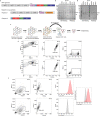
Extended Data Fig. 1. Screening strategy, reporter virus particle system, and gating strategy.
a, Ross River (RRV) reporter virus particle (RVP) system. Cells are transfected with two plasmids. CD20 or GFP is included as a reporter downstream of the capsid (C) after a 2A peptide sequence. The arrow indicates a subgenomic promoter. b, SDS-PAGE gel of purified RVPs imaged with a stain-free imaging system. The experiment was performed twice independently, and a representative gel image is shown. c, Screening strategy. HEK 293T-Cas9 cells are first transduced with the guide (sgRNA) library using vesicular stomatitis virus (VSV) glycoprotein G pseudotyped lentiviruses and are then infected with RVPs expressing CD20. Infected cells are depleted using magnetic beads against CD20. Selection is repeated iteratively to improve the signal-to-noise ratio of the screen. Non-infected, CD20 negative cells are sequenced using next generation sequencing at the final step. See Methods for additional details. d, Coomassie-stained SDS-PAGE gel of purified virus-like particles (VLPs). The experiment was performed twice independently, and a representative gel image is shown. e, Flow cytometry gating strategy for quantification of GFP-expressing cells after RVP infection. K562 cells expressing human VLDLR (top panels) or wild-type (WT) K562 cells (bottom panels) were infected with GFP-expressing SFV RVPs. The percentage of cells falling within each gate is shown. The example is from an experiment shown in Fig. 4e. f, Flow cytometry gating strategy for detection of receptor cell surface staining. K562 cells overexpressing VLDLR (top panels) or WT K562 cells (bottom panels) were stained with RAPFLAG and a FLAG-APC antibody was used for detection. In the rightmost panel, the staining of each cell type is overlaid to allow for comparison. The example is from an experiment shown in Extended Data Fig. 3c. M: molecular weight marker. For gel source data, see Supplementary Fig. 1.
Extended Data Fig. 2
Extended Data Fig. 2. Knockout cell line validation and antibody blocking of SFV E2–E1-mediated entry into multiple cell lines.
a, Genotyping DNA gel (left panel) and anti-VLDLR (α-VLDLR) antibody cell surface staining of WT HEK 293T (middle panel) or HEK 293T VLDLR clonal knockout (K.O.) cells (right panel) as monitored by FACS. The experiment was performed at least twice independently, and a representative gel image is shown. b, Anti-VLDLR (α-VLDLR) cell surface staining of WT HEK 293T, HEK 293T VLDLR K.O., and HEK 293T VLDLR K.O. cells transiently transfected with cDNA encoding VLDLR-Flag (VLDLRFLAG) as monitored by FACS. c, α-VLDLR cell surface staining of the indicated cell types as monitored by FACS. d, The indicated cell types were infected with GFP-expressing SFV single-cycle RVPs in the presence or absence of a α-VLDLR or an anti-HLA control antibody (α-HLA) and infection was measured by FACS. Means ± standard deviation from two experiments performed in triplicate (n = 6) are shown. One-way ANOVA with Tukey’s multiple comparisons test, ****P < 0.0001 (d). For gel source data, see Supplementary Fig. 1. Source data
Extended Data Fig. 3
Extended Data Fig. 3. Immunostaining to monitor cell surface receptor expression.
a, Anti-FLAG (α-FLAG) and anti-MXRA8 (α-Mxra8), staining of WT K562 cells or K562 cells expressing the indicated constructs as monitored by FACS. b, Anti-ApoER2 (α-ApoER2) and anti-LDLR (α-LDLR) staining of the indicated cell types as monitored by FACS. c, RAPFLAG staining of WT K562 cells or K562 cells transduced with the indicated constructs as monitored by ɑ-FLAG-tag staining and FACS.
Extended Data Fig. 4
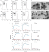
Extended Data Fig. 4. VLDLR and ApoER2 ligand binding domains directly bind alphavirus E2–E1 proteins.
a, Size exclusion chromatography traces of the indicated purified proteins. Insets are SDS-PAGE gels of the peak fraction. Molecular weight markers are indicated. Each experiment was performed at least twice, and representative traces are shown. b, Electron micrographs of negatively stained purified VLPs. Scale bar is 100 nm. The experiment was performed twice, and representative micrographs are shown. c, Sensorgrams for binding of the indicated alphavirus VLPs to tips coated with VLDLRLBD-Fc, ApoER2LBDiso1-Fc, or Mxra8ect-Fc fusion proteins as measured by biolayer interferometry. Fc fusion protein coated sensor-tips surfaces were incubated with RAP or transferrin, or kinetic buffer alone, and VLPs were associated followed by dissociation. The experiment was performed twice and representative results from one experiment are shown. Source data
Extended Data Fig. 5
Extended Data Fig. 5. Role of VLDLR and ApoER2 in E2–E1-mediated cellular infection by divergent alphaviruses.
a, Wild-type (WT) or clonal VLDLR knockout (K.O.) HEK 293T cells were infected with GFP-expressing single-cycle alphavirus RVPs with relative infection measured by FACS. EEEV RVPs more efficiently entered VLDLR K.O. cells, which we suspect could be related to clonal variability, as the cell line was generated by clonal dilution. b, Vero cells were infected with GFP-expressing alphavirus single-cycle RVPs in the presence of the indicated antibodies with relative infection measured by FACS. c, Infection of WT or transduced K562 cells with GFP-expressing single-cycle RVPs. Cells were imaged by fluorescence microscopy. Scale bar is 100 μm. The experiment was performed twice, and representative images are shown. d, Infection of WT or transduced K562 cells with GFP-expressing single-cycle RVPs measured by FACS. NRP2 is a control membrane protein. e, K562 cells expressing VLDLR or ApoER2iso2 were infected with the indicated single-cycle RVPs in the presence of RAP, soluble VLDLR LBD (sVLDLRLBD), or a control protein (transferrin, Tf) with infection measured by FACS. f, WT or transduced K562 cells were infected with the indicated GFP-expressing single-cycle RVPs with infection measured by FACS. g, SFV A774 plaque reduction neutralization test with the indicated proteins performed on Vero cells. h, WT K562 cells or K562 cells transduced to express LDLRAD3 were infected with the indicated GFP-expressing single-cycle RVPs with infection measured by FACS. Means ± standard deviation from an experiment performed once in triplicate (n = 3) (a), or experiments performed twice in triplicate (n = 6) with similar results (b, dh). One-way ANOVA with Tukey’s multiple comparisons test, ****P < 0.0001 (a, b, dh). Two-way ANOVA with Šídák’s multiple comparison test, ****P < 0.0001 (g). Cell surface expression of constructs used in (c), (d), and (f) was confirmed with immunostaining (see Extended Data Fig. 3). Source data
Extended Data Fig. 6
Extended Data Fig. 6. Ligand-binding domain sequence alignment and domain organization of ApoER2 constructs.
a, Sequence alignment of the Homo sapiens, Mus musculus, Equus caballus, and Sturnus vulgaris ApoER2 ligand binding domains. The LDLR class A (LA) repeats contained in each protein are shown in parentheses. The domain numbering is based on the human sequence shown. b, Schematic representation of the ectodomains of ApoER2 constructs used in this study. In mammals, exon regions encoding LA repeats 4-6 are almost exclusively spliced out, while the predominant avian isoforms retain these repeats. Panel (a) was generated using ESPrit 3.0.
Extended Data Fig. 7
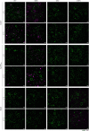
Extended Data Fig. 7. Representative confocal microscopy images for virus-like particle cell binding and internalization.
K562 cells transduced with human VLDLR, human ApoER2iso2, or human MXRA8 were incubated with fluorescently labeled VLPs at 4 °C or 37 °C and then imaged by live cell confocal microscopy. WGA: wheat germ agglutinin. Scale bar is 10 μm. The experiment was performed twice independently, and representative images are shown.
Extended Data Fig. 8
Extended Data Fig. 8. Workflow diagram of the 3-dimensional quantification of virus-like particle cell surface membrane binding and internalization.
a, 3D analysis of multi-colored stacks (pink, VLPs; green, cell membranes) using Arivis 4DFusion. Two custom-made pipelines were used to detect VLPs and cellular compartments. b, VLPs: left panel shows 3D rendering of VLP stacks, and right panel shows 3D rendering of detected VLPs. c, Cellular compartments: left panel shows 3D rendering of cellular membranes stacks; right, top panel shows 3D rendering of the detected cytoplasms (red) overlayed with an enhanced-membrane filter (white); right, bottom panel shows 3D rendering of the detected membranes (yellow). Objects obtained in each pipeline where combined to quantify the number of VLPs in each cellular compartment. d, Top: single plane representation of the detected objects, showing VLPs in the cytoplasm and the membrane. Bottom: 3D-view of the same cell. Related to Fig. 3c and 3d.
Extended Data Fig. 9
Extended Data Fig. 9. Effects of VLDLRLBD-Fc and RAP on E2–E1-mediated neuron infection and viral replication assays.
a, Infection of human neurons derived from induced pluripotent stem cell (iPSCs) with GFP-expressing SFV single-cycle RVPs in the presence of the indicated proteins. Cells were imaged by fluorescence microscopy. The experiment was performed twice with representative images shown. b, Quantification of single-cycle SFV RVP infection of human iPSC-derived neurons for the experiment shown in (a) using a live cell imaging system (see Methods for additional details) . c, Merged phase contrast and fluorescent microscopy for the experiment with mouse cortical neurons shown in Fig. 4a. Scale bars are 100 μm. Magnification is 20X. d, Merged phase contrast and fluorescent microscopy images for the experiment with human neurons shown in (a). Scale bars are 100 μm. Magnification is 10X. e, Viral replication curve for SFV, EEEV, and SINV strains in transduced K562 cells. Means ± standard deviation from two experiments done in triplicate (n = 6) with one-way ANOVA with Tukey’s multiple comparisons test, ****P < 0.0001 (b). Means ± standard deviation from two experiments done in triplicate (n = 6) with two-way ANOVA with Tukey’s multiple comparisons test, *P = 0.0233, ****P < 0.0001 (e). Source data
Extended Data Fig. 10
Extended Data Fig. 10. Sequence alignment and domain organization of VLDLR constructs and summary of observed effects with alphavirus RVPs.
a, Sequence alignment of the Homo sapiens, Mus musculus, Equus caballus, Sturnus vulgaris, Aedes aegypti, Aedes albopictus, and C. elegans VLDLR ortholog ligand binding domains. The LDLR class A (LA) repeats contained in each protein are shown in parentheses. The domain numbering is based on the human sequence shown. b, Schematic representation of the ectodomains of VLDLR constructs used in this study. c, Summary of effects observed with GFP-expressing RVP infection of K562 cells transduced to express various VLDLR or ApoER2 orthologs derived from data shown in Extended Data Fig. 5d and Fig. 4e and 4f. +++: RVP infection with greater than 50% GFP positive cells achieved with overexpression. ++: RVP infection with 20–50% GFP positive cells achieved with overexpression. +: RVP infection with 5–20% GFP positive cells achieved with overexpression. +/-: RVP infection with less than 5% GFP positive cells of unclear biological significance. -: no enhancement. Panel (a) was generated using ESPrit 3.0.

Comment in

Similar articles

Cited by

References

    1. Rose PP, et al. Natural resistance-associated macrophage protein is a cellular receptor for Sindbis virus in both insect and mammalian hosts. Cell Host Microbe. 2011;10:97–104. doi: 10.1016/j.chom.2011.06.009. - DOI - PMC - PubMed
    1. Zhang R, et al. Mxra8 is a receptor for multiple arthritogenic alphaviruses. Nature. 2018;557:570–574. doi: 10.1038/s41586-018-0121-3. - DOI - PMC - PubMed
    1. Ma H, et al. LDLRAD3 is a receptor for Venezuelan equine encephalitis virus. Nature. 2020;588:308–314. doi: 10.1038/s41586-020-2915-3. - DOI - PMC - PubMed
    1. Cheng RH, et al. Nucleocapsid and glycoprotein organization in an enveloped virus. Cell. 1995;80:621–630. doi: 10.1016/0092-8674(95)90516-2. - DOI - PMC - PubMed
    1. Paredes AM, et al. Three-dimensional structure of a membrane-containing virus. Proc. Natl Acad. Sci. USA. 1993;90:9095–9099. doi: 10.1073/pnas.90.19.9095. - DOI - PMC - PubMed

Publication types

LinkOut - more resources