

Small bowel intussusception and concurrent jejunal polyp with neoplastic transformation: a new diagnosis of Peutz-Jeghers syndrome
- PMID: 34928720
- PMCID: PMC10335009
- DOI: 10.1308/rcsann.2021.0142
Small bowel intussusception and concurrent jejunal polyp with neoplastic transformation: a new diagnosis of Peutz-Jeghers syndrome
Abstract
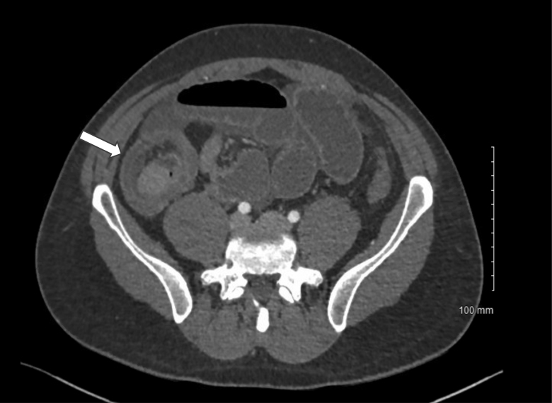
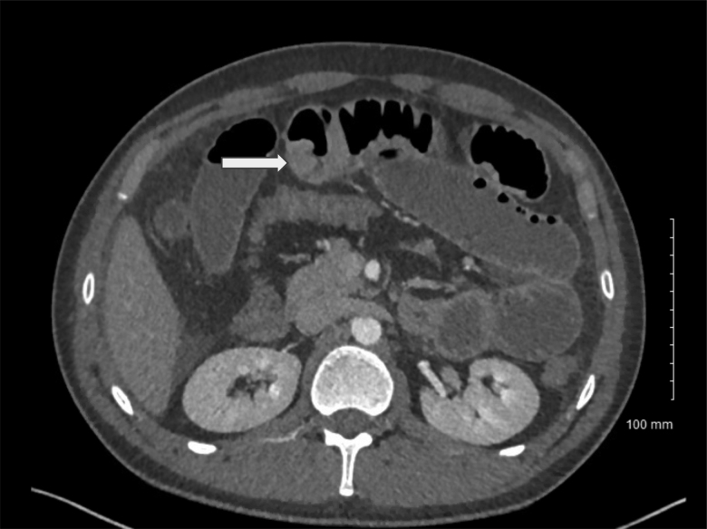
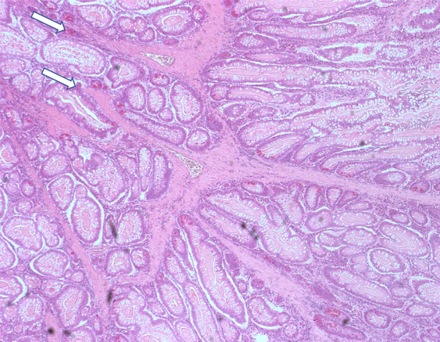
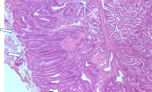
Peutz-Jeghers syndrome (PJS) is a rare hereditary disease characterised by hyperpigmentation of the oral mucosa and gastrointestinal hamartomatous polyps. We report a case of a 27-year-old man who presented with a 5-day history of epigastric pain and rectal bleeding. Computed tomography suggested small bowel obstruction secondary to ileocolic intussusception and an incidental polyp in the mid jejunum. The patient underwent exploratory laparotomy during which right hemicolectomy and small bowel resection were performed. Histology from surgical specimens revealed Peutz-Jeghers polyps, one of which had low-grade dysplasia. This case emphasises that although rare, adults with PJS can present with intussusception. Also illustrated is the extremely rare possibility of concurrent polyps occurring in different parts of the bowel with neoplastic transformation. Intussusception is a challenge to diagnose because the presentation is often non-specific. Clinical history-taking and physical examination along with prompt axial imaging is important for the diagnosis. Careful examination of the bowel and polypectomy during laparotomy may prevent neoplastic transformation and short bowel syndrome.
Keywords: Abdominal pain; Dysplasia; Intussusception; Neoplasm; Per rectum bleeding; Peutz–Jeghers; Polyp.
Figures
Similar articles
-
A case of jejunal solitary Peutz-Jeghers polyp with intussusception identified by double-balloon enteroscopy.Clin J Gastroenterol. 2020 Dec;13(6):1129-1135. doi: 10.1007/s12328-020-01197-2. Epub 2020 Aug 10. Clin J Gastroenterol. 2020. PMID: 32779147
-
An unusual presentation revealing Peutz-Jeghers syndrome in adult.Ann Med Surg (Lond). 2020 Sep 1;58:87-90. doi: 10.1016/j.amsu.2020.08.034. eCollection 2020 Oct. Ann Med Surg (Lond). 2020. PMID: 32953105 Free PMC article.
-
Massive intussusception caused by a solitary Peutz-Jeghers type hamartomatous polyp.Ann R Coll Surg Engl. 2018 Apr;100(4):e91-e93. doi: 10.1308/rcsann.2018.0019. Epub 2018 Feb 27. Ann R Coll Surg Engl. 2018. PMID: 29484932 Free PMC article.
-
Solitary Peutz-Jeghers Type Polyp of Jejunum with Gastric Fundic and Antral Gland Lining Mucosa: A Case Report and Review of Literature.Int J Surg Pathol. 2022 Aug;30(5):539-542. doi: 10.1177/10668969211067760. Epub 2021 Dec 27. Int J Surg Pathol. 2022. PMID: 34955063 Review.
-
Clinics in diagnostic imaging. 159. Jejunal intussusception due to Peutz-Jeghers syndrome.Singapore Med J. 2015 Feb;56(2):81-5; quiz 86. doi: 10.11622/smedj.2015022. Singapore Med J. 2015. PMID: 25715854 Free PMC article. Review.
References
Publication types
MeSH terms
LinkOut - more resources
Full Text Sources
Medical