. 2021 Nov 27;7(12):e08490.
doi: 10.1016/j.heliyon.2021.e08490. eCollection 2021 Dec.

TRPM7 N-terminal region forms complexes with calcium binding proteins CaM and S100A1

Affiliations

TRPM7 N-terminal region forms complexes with calcium binding proteins CaM and S100A1

Kristyna Bousova et al. Heliyon. .

Abstract

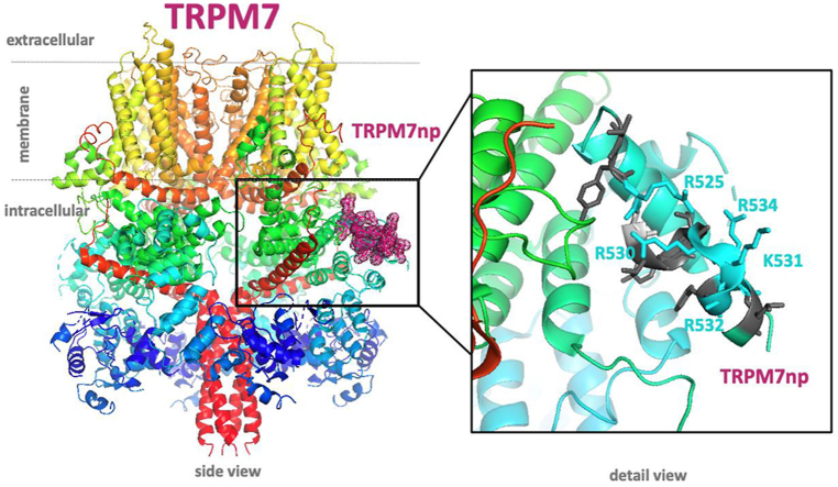
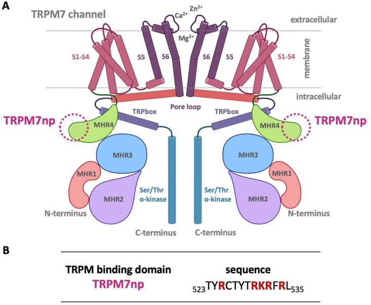
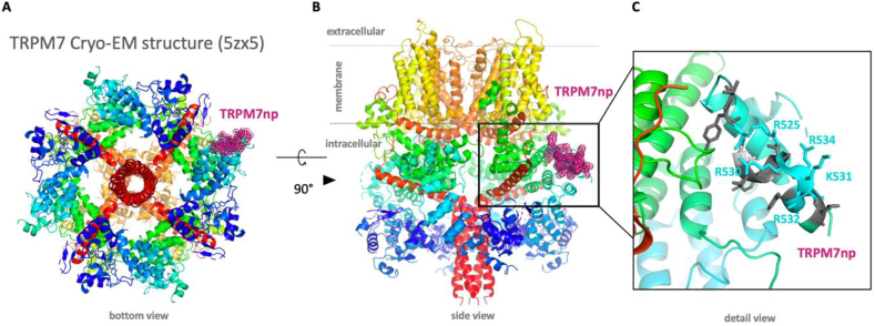
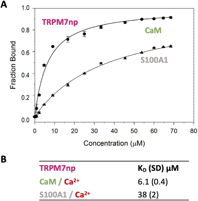
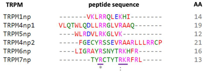
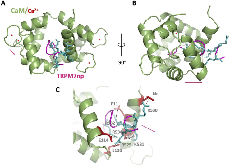
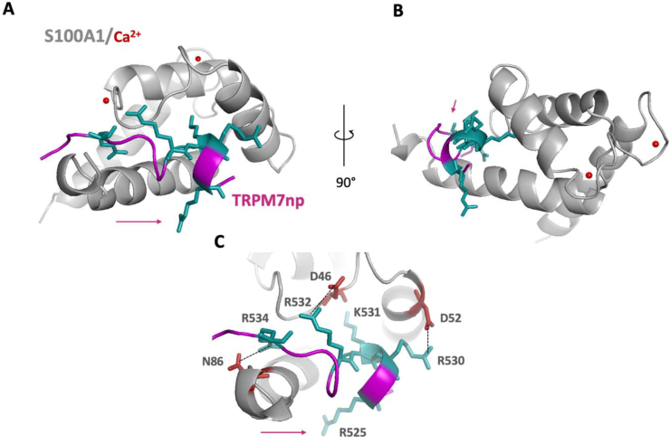
Transient receptor potential melastatin 7 (TRPM7) represents melastatin TRP channel with two significant functions, cation permeability and kinase activity. TRPM7 is widely expressed among tissues and is therefore involved in a variety of cellular functions representing mainly Mg2+ homeostasis, cellular Ca2+ flickering, and the regulation of DNA transcription by a cleaved kinase domain translocated to the nucleus. TRPM7 participates in several important biological processes in the nervous and cardiovascular systems. Together with the necessary function of the TRPM7 in these tissues and its recently analyzed overall structure, this channel requires further studies leading to the development of potential therapeutic targets. Here we present the first study investigating the N-termini of TRPM7 with binding regions for important intracellular modulators calmodulin (CaM) and calcium-binding protein S1 (S100A1) using in vitro and in silico approaches. Molecular simulations of the discovered complexes reveal their potential binding interfaces with common interaction patterns and the important role of basic residues present in the N-terminal binding region of TRPM.

Keywords: Binding region; CaM; Calcium; Fluorescence anisotropy; S100A1; TRPM7.


Conflict of interest statement

The authors declare no conflict of interest.

Figures

Image 1
Graphical abstract
Figure 1
Figure 1
Shared CaM and S100A1 binding region on TRPM7. (A) Common membrane topology of the TRPM7 (homodimer in scheme) with the location of the TRPM7np binding epitope (purple dashed circle) shown in both monomeric units; the biological formation of TRPM7 channel appears commonly in homo-tetramer formation. TRP channels consist of six transmembrane helices (S1–S6) with a pore loop and a pore helix between S5 and S6. The long cytoplasmic N-terminus contains melastatin homology regions (MHR 1–4), the C-terminus contains TRP box, Ser/Thr and alpha-kinase regions. The cytosolic regions often contain specific binding regions for modulatory molecules. (B) Amino acid sequence of the TRPM7np (UniProtKB/SwissProt: Q96QT4, position T523-L535) binding epitope with red highlighted basic residues investigated as novel CaM/S100A1 binding sites.
Figure 2
Figure 2
TRPM7np localization in the TRPM7 channel (PDB: 5ZX5). (A) Bottom view of the whole TRPM7 structure with the purple TRPM7np localization on the right. (B) Side view of the whole TRPM7 structure. The frame localizes TRPM7np binding site. (C) The corresponding region of TRPM7 known to bind CBDs in TRPM6. Detail of the TRPM7np potential binding site for CaM and S100A1 displaying the accessible amino acid side chains of TRPM7np potentially significant for the interactions. Grey sticks indicate the hydrophobic amino acids and blue sticks indicate the basic amino acids R525, R530, K531, R532, R534 predicted to be responsible for the interactions with ligands.
Figure 3
Figure 3
TRPM7np interacts with CaM and S100A1. The bound fractions (FB) of (A) TRPM7np as a function of CaM and S100A1 concentration were obtained from steady-state fluorescence anisotropy experiments. Binding isotherms (solid lines) were generated from the best fit using Eq. (2) (Materials and Methods). Five independent measurements were performed for each gradually increasing CaM/S100A1 concentrations (stock solutions c (CaM) = 118 μM, c (S100A) = 112 μM) and the calculated SD is represented by error bars. (B) The equilibrium dissociation constants (KD) of CaM/S100A1 interactions with TRPM7np together with corresponding standard deviations (SD).
Figure 4
Figure 4
Multiple alignment of selected CaM binding regions in TRPM channels. CLUSTAL 1.2.4 multiple sequence alignment of CaM/S100A1 binding regions of TRPMs N-termini [15, 25, 37, 38, 40, 41]. The sequence alignment shows a first perfect match (indicated by an asterisk) of arginine residues and second almost accurate match (indicated by dots) of basic residues across the TRPM sequences. Red colour stands for hydrophobic, pink for basic, blue for acidic amino acids; all others amino acids stand for green. The number on the right side of the sequence marks its amino acid length.
Figure 5
Figure 5
In silico analysis of the TRPM7np/CaM complex interface. (A) Top view of the TRPM7np/CaM-Ca2+ complex representation 1:1. TRPM7np (backbone in violet) and CaM (backbone in green, red balls represent calcium ions) composing the binding interface, the blue side chains represent basic amino acids of TRPM7np interacting with CaM. (B) Side view of TRPM7np/CaM complex ribbon representation with the same binding interface. (C) Detailed representation of TRPM7np positively charged residues R525, R530, K531, R532 and R534 (blue sticks) with negatively charged residues of CaM E6, E11, E14, E114 and E120 (red sticks) predicted to be involved in the non-covalent bonding. The colour convention was used same as in the A representation. The violet arrow symbolizes the direction of the helix (pseudo-helix) for a clear visualization of the orientation of the TRPM7np in the complex.
Figure 6
Figure 6
In silico analysis of the TRPM7np/S100A1 complex interface. (A) Top view of the TRPM7np/S100A1-Ca2+ complex 1:1. TRPM7np (backbone in violet) and S100A1 (backbone in grey, red balls represent calcium ions) composing the binding interface, the blue side chains represent basic amino acids of the TRPM7np interacting with S100A1. (B) Side view of TRPM7np/CaM complex ribbon representation with the same binding interface. (C) Detailed representation of TRPM7np positively charged residues R525, R530, K531, R532 and R534 (blue sticks) with negatively charged residues of S100A1 D46, D52 and N86 (red sticks) predicted to be involved in the non-covalent bonding. The colour convention was used as in the A representation. The violet arrow symbolizes the direction of the helix (pseudo-helix) for a clear visualization of the orientation of the TRPM7np in the complex.

Similar articles

Cited by

References

    1. Zou Z.G., Rios F.J., Montezano A.C., Touyz R.M. TRPM7, magnesium, and signaling. Int. J. Mol. Sci. 2019;20(8) - PMC - PubMed
    1. Abumaria N., Li W., Clarkson A.N. Role of the chanzyme TRPM7 in the nervous system in health and disease. Cell. Mol. Life Sci. 2019;76(17):3301–3310. - PMC - PubMed
    1. Ryazanova L.V., Dorovkov M.V., Ansari A., Ryazanov A.G. Characterization of the protein kinase activity of TRPM7/ChaK1, a protein kinase fused to the transient receptor potential ion channel. J. Biol. Chem. 2004;279(5):3708–3716. - PubMed
    1. Kim T.Y., Shin S.K., Song M.-Y., Lee J.E., Park K.-S. Identification of the phosphorylation sites on intact TRPM7 channels from mammalian cells. Biochem. Biophys. Res. Commun. 2012;417(3):1030–1034. - PubMed
    1. Clark K., Middelbeek J., Morrice N.A., Figdor C.G., Lasonder E., van Leeuwen F.N. Massive autophosphorylation of the Ser/Thr-rich domain controls protein kinase activity of TRPM6 and TRPM7. PLoS One. 2008;3(3) - PMC - PubMed

LinkOut - more resources