. 2021 Dec 9;4(1):1379.
doi: 10.1038/s42003-021-02902-8.

Energetics of lipid transport by the ABC transporter MsbA is lipid dependent

Affiliations

Energetics of lipid transport by the ABC transporter MsbA is lipid dependent

Dawei Guo et al. Commun Biol. .

Abstract

The ABC multidrug exporter MsbA mediates the translocation of lipopolysaccharides and phospholipids across the plasma membrane in Gram-negative bacteria. Although MsbA is structurally well characterised, the energetic requirements of lipid transport remain unknown. Here, we report that, similar to the transport of small-molecule antibiotics and cytotoxic agents, the flopping of physiologically relevant long-acyl-chain 1,2-dioleoyl (C18)-phosphatidylethanolamine in proteoliposomes requires the simultaneous input of ATP binding and hydrolysis and the chemical proton gradient as sources of metabolic energy. In contrast, the flopping of the large hexa-acylated (C12-C14) Lipid-A anchor of lipopolysaccharides is only ATP dependent. This study demonstrates that the energetics of lipid transport by MsbA is lipid dependent. As our mutational analyses indicate lipid and drug transport via the central binding chamber in MsbA, the lipid availability in the membrane can affect the drug transport activity and vice versa.


Conflict of interest statement

The authors declare no competing interests.

Figures
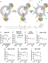
Fig. 1
Fig. 1. PE transport by MsbA.
a Schematic showing proteoliposomes containing headgroup-labelled biotin-PE (headgroup depicted as red circle with two C18 acyl chains in grey) in the inner and outer leaflet of the membrane, and the MsbA homodimer inserted in an inside-out fashion. Step 1, the PE transport reaction (red arrow) by MsbA is initiated by the addition of Mg-ATP in the external buffer and the imposition of a ΔpH (interior acidic) across the membrane. ATP binding and hydrolysis (brown arrow) at the nucleotide-binding domain dimer (in orange and slate blue) and simultaneous proton conduction (blue arrow) by the membrane domains (MD) (in light orange and light blue) provide metabolic energy for the transport of PE from the outer leaflet of the membrane to the inner leaflet. Step 2, following transport activity, the remaining amount of biotin-PE in the outer leaflet is quantified from the fluorescence emission (yellow lightning bolts) of fluorescence-tagged avidin (blue concave rectangles) when a bound quencher (green circles) is displaced by the binding of the biotin moiety. b Structural formula of the 1,2-dioleoyl (C18) biotin-PE in which the orange bracket highlights the phosphodiester moiety. c, d Biotin-PE transport assays with MsbA-WT before (c) or after (d) the addition of 1% (v/v) detergent Triton X-100 at the end of the transport reaction. Note the change in the fluorescence scale in d. Data refer to different provisions of metabolic energy: (i) Control (no metabolic energy, pHin 6.8/pHout 6.8) (set at 100%), (ii) imposed ΔpH (pHin 6.8/pHout 8.0), (iii) ATP (pHin 6.8/pHout 6.8), (iv) imposed ΔpH (pHin 6.8/pHout 8.0) plus ATP, or (v) ATP 8/8 (pHin 8.0/pHout 8.0). Control 2 in d refers to the Control after Triton X-100 addition. The MsbA inhibitor G907 was used in c. e Biotin-PE transport assays with liposomes without MsbA proteins. fi Biotin-PE transport assays with ATPase-deficient MsbA-ΔK382 (f) and substrate binding chamber mutants MsbA-TripRA (R78A R148A R296A) (g), MsbA-K299A (h) and MsbA-DED (D41N E149Q D252N) (i). Data represent observations in three experiments (n = 3) with independently prepared batches of proteoliposomes. Lines and error bars in scatter dot plots refer to mean ± s.e.m. (one-way analysis of variance; ****P < 0.0001). Asterisks above the square brackets refer to comparisons with the control without metabolic energy.
Fig. 2
Fig. 2. Lipid-A transport by MsbA.
a Structural formula of biotin-Lipid-A containing four primary hydroxymyristate (C14) acyl chains and one secondary myristate and laurate (C12) acyl chain. The orange brackets highlight the two phosphate groups in the glucosamine moieties of the lipid. b Biotin-Lipid-A assays with MsbA-WT and substrate binding chamber mutant MsbA-TripRA (R78A R148A R296A). Data refer to different provisions of metabolic energy: (i) Control (no metabolic energy, pHin 6.8/pHout 6.8), (ii) ATP (pHin 6.8/pHout 6.8) or (iii) imposed ΔpH (pHin 6.8/pHout 8.0) plus ATP. Data represent observations in three experiments (n = 3) with independently prepared batches of proteoliposomes. Lines and error bars in the scatter dot plot refer to mean ± s.e.m. (one-way analysis of variance; ****P < 0.0001). Asterisks above the horizontal line refer to comparisons with the control without metabolic energy.
Fig. 3
Fig. 3. Structure models of inward-facing MsbA with bound Lipid-A and outward-facing MsbA without the lipid.
a Ribbon diagram of inward-facing MsbA with bound Lipid-A (PDB-ID: 5TV4) shows the location of arginine residues R78, R148, R296, K299 and carboxyl residues D41, E149 and D252 (referred to as the ‘ring’ residues) in the substrate binding chamber near the centre of phospholipid bilayer. View in the plane of the plasma membrane with the two half-transporters in light blue and light orange. b Close-up view from the external face of the membrane shows how the arginine and carboxyl side chains (in blue and red stick representation) in the MD of each half-transporter form a ring of hydrophilic interactions with the 1 and 4′ phosphate group (in orange spheres) and the glucosamine, ester and amide groups connecting the acyl chains in Lipid-A (in green stick representation). The colours of the residue numbers refer to the half-transporters in which the residues are located. The biotin tag in the disaccharide core of our biotin-Lipid-A (Fig. 2a) does not interfere with the interactions of Lipid-A with the ring residues. c Ribbon diagram of outward-facing MsbA (PDB-ID: 3B60) from which Lipid-A has dissociated, shown with the same arginine and carboxyl residues as in a. d Close-up view from the external face of the membrane shows that R78-D41 and R296-E149 form salt-bridges within each half-transporter (indicated by circles with distance in Å). Figure was generated in PyMol v2.4.1.
Fig. 4
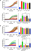
Fig. 4. Lipid-A and phospholipid transport by MsbA proteins in E. coli cells.
E. coli WD2 expresses a genome-encoded temperature-sensitive MsbA protein containing an alanine to threonine substitution at position 270 in TMH5. At non-permissive temperature (44 °C), this MsbA mutant rapidly inactivates. Due to a deficiency in the MsbA-mediated transport of phospholipids and Lipid-A to the outer membrane, the biogenesis of the outer membrane is impaired, prohibiting cell growth and division,. In contrast, E. coli WD2 cells exhibit normal growth at 30 °C. To test whether the expression of plasmid-encoded MsbA proteins can rescue the E. coli WD2 cells at 44 °C, the cells were transformed with pBAD24-based plasmids containing wild-type or mutant msbA genes. Medium without cells (Medium) (blue) and cells with empty plasmid (Control) (red) served as controls. a Expression of MsbA-WT (green), single substrate binding chamber mutants R78A (magenta), R148A (orange) or R296A (black), ATPase-deficient MsbA-ΔK382 (light brown) or drug transport-active MsbA-MD lacking the nucleotide-binding domain (dark blue). b Expression of MsbA-WT (green), double mutants R78A R148A (magenta), R148A R296A (orange) or R78A R296A (black), MsbA-TripRA (R78A R148A R296A) (light brown) or triple substrate-binding chamber mutant MsbA-DED (D41N E149Q D252N) (dark blue). Histograms show OD600 levels near 375 min; bar colours match those in the traces. The results demonstrate that ATP hydrolysis at the nucleotide-binding domains and arginine and carboxyl side chains in the central binding cavity are important for in vivo lipid transport by MsbA. All mutants were equally well expressed as MsbA-WT in the plasma membrane (Supplementary Figs. 3 and 4). Data points represent observations in three experiments (n = 3) with independently prepared batches of cells. Values in histograms show mean ± s.e.m. (one-way analysis of variance; **P < 0.01; ***P < 0.001; ****P < 0.0001). In the data for 44 °C, the asterisks above bars refer to comparison with the non-expressing control, whereas the asterisks above the square brackets refer to comparisons with MsbA-WT.
Fig. 5
Fig. 5. Ethidium transport by MsbA in L. lactis.
a ATP-depleted cells were preloaded with 2 µM ethidium after which ethidium efflux was initiated by the addition of glucose (+Glc) as a source of metabolic energy,. Active efflux by MsbA-WT was abolished in the presence of 2.5 µM of the MsbA inhibitor G907 (0.7x the reported in-vivo IC50 in cells). MsbA-mediated ethidium efflux was inhibited by the triple DED mutations in the substrate binding chamber and was enhanced by K299A and sequential R-to-A replacements in the chamber. Fluorescence traces are the mean of three independent experiments. Values in histogram show significance of fluorescence levels near 600 s. All mutants were equally well expressed as MsbA-WT in the plasma membrane (Supplementary Figs. 5 and 6). b Effect of R-to-A mutations on the apparent affinity (Km) of MsbA for ethidium in the transport reaction. Histogram data represent observations in three experiments (n = 3) with independently prepared batches of cells and are expressed as mean ± s.e.m. (one-way analysis of variance; *P <0.05; **P < 0.01; ****P < 0.0001). Asterisks above the square brackets refer to comparisons with the MsbA-WT, whereas asterisks above the horizontal line refer to comparisons with the non-expressing control.
Fig. 6
Fig. 6. Schematic on the energetics of lipid and drug transport by MsbA.
a In Lipid-A flopping, the arginine side chains of R78, R148 and R296 (indicated by blue B) and interspersed acidic side chains of D41, E149 and D252 (indicated by red C) in the central binding chamber (indicated by half blue ovals) coordinate a multitude of polar interactions with the two glucosamine and phosphate moieties of Lipid-A (in cyan blue). The sequence of reaction steps 1–4, underlying the alternating access of the binding chamber, is dependent on ATP binding and hydrolysis in our experiments and is based on structures for inward-open apo MsbA (PDB-ID: 3B5W), inward-facing G907-Lipid-A-bound MsbA (PDB-ID: 6BPL) and outward-closed ADP-vanadate-bound MsbA (PDB-ID: 5TTP) from E. coli and outward-facing AMP-PNP-bound MsbA from Salmonella typhimurium (PDB-ID: 3B60). Out and in refer to the outside and inside of the plasma membrane. b Ethidium transport is ATP binding and hydrolysis and ΔpH (interior alkaline)-dependent. Deprotonation of the C residues in inward-facing conformation in Step 1′ is followed by ethidium (Et+) binding near these residues. The B residues are not essential in this reaction and are omitted in the schematic. Step 2′, Transition to outward-facing state. Step 3′, Ethidium release and protonation of C residues. Step 4′, ΔpH-dependent transition to inward-facing state. c In PE flopping, Steps 1”–4” represent analogous ATP and ΔpH-dependent reactions as shown for ethidium, in which the B residues coordinate the binding of the phosphodiester group in PE. As the C residues are not essential in our PE transport measurements, proton coupling will involve protonatable groups (indicated by [nH+]) other than D41, E149 and D252.

Similar articles

Cited by

References

    1. Kornberg RD, McConnell HM. Inside-outside transitions of phospholipids in vesicle membranes. Biochemistry. 1971;10:1111–1120. doi: 10.1021/bi00783a003. - DOI - PubMed
    1. Nakano M, et al. Flip-flop of phospholipids in vesicles: kinetic analysis with time-resolved small-angle neutron scattering. J. Phys. Chem. B. 2009;113:6745–6748. doi: 10.1021/jp900913w. - DOI - PubMed
    1. Bethel NP, Grabe M. Atomistic insight into lipid translocation by a TMEM16 scramblase. Proc. Natl Acad. Sci. USA. 2016;113:14049–14054. doi: 10.1073/pnas.1607574113. - DOI - PMC - PubMed
    1. Vestergaard AL, et al. Critical roles of isoleucine-364 and adjacent residues in a hydrophobic gate control of phospholipid transport by the mammalian P4-ATPase ATP8A2. Proc. Natl Acad. Sci. USA. 2014;111:E1334–E1343. doi: 10.1073/pnas.1321165111. - DOI - PMC - PubMed
    1. Olsen JA, Alam A, Kowal J, Stieger B, Locher KP. Structure of the human lipid exporter ABCB4 in a lipid environment. Nat. Struct. Mol. Biol. 2020;27:62–70. doi: 10.1038/s41594-019-0354-3. - DOI - PubMed

Publication types

MeSH terms

Substances

LinkOut - more resources