. 2021 Dec 8;11(1):23600.
doi: 10.1038/s41598-021-03160-8.

RGEN-seq for highly sensitive amplification-free screen of off-target sites of gene editors

Affiliations

RGEN-seq for highly sensitive amplification-free screen of off-target sites of gene editors

Alexander Kuzin et al. Sci Rep. .

Abstract

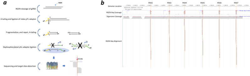
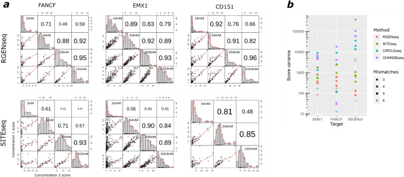
Sensitive detection of off-target sites produced by gene editing nucleases is crucial for developing reliable gene therapy platforms. Although several biochemical assays for the characterization of nuclease off-target effects have been recently published, significant technical and methodological issues still remain. Of note, existing methods rely on PCR amplification, tagging, and affinity purification which can introduce bias, contaminants, sample loss through handling, etc. Here we describe a sensitive, PCR-free next-generation sequencing method (RGEN-seq) for unbiased detection of double-stranded breaks generated by RNA-guided CRISPR-Cas9 endonuclease. Through use of novel sequencing adapters, the RGEN-Seq method saves time, simplifies workflow, and removes genomic coverage bias and gaps associated with PCR and/or other enrichment procedures. RGEN-seq is fully compatible with existing off-target detection software; moreover, the unbiased nature of RGEN-seq offers a robust foundation for relating assigned DNA cleavage scores to propensity for off-target mutations in cells. A detailed comparison of RGEN-seq with other off-target detection methods is provided using a previously characterized set of guide RNAs.


Conflict of interest statement

The authors declare no competing interests.

Figures

Figure 1
Figure 1
Overview of RGEN-seq workflow. (a), Schematic workflow illustrating preparation of the RGEN-seq library. The p7L-adapter, which is an Illumina Y-type index adapter (orange), is missing the p5 site; the p5L-adapter (green), which is a truncated Illumina Y-adapter, lacks the p7 site. Ligation of the latter adapter to both sides of free fragmented DNA prevents “background” library molecules from binding to the flowcell and generating clusters. Target and off-targets cut sites are detected by spotting the characteristic read alignment pattern around cleavage sites. (b) Representative IGV image of alignment around six cut sites (RNA2-RNA7) obtained using RGEN-seq with pooled six RGENs targeting an E. coli insertion sequence in the CHO-K1 genome. Red lines indicate target cut sites. Note characteristic alignment pattern falling to the left and right sides of the cut sites. For comparison, coverage in the same genomic region generated by the DIGENOME-seq protocol is shown.
Figure 2
Figure 2
Optimization of RGEN-seq: effect of end repair. (a), UpSet plot showing effects of different treatment combinations on the output of productive reads as percentage of total mapped reads. TdT, terminal transferase; CIP, calf intestinal phosphatase; ER, DNA end repair after RGEN cleavage; nxCleanups, number of clean-up steps in a protocol variant (see Supplementary Protocol for details). (b), Leveraging skewed read coverage of the proximal and distal sequences of RGEN clevage sites. Violin plots demonstrate the effect of end repair of the cleaved sites. Coverage skewness is defined as follows: %skewness = ((|Lcov-Rcov|)/(Lcov + Rcov)) × 100, where Lcov is the number of reads mapped to the proximal side of a cut site, and Rcov is the number of reads mapped to the distal side of the same cut site.. ER, end repair; AT- A-tailing.
Figure 3
Figure 3
Optimization of RGEN-seq: effect of number of reads and RGEN concentration on cleavage sites recovery. (a) Scatterplots of RGEN-seq off-target scores between two independent libraries prepared from the same source of genomic DNA using six multiplexed sgRNAs. A Chinese hamster CHO-K1 clone bearing a 180 kb E. coli insert (clone AD49ZG30) was the source of gDNA; sgRNAs were designed to target the 180 kb E. coli insert sequence in the AD49ZG genome. Cleavage sites were called with BLENDER22 using two million reads unless specified otherwise. (b) The number of recovered RGEN-seq cut sites for seven sgRNAs is directly proportional to the number of total mapped reads. R2 sets for each guide RNA containing 1.25 M to 20 M reads from the same sequencing experiment were mapped and analyzed by BLENDER. Number of identified off-target sites in each set was plotted as a function of number of reads in the set. Genomic DNA was the same as in (a). (c), Effect of RGEN concentrations on the number of recovered off-targets using HEK293 gDNA and sgRNAs previously characterized by other off-target detection methods. (d), Boxplot representation of the cleavage sites score distributions as a function of the EMX1 RGEN concentration.
Figure 4
Figure 4
Comparison of intersecting and distinct Cas9 cut sites across different biochemical methods. (a) UpSet plots of intersecting (lower plot) and distinct (upper plot) off-target sites of sgRNAs against FANCF and VEGFA genes produced by five methods, and (b) UpSet plots of off-target sites of six sgRNAs detected by four methods. A distinct mode corresponds to exclusive intersections that contain the elements of the sets represented by the colored circles, but not of the others. Method combinations shown at the bottom of the plots are represented by solid circles colored according to the number of selected methods in the combination. (c) Venn diagrams showing intersecting off-target sites produced by RGEN-seq and SITE-seq using sgRNAs targeted against CD151 and XRCC5 genes. In all methods except DIGENOME, 2 million reads were used for analysis and off-target sites were identified using BLENDER with default parameters. In case of SITE-seq, CIRCLE-seq, and CHANGE-seq sequencing reads were downloaded from the NCBI’s SRA as reported in the original publications and analyzed in the same way as RGEN-seq data. Genomic DNA in (a) used in CIRCLE-seq and CHANGE-seq was from K562 and U2OS cell lines, respectively, while in RGEN-seq, SITE-seq, and DIGENOME it was from HEK293 cells. In (b) genomic DNA was from HEK293 cell lines in all methods in S1-S4 panels, in RGEN-seq and SITE-seq in the EMX1 panel, and in DIGENOME in the RNF2 panel. In CHANGE-seq in EMX1 and RNF2 panels DNA was from U2OS cells, in RGEN-seq and CIRCLE-seq in the RNF2 panel DNA was from K562 cells. In (c) DNA was from HEK293 cells.
Figure 5
Figure 5
Off-target score correlation and score variance for different methods. (a) RGEN-seq vs SITE-seq: score correlations between shared cut sites from experiments performed on the same cellular source of genomic DNA (HEK293 gDNA) but at four different Cas9 RGEN concentrations using sgRNAs against FANCF, EMX1, and CD151 gene targets. Correlation between four RGEN concentrations was calculated using Pearson’s correlation coefficient (b) Comparison of score variance (log scale) between four methods. Off-target sites are grouped according to number of mismatches (the 3-mismatch group includes sites with 0–3 mismatches).

Similar articles

Cited by

References

    1. Doudna, J. A. & Charpentier, E. The new frontier of genome engineering with CRISPR-Cas9. Science346, (2014). - PubMed
    1. Hsu PD, Lander ES, Zhang F. Development and applications of CRISPR-Cas9 for genome engineering. Cell. 2014;157:1262–1278. - PMC - PubMed
    1. Komor AC, Badran AH, Liu DR. CRISPR-based technologies for the manipulation of eukaryotic genomes. Cell. 2017;168:20–36. - PMC - PubMed
    1. Dai W-J, et al. CRISPR-Cas9 for in vivo gene therapy: promise and hurdles. Mol. Ther. Nucleic Acids. 2016;5:e349. - PMC - PubMed
    1. Fellmann C, Gowen BG, Lin P-C, Doudna JA, Corn JE. Cornerstones of CRISPR–Cas in drug discovery and therapy. Nat. Rev. Drug Discov. 2017;16:89–100. - PMC - PubMed