N-glycosylation profiles of the SARS-CoV-2 spike D614G mutant and its ancestral protein characterized by advanced mass spectrometry
- PMID: 34876606
- PMCID: PMC8651636
- DOI: 10.1038/s41598-021-02904-w
N-glycosylation profiles of the SARS-CoV-2 spike D614G mutant and its ancestral protein characterized by advanced mass spectrometry
Abstract
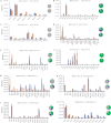
N-glycosylation plays an important role in the structure and function of membrane and secreted proteins. The spike protein on the surface of the severe acute respiratory syndrome coronavirus 2 (SARS-CoV-2), the virus that causes COVID-19, is heavily glycosylated and the major target for developing vaccines, therapeutic drugs and diagnostic tests. The first major SARS-CoV-2 variant carries a D614G substitution in the spike (S-D614G) that has been associated with altered conformation, enhanced ACE2 binding, and increased infectivity and transmission. In this report, we used mass spectrometry techniques to characterize and compare the N-glycosylation of the wild type (S-614D) or variant (S-614G) SARS-CoV-2 spike glycoproteins prepared under identical conditions. The data showed that half of the N-glycosylation sequons changed their distribution of glycans in the S-614G variant. The S-614G variant showed a decrease in the relative abundance of complex-type glycans (up to 45%) and an increase in oligomannose glycans (up to 33%) on all altered sequons. These changes led to a reduction in the overall complexity of the total N-glycosylation profile. All the glycosylation sites with altered patterns were in the spike head while the glycosylation of three sites in the stalk remained unchanged between S-614G and S-614D proteins.
© 2021. The Author(s).
Conflict of interest statement
The authors declare no competing interests.
Figures


Similar articles
-
Analysis of the N-glycosylation profiles of the spike proteins from the Alpha, Beta, Gamma, and Delta variants of SARS-CoV-2.Anal Bioanal Chem. 2023 Aug;415(19):4779-4793. doi: 10.1007/s00216-023-04771-y. Epub 2023 Jun 24. Anal Bioanal Chem. 2023. PMID: 37354227 Free PMC article.
-
Distinct shifts in site-specific glycosylation pattern of SARS-CoV-2 spike proteins associated with arising mutations in the D614G and Alpha variants.Glycobiology. 2022 Feb 26;32(1):60-72. doi: 10.1093/glycob/cwab102. Glycobiology. 2022. PMID: 34735575 Free PMC article.
-
Analysis of Glycosylation and Disulfide Bonding of Wild-Type SARS-CoV-2 Spike Glycoprotein.J Virol. 2022 Feb 9;96(3):e0162621. doi: 10.1128/JVI.01626-21. Epub 2021 Nov 24. J Virol. 2022. PMID: 34817202 Free PMC article.
-
Glycosylation of SARS-CoV-2: structural and functional insights.Anal Bioanal Chem. 2021 Dec;413(29):7179-7193. doi: 10.1007/s00216-021-03499-x. Epub 2021 Jul 7. Anal Bioanal Chem. 2021. PMID: 34235568 Free PMC article. Review.
-
Structural basis of severe acute respiratory syndrome coronavirus 2 infection.Curr Opin HIV AIDS. 2021 Jan;16(1):74-81. doi: 10.1097/COH.0000000000000658. Curr Opin HIV AIDS. 2021. PMID: 33186231 Review.
Cited by
-
Analysis of the N-glycosylation profiles of the spike proteins from the Alpha, Beta, Gamma, and Delta variants of SARS-CoV-2.Anal Bioanal Chem. 2023 Aug;415(19):4779-4793. doi: 10.1007/s00216-023-04771-y. Epub 2023 Jun 24. Anal Bioanal Chem. 2023. PMID: 37354227 Free PMC article.
-
Analysis of memory B cells identifies conserved neutralizing epitopes on the N-terminal domain of variant SARS-Cov-2 spike proteins.Immunity. 2022 Jun 14;55(6):998-1012.e8. doi: 10.1016/j.immuni.2022.04.003. Epub 2022 Apr 7. Immunity. 2022. PMID: 35447092 Free PMC article.
-
Protein complex heterogeneity and topology revealed by electron capture charge reduction and surface induced dissociation.bioRxiv [Preprint]. 2024 Jul 17:2024.03.07.583498. doi: 10.1101/2024.03.07.583498. bioRxiv. 2024. Update in: ACS Cent Sci. 2024 Jul 26;10(8):1537-1547. doi: 10.1021/acscentsci.4c00461. PMID: 38496594 Free PMC article. Updated. Preprint.
-
Molecular evolution of human coronavirus-NL63, -229E, -HKU1 and -OC43 in hospitalized children in China.Front Microbiol. 2022 Nov 2;13:1023847. doi: 10.3389/fmicb.2022.1023847. eCollection 2022. Front Microbiol. 2022. PMID: 36406425 Free PMC article.
-
Inclusion of deuterated glycopeptides provides increased sequence coverage in hydrogen/deuterium exchange mass spectrometry analysis of SARS-CoV-2 spike glycoprotein.Rapid Commun Mass Spectrom. 2024 Mar 15;38(5):e9690. doi: 10.1002/rcm.9690. Rapid Commun Mass Spectrom. 2024. PMID: 38355883
References
MeSH terms
Substances
Supplementary concepts
LinkOut - more resources
Full Text Sources
Miscellaneous

