. 2021 Dec 7;7(1):377.
doi: 10.1038/s41420-021-00767-8.

Corticotropin releasing hormone promotes inflammatory bowel disease via inducing intestinal macrophage autophagy

Affiliations

Corticotropin releasing hormone promotes inflammatory bowel disease via inducing intestinal macrophage autophagy

Sheng-Bing Zhao et al. Cell Death Discov. .

Abstract

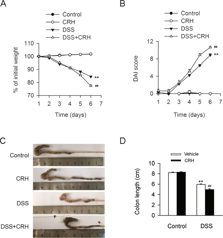
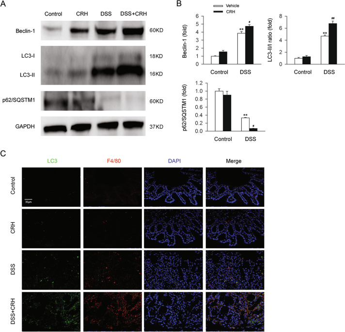
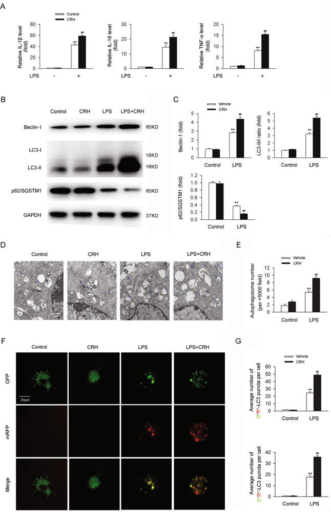
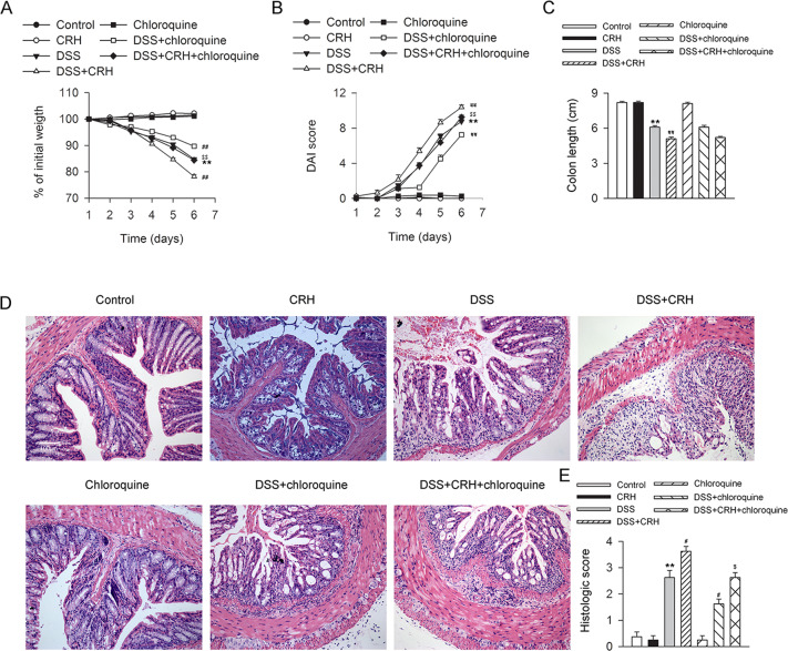
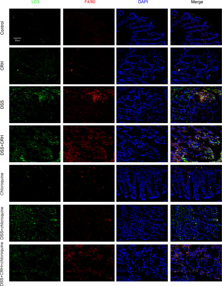
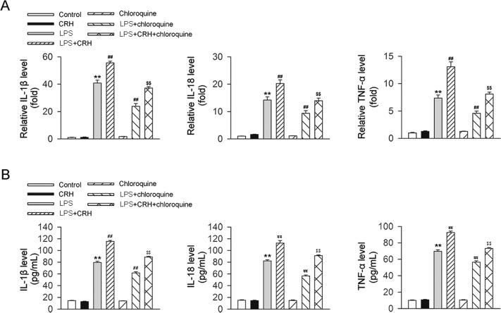
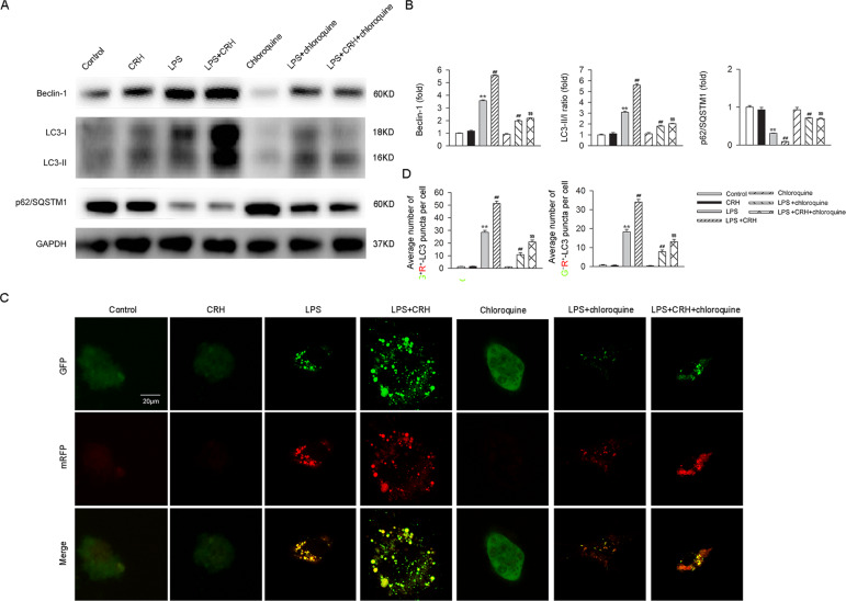
Psychosocial stress is a vital factor contributing to the pathogenesis and progression of inflammatory bowel disease (IBD). The contribution of intestinal macrophage autophagy to the onset and development of IBD has been widely studied. Herein, we investigated the underlying mechanism of psychosocial stress in an IBD mouse model pertaining to macrophage autophagy. Corticotropin releasing hormone (CRH) was peripherally administrated to induce psychosocial stress. For in vivo studies, dextran sulfate sodium (DSS) was used for the creation of our IBD mouse model. For in vitro studies, lipopolysaccharide (LPS) was applied on murine bone marrow-derived macrophages (BMDMs) as a cellular IBD-related challenge. Chloroquine was applied to inhibit autophagy. We found that CRH aggravated the severity of DSS-induced IBD, increasing overall and local inflammatory reactions and infiltration. The levels of autophagy in intestinal macrophages and murine BMDMs were increased under these IBD-related inflammatory challenges and CRH further enhanced these effects. Subsequent administration of chloroquine markedly attenuated the detrimental effects of CRH on IBD severity and inflammatory reactions via inhibition of autophagy. These findings illustrate the effects of peripheral administration of CRH on DSS-induced IBD via the enhancement of intestinal macrophage autophagy, thus providing a novel understanding as well as therapeutic target for the treatment of IBD.


Conflict of interest statement

The authors declare no competing interests.

Figures

Fig. 1
Fig. 1. Peripheral administration of CRH contributed to the deterioration of IBD.
C57BL/6 mice were administered DSS (3%) for 6 days (and a control group was provided with water only for comparison). For the CRH treatment group, CRH (or vehicle only) was intraperitoneally injected at a dose of 50 μg/kg body weight from day 1 to day 6 using saline as the vehicle. Body weight loss, stool consistency, occult or gross blood per rectum, and colon length were then recorded by two researchers who were blinded to the treatments. AD In comparison with the DSS + vehicle group, mice in the DSS + CRH group demonstrated a significant aggravation of IBD-associated changes in body weight, DAI score, and colon length (n = 8 per group). **P < 0.01 vs. the control group; ##P < 0.01 vs. the DSS + vehicle group.
Fig. 2
Fig. 2. CRH contributed to the increase of inflammation in IBD mice.
C57BL/6 mice were administered DSS (3%) for 6 days (and a control group was provided with water only for comparison). For the CRH treatment group, CRH (or vehicle only) was intraperitoneally injected at a dose of 50 μg/kg body weight from day 1 to day 6 using saline as the vehicle. A In comparison with the DSS + vehicle group, mice in the DSS + CRH group demonstrated increased levels of TNF-α and IL-18 in serum (n = 6 per group). **P < 0.01 vs. the control group; #P < 0.05 vs. the DSS + vehicle group; ##P < 0.01 vs. the DSS + vehicle group. B, C The left edge of the left colon was separated and fixed, and then H&E staining was used for the detection of inflammatory infiltration (which was assessed using a histological score). In comparison with DSS-induced colitis mice, mice in the DSS + CRH group demonstrated a significant aggravation of inflammation in the left colon (n = 8 per group). **P < 0.01 vs. the control group; #P < 0.05 vs. the DSS + vehicle group. D IL-1β, IL-18, and TNF-α mRNA levels in left colon tissues were analyzed by real-time qPCR. CRH administration also further enhanced levels of these proinflammatory factors in DSS-induced mice (n = 6 per group). **P < 0.01 vs. the control group; ##P < 0.01 vs. the DSS + vehicle group.
Fig. 3
Fig. 3. Intestinal autophagy in IBD was further enhanced by CRH.
C57BL/6 mice were administered DSS (3%) for 6 days (and a control group was provided with water only for comparison). For the CRH treatment group, CRH (or vehicle only) was intraperitoneally injected at a dose of 50 μg/kg body weight from day 1 to day 6 using saline as the vehicle. A, B The levels of autophagy-related proteins, including Beclin-1, the LC3-II/I ratio, and p62/SQSTM1, were detected by western blot. In comparison with the DSS + vehicle group, mice in the DSS + CRH group exhibited significantly increased levels of Beclin-1 and the LC3-II/I ratio, but a decrease of p62 (n = 5 per group). **P < 0.01 vs. the control group; #P < 0.05 vs. the DSS + vehicle group; ##P < 0.01 vs. the DSS + vehicle group. C The left edge of the left colon was separated and fixed and then H&E staining was used for the detection of inflammatory infiltration (which was assessed using a histological score). An increase was observed in the number of LC3 dots and the number of macrophages (which were colocalized).
Fig. 4
Fig. 4. Autophagy in BMDMs challenged with LPS was aggravated by CRH.
BMDMs from C57BL/6 mice were isolated and treated with/without LPS (100 ng/mL)/CRH (10−8 M) for 24 h. IL-1β, IL-18, and TNF-α mRNA levels were analyzed by real-time qPCR. The levels of autophagy-related proteins were analyzed in western blots. Autophagosome number was evaluated using transmission electron microscopy. Autophagy flux was assessed using the mRFP-GFP-LC3 plasmid. A In comparison with LPS-treated BMDMs, BMDMs treated with LPS + CRH exhibited a significant increase in the mRNA levels of proinflammatory cytokines (n = 6 per group). **P < 0.01 vs. the control group; ##P < 0.01 vs. the LPS + vehicle group. B, C In comparison with the LPS + vehicle group, BMDMs in the LPS + CRH group demonstrated an increase in Beclin-1, an increase in the LC3-II/I ratio, and a decrease in p62/SQSTM1 (n = 5 per group). **P < 0.01 vs. the control group; ##P < 0.01 vs. the LPS + vehicle group. An increase in the number of autophagosomes (D, E) (n = 5 per group) and an increase in autophagy flux (F, G) were also observed (n = 6 per group). **P < 0.01 vs. the control group; ##P < 0.01 vs. the LPS + vehicle group.
Fig. 5
Fig. 5. Chloroquine attenuated CRH-induced colonic damage in IBD mice.
C57BL/6 mice were administered DSS (3%) for 6 days (and a control group was provided with water only for comparison). For the CRH, chloroquine, and CRH-chloroquine groups, CRH (50 μg/kg body weight) and/or chloroquine (60 mg/kg body weight) was intraperitoneally administrated from day 1 through day 6 (using saline as a vehicle). Initial body weight, DAI score, and colon length were subsequently determined by two researchers blinded to the treatment groups. In addition, inflammation in the left colon was assessed by HE staining. AC In comparison with the DSS + vehicle group, mice in the DSS + CRH group exhibited significant increases in body weight loss, DAI scores, and colon shortening. In contrast, coadministration of chloroquine largely attenuated the aggravation effects of CRH on body weight loss and DAI Score, but no significant change in colon length (n = 8 per group). **P < 0.01 vs. the control group; ##P < 0.01 vs. the DSS + vehicle group; $$P < 0.01 vs. the DSS + CRH group. D, E H&E staining was used to assess histological scores in the left colon. In comparison with the DSS + vehicle group, mice in the DSS + CRH group demonstrated an aggravation of inflammatory infiltration. In contrast, coadministration of chloroquine largely alleviated the impact of CRH (n = 8 per group). **P < 0.01 vs. the control group; #P < 0.05 vs. the DSS + vehicle group; $P < 0.05 vs. the DSS + CRH group.
Fig. 6
Fig. 6. Chloroquine attenuated the increase in autophagy and number of macrophages in IBD mice.
C57BL/6 mice were administered DSS (3%) for 6 days (and a control group was provided with water only for comparison). For the CRH and CRH-rapamycin groups, CRH (50 μg/kg body weight) and/or rapamycin (1.25 mg/kg body weight) was intraperitoneally injected from day 1 through day 6 using saline as a vehicle. The left edge of the colon was isolated and fixed, and immunofluorescence staining was used to assess the number of macrophages (LC3 dots) and the level of autophagy (F4/80 dots). In comparison with the DSS + vehicle group, mice in the DSS + CRH group demonstrated a significant increase in the numbers of LC3 dots and F4/80 dots. This increase was attenuated following chloroquine administration. Notably, LC3 dots colocalized obviously with the F4/80 dots.
Fig. 7
Fig. 7. Chloroquine alleviated the CRH-induced enhancement of inflammation in BMDMs under the challenge of LPS.
Murine BMDMs were isolated and subsequently stimulated with LPS (100 ng/mL), CRH (10−8 M), and/or chloroquine (10 μM). Real-time qPCR and ELISA were then used to assess the levels of proinflammatory factors. In comparison with the LPS + vehicle group, CRH administration further increased the levels of IL-1β, TNF-α, and IL-18 mRNA (A) and protein (B). (n = 6 per group.) **P < 0.01 vs. the control group; ##P < 0.01 vs. the chloroquine + vehicle group; $$P < 0.01 vs. the chloroquine + CRH group.
Fig. 8
Fig. 8. Chloroquine attenuated the CRH-induced increase in autophagy in BMDMs under the challenge of LPS.
BMDMs were treated with LPS (100 ng/mL), CRH (10−8 M), and/or chloroquine (10 μM). Representative images of mRFP-GFP-LC3-transfected BMDMs were then assessed by immunofluorescence. A, B In comparison with the LPS + vehicle group, mice in the LPS + CRH group exhibited a significant increase in Beclin-1 and the LC3-II/I ratio, but a decrease in p62. These effects of CRH were blocked by chloroquine administration. (n = 5 per group). **P < 0.01 vs. the control group; ##P < 0.01 vs. the LPS + vehicle group; $$P < 0.01 vs. the LPS + CRH group. C While CRH significantly elevated autophagy levels under the challenge of LPS, chloroquine administration attenuated the effects of CRH. D Quantitative analysis of the number of yellow autophagosomes and red autolysosomes. (n = 6 per group). **P < 0.01 vs. the control group; ##P < 0.01 vs. the LPS + vehicle group; $$P < 0.01 vs. the LPS + CRH group.

Similar articles

Cited by

References

    1. Palmela C, Chevarin C, Xu Z, Torres J, Sevrin G, Hirten R, et al. Adherent-invasive Escherichia coli in inflammatory bowel disease. Gut. 2018;67:574–87.. - PubMed
    1. Singh S, Al-Darmaki A, Frolkis AD, Seow CH, Leung Y, Novak KL, et al. Postoperative mortality among patients with inflammatory bowel diseases: a systematic review and meta-analysis of population-based studies. Gastroenterology. 2015;149:928–37. - PubMed
    1. Regueiro M, Greer JB, Szigethy E. Etiology and treatment of pain and psychosocial issues in patients with inflammatory bowel diseases. Gastroenterology. 2017;152:430–9.e4. - PubMed
    1. Ventham NT, Kennedy NA, Nimmo ER, Satsangi J. Beyond gene discovery in inflammatory bowel disease: the emerging role of epigenetics. Gastroenterology. 2013;145:293–308. - PMC - PubMed
    1. Leber A, Hontecillas R, Tubau-Juni N, Zoccoli-Rodriguez V, Abedi V, Bassaganya-Riera J. NLRX1 modulates immunometabolic mechanisms controlling the host-gut microbiota interactions during inflammatory bowel disease. Front Immunol. 2018;9:363. - PMC - PubMed