Review
. 2022 Apr 11;61(16):e202110855.
doi: 10.1002/anie.202110855. Epub 2022 Mar 30.

Synthetic Cells: From Simple Bio-Inspired Modules to Sophisticated Integrated Systems

Affiliations
Review

Synthetic Cells: From Simple Bio-Inspired Modules to Sophisticated Integrated Systems

Camila Guindani et al. Angew Chem Int Ed Engl. .

Abstract

Bottom-up synthetic biology is the science of building systems that mimic the structure and function of living cells from scratch. To do this, researchers combine tools from chemistry, materials science, and biochemistry to develop functional and structural building blocks to construct synthetic cell-like systems. The many strategies and materials that have been developed in recent decades have enabled scientists to engineer synthetic cells and organelles that mimic the essential functions and behaviors of natural cells. Examples include synthetic cells that can synthesize their own ATP using light, maintain metabolic reactions through enzymatic networks, perform gene replication, and even grow and divide. In this Review, we discuss recent developments in the design and construction of synthetic cells and organelles using the bottom-up approach. Our goal is to present representative synthetic cells of increasing complexity as well as strategies for solving distinct challenges in bottom-up synthetic biology.

Keywords: artificial cells; artificial organelles; cell mimics; microreactors; synthetic biology.


Conflict of interest statement

The authors declare no conflict of interest.

Figures
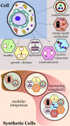
Figure 1
Figure 1
Modular approach for building synthetic cells with cell‐like properties. The integration of functional modules creates synthetic cells with increasing complexity.
Figure 2
Figure 2
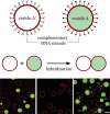
Formation of a polymer network. Hybridization of complementary DNA strands present on the surface of the polymer vesicles promotes directed adhesion. Scale bar: 100 μm. Adapted from Ref. . Creative Commons CC BY license 2020.
Figure 3
Figure 3
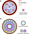
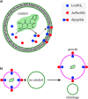
Multicompartmentalized hybrid systems. a) Synthetic cells obtained from nanopolymersomes in membranized coacervates. b) Synthetic organelles obtained from nanoliposomes arranged in layers within a polymeric capsule. Redrawn from Ref.  and Ref. [27b].
Figure 4
Figure 4
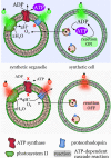
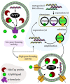
Energy self‐sufficient synthetic cells. Top left: Synthetic organelles featuring transmembrane enzymes for light‐controlled proton generation and ATP synthesis. Top right: Synthetic cells with encapsulated synthetic organelles capable of ATP generation when exposed to green light. The internal ATP drives reactions inside the synthetic cells. Bottom: synthetic organelles and cells do not operate if illuminated with red light. Redrawn from Ref. .
Figure 5
Figure 5
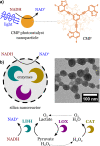
Regeneration of NAD with nanoparticles. a) Photocatalysis: a conjugated microporous polymer nanoparticle exposed to visible light oxidizes NADH to NAD. Adapted from Ref. . b) Enzymatic cascade reaction: enzymes encapsulated in porous silica nanoparticles recycle NAD in a continuous manner. The nanoreactor contains three enzymes: lactate dehydrogenase (LDH), lactate oxidase (LOX), and catalase (CAT). Adapted from Ref. . Creative Commons CC BY license 2021.
Figure 6
Figure 6
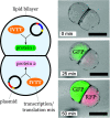
Compartmentalized protein synthesis in a synthetic cell with two fused lipid compartments. Each compartment carries the components necessary to synthesize two distinct proteins (GFP and RFP). The bright‐field fluorescent composite images show the synthesis of the compartmentalized cell‐free protein. Scale bar: 200 μm. Adapted from Ref.  with permission from the PCCP Owner Societies.
Figure 7
Figure 7
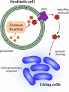
Communication between a synthetic cell and bacteria (Vibrio harveyi). Products from the encapsulated formose reaction escape from the interior of the synthetic cells through a transmembrane protein pore. The bacteria sense the low molecular weight signaling molecules, which triggers a bioluminescence response. Redrawn from Ref. .
Figure 8
Figure 8
Communication between synthetic cells. a) Signaling mediated by cell‐free expression of a pore protein. The sender cell contains the components necessary for the synthesis of the membrane porin α‐hemolysin. The porin inserts itself into the membrane of the sender. The sender releases a signal molecule (glucose) through the pore, which reaches the receiver cells. There, the signal initiates an internal cascade reaction that produces the fluorescent product (resorufin). Redrawn from Ref. [48a]. b) Signaling mediated by allosteric triggers. The sender cell produces an internal signal molecule (AMP) that diffuses to the receiver cell. Inside the receiver, AMP triggers a response cascade reaction through the allosteric activation of the first enzyme in the cascade. Adapted from Ref. [9b]. Scale bar: 30 μm. Creative Commons CC BY license 2020.
Figure 9
Figure 9
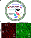
DNA replication, transcription, and translation in a synthetic cell. DNA‐replication module plus gene‐expression module. a) Scheme showing the internal composition and synthetic processes (replication and expression) of the synthetic cell. b) Fluorescent confocal micrographs of liposomes labeled with a membrane dye (red) or acridine orange (DNA dye). Scale bar: 20 μm. Adapted from Ref. . Creative Commons CC BY license 2018.
Figure 10
Figure 10
Synthetic cell growth promoted by catalytic self‐synthesis and self‐assembly of phospholipids. a) Growth based on a self‐reproducing oligotriazole‐Cu+ catalyst. Redrawn from Ref. [56d]. b) Growth based on Cu+‐mediated catalysis. Scale bar: 10 μm. Adapted from Ref. . Creative Commons CC BY license 2019.
Figure 11
Figure 11
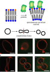
Control of vesicle shape by protein adsorption. The scheme shows His‐tagged GFP molecules binding to lipid anchors (orange) in the membrane. The degree of coverage by GPF controls the membrane curvature and morphology. Scale bars: 5 μm. Adapted from Ref. [9c]. Creative Commons CC BY license 2020.
Figure 12
Figure 12
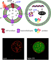
Synthetic cell showing protein synthesis powered by self‐produced ATP. Vesicle diameter ca. 10 μm. Internal synthetic organelles synthesize ATP on the surface of lipid compartments carrying light‐driven proton pumps (bacteriorhodopsin) and ATP synthase embedded in the lipid membrane. Adapted from Ref. . Creative Commons CC BY license 2019.
Figure 13
Figure 13
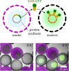
Gene expression and intercellular communication between synthetic cells. Sender lipid‐based cells synthesize fusion proteins (TetR‐sfGFP) that reach the receiver cells by molecular diffusion. The protein accumulates only in the receiver cells by interaction between the protein and tetO array plasmids, which bind to TetR. Adapted from Ref. [9a]. Creative Commons CC BY license 2018.
Figure 14
Figure 14
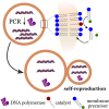
DNA replication and self‐reproduction in synthetic cells. Self‐reproduction is characterized by the partition of internal content between parent and daughter cells during the process. Redrawn from Ref. [6c].
Figure 15
Figure 15
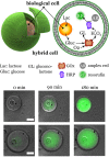
Synthetic hybrid cells. Encapsulation of biological cells into lipid vesicles creates hybrid synthetic cells. The micrographs show the synthesis of resorufin, the final product of an enzymatic cascade reaction that uses the glucose produced by the internalized biological cell. Scale bar: 25 μm. Adapted from Ref. . Creative Commons CC BY license 2018.
Figure 16
Figure 16
Fabrication of molecular factories. Cell blebbing releases giant plasma membrane vesicles that become independent hybrid cells. Synthetic organelles initially internalized by the biological cells become part of the hybrid cells along with other cytosolic components. Redrawn from Ref. .
Figure 17
Figure 17
Evolution of a pore‐membrane protein in synthetic cells. The method involves the cell‐free synthesis of α‐hemolysin in liposomes. The performance of the pores was evaluated by the diffusion of a tracer (AF488) present inside the liposomes. Redrawn from Ref. .
Figure 18
Figure 18
Competitive growth in synthetic cells. Lipid vesicles carrying a dipeptide catalyst produce a hydrophobic dipeptide from precursors inside the vesicles. The hydrophobic dipeptides accumulate in the vesicle membrane. b) Membranes with embedded dipeptides show an enhanced uptake of lipids and can grow by consuming lipids from vesicles that do not carry the dipeptide catalyst. Redrawn from Ref. .
Figure 19
Figure 19
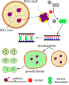
Morphogenesis in coacervate‐based synthetic cells. Artificial metalloenzymes within coacervate microdroplets catalyze the internal production of a DNA intercalator from an inactive precursor. The intercalator dynamizes the coacervates by weakening the DNA interactions, thereby resulting in morphological responses such as fusion and growth. Redrawn from Ref. .

Similar articles

Cited by

References

    1. None
    1. Noireaux V., Maeda Y. T., Libchaber A., Proc. Natl. Acad. Sci. USA 2011, 108, 3473–3480; - PMC - PubMed
    1. Xu C., Hu S., Chen X. Y., Mater. Today 2016, 19, 516–532. - PMC - PubMed
    1. None
    1. Elani Y., Angew. Chem. Int. Ed. 2021, 60, 5602–5611; - PMC - PubMed
    2. Angew. Chem. 2021, 133, 5662–5671; - PMC - PubMed

Publication types

MeSH terms

LinkOut - more resources