. 2022 Feb 1;33(2):ar14.
doi: 10.1091/mbc.E21-09-0432. Epub 2021 Dec 1.

Paxillin promotes breast tumor collective cell invasion through maintenance of adherens junction integrity

Affiliations

Paxillin promotes breast tumor collective cell invasion through maintenance of adherens junction integrity

Weiyi Xu et al. Mol Biol Cell. .

Abstract

Distant organ metastasis is linked to poor prognosis during cancer progression. The expression level of the focal adhesion adapter protein paxillin varies among different human cancers, but its role in tumor progression is unclear. Herein we utilize a newly generated PyMT mammary tumor mouse model with conditional paxillin ablation in breast tumor epithelial cells, combined with in vitro three-dimensional (3D) tumor organoids invasion analysis and 2D calcium switch assays, to assess the roles for paxillin in breast tumor cell invasion. Paxillin had little effect on primary tumor initiation and growth but is critical for the formation of distant lung metastasis. In paxillin-depleted 3D tumor organoids, collective cell invasion was substantially perturbed. The 2D cell culture revealed paxillin-dependent stabilization of adherens junctions (AJ). Mechanistically, paxillin is required for AJ assembly through facilitating E-cadherin endocytosis and recycling and HDAC6-mediated microtubule acetylation. Furthermore, Rho GTPase activity analysis and rescue experiments with a RhoA activator or Rac1 inhibitor suggest paxillin is potentially regulating the E-cadherin-dependent junction integrity and contractility through control of the balance of RhoA and Rac1 activities. Together, these data highlight new roles for paxillin in the regulation of cell-cell adhesion and collective tumor cell migration to promote the formation of distance organ metastases.


Figures
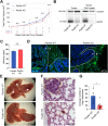
FIGURE 1:
FIGURE 1:
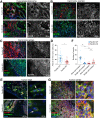
Paxillin KO breast tumor mouse develops fewer lung metastases. (A) Paxillin expression does not significantly affect total tumor size; n = number of animals. (B) Western blot showing paxillin is absent in KO tumor and isolated tumor cell lysates. (C) Paxillin does not affect tumor latency; n (animals) (paxillin WT) = 39, n (paxillin KO) = 36. (D) Paxillin is depleted only in KO tumor cells and not stroma cells as visualized by immunofluorescence staining of tumor sections. T, tumor; S, stroma. (E) Whole lungs. White arrows show surface metastases. (F) H&E staining of lung sections. Lung metastases are highlighted with dashed circles. (G) Quantification of total number of lung metastases; n = 6 animals per genotype. A Student’s t test was performed for statistical analysis. Data represent mean ± SEM *< 0.05.
FIGURE 2:
FIGURE 2:
Paxillin KO primary tumors are less invasive. Representative tumor sections from (A) early stage (hyperplasia or adenoma) tumors and the center (B) and the edge (C) of late stage (carcinoma) tumors stained for the tumor marker EpCAM and invasiveness marker ck14. The gray scale images show EpCAM-positive tumor cells. T, tumor; S, stroma. (D) Quantification of area of ck14 positive tumor cells; n (paxillin WT) = 4 animals, n (paxillin KO) = 5 animals. Each dot represents one image. (E) Paxillin WT tumor cells invade into the stroma collectively at the tumor edge (arrow top); few paxillin KO tumor cells invade into the stroma, and these were individual cells (arrow bottom). (F) Quantification of multicell, collective invasion and single cell invasion. Each string/group of tumor cells or a single cell in the stroma was counted as an event; n = 3 animals per genotype. (G) E-cadherin-mediated cell–cell junction organization is not affected in the tumor center of either paxillin WT or paxillin KO animals. A Student’s t test was performed for statistical analysis. Data represent mean ± SEM *< 0.05, **< 0.01.
FIGURE 3:
FIGURE 3:
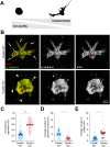
Paxillin KO tumor organoids are less invasive. (A) Illustration of the Fiji-generated masks of tumor organoids. A high degree of circularity of the organoid correlates with low invasiveness. (B) Representative confocal images of tumor organoids. Arrows point to collective cell protrusions in paxillin WT organoids; arrowheads point to individual tumor cells in paxillin KO organoids. The asterisk indicates a hyper mesenchymal cell in a paxillin KO organoid. (C) Quantification of organoid circularity. Three animals per genotype; n (paxillin WT) = 172 organoids, n (paxillin KO) = 215 organoids. (D) Quantification of the average number of collective protrusions per organoid; n = 3 animals per genotype. (E) Quantification of the average number of single cells per field; n = 5 animals per genotype. A Student’s t test was performed for statistical analysis for C, D. A Mann–Whitney U test was performed for E. Data represent mean ± SEM *< 0.05, **< 0.01, ***< 0.001.
FIGURE 4:
FIGURE 4:
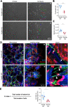
Paxillin KO tumor cells have defective collective migration on a 2D substrate. (A) Still images from phase-contrast movies of tumor cell migration for 14 h postwound. Colored lines indicate the tracks of individual cells at the wound edge from initial position (0 h) to final position (14 h). Asterisk indicates the wound area. (B) Directionality of cells at the wound edge; n = 3 animals per genotype. A minimum of five movies per animal, five cells per movie. (C) Area of cells migrated into the wound; n = 3 animals per genotype. A minimum of five movies per animal, five cells per movie. (D) Tumor cells stained for E-cadherin, vinculin, and F-actin after 8 h of migration. Representative images of AJs in the monolayer or at the leading edge. Arrows point to mature AJs; arrowheads point to disorganized AJs. Asterisk indicates the wound area. (E) AJ index was defined as the total number of mature AJs vs. the total number of cells at the wound edge; n = 3 animals per genotype. Each dot represents one image. A Student’s t test was performed for statistical analysis. Data represent mean ± SEM *< 0.05, **< 0.01.
FIGURE 5:
FIGURE 5:
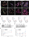
Paxillin is required for AJ formation involving endocytosis and recycling of junctional E-cadherin. (A) A calcium switch assay was used to monitor recovery of AJ integrity in cell islands. Cell AJ recovery was monitored following addition of calcium for time indicated. Cells were stained for E-cadherin, vinculin, and F-actin. (B) Quantification of the number of mature AJs per cell at indicated time points after calcium restoration; n = 3 animals per genotype. Each dot represents one image of a cell island. (C) Endocytosed biotin-labeled E-cadherin in paxillin WT or paxillin KO tumor cells during a calcium switch assay. (D) Biotin-labeled E-cadherin recycled to the cell surface during AJ formation during a calcium switch assay. (E) The internalized E-cadherin levels were normalized to total surface E-cadherin for each genotype; n = 3 animals per genotype. (F) Recycled E-cadherin to the cell surface at different time points was normalized to 0 min for each genotype; n = 5 animals per genotype. A Student’s t test was performed for statistical analysis for B, E, and F; a Mann–Whitney U test was performed for B 30 min and E 5 min. Data represent mean ± SEM *< 0.05, **< 0.01, ***< 0.001.
FIGURE 6:
FIGURE 6:
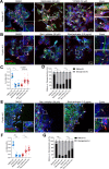
MT acetylation is important for AJ organization in tumor cell islands and for tumor organoid invasion. (A) Representative images of paxillin WT and paxillin KO tumor cells in 2D treated with tubacin (2 μM) and stained for E-cadherin, vinculin, and F-actin. (B) Quantification of the percentage of mature AJs and disorganized AJs; n = 3 animals per genotype. (C) Representative confocal images of 3D tumor organoids treated with DMSO or tubacin (2 μM). Both paxillin WT and paxillin KO organoids showed high levels of acetylated-tubulin after tubacin treatment. Tubacin-treated paxillin KO organoids showed increased collective cell invasion as compared with the DMSO-treated paxillin KO organoids. Arrows point to collective cell invasion. (D) Quantification of organoid circularity; n = 3 animals per genotype. Each dot represents one organoid. A one-way ANOVA analysis with Tukey’s multiple comparison was performed for statistical analysis. Data represent mean ± SEM *< 0.05, **< 0.01, ***< 0.001.
FIGURE 7:
FIGURE 7:
Paxillin maintains AJs in part through regulation of Rho GTPase activity. (A) Paxillin WT cells were treated with Myosin II inhibitor blebbistatin (50 μM) or ROCK inhibitor Y27632 (50 μM) for 4 h and then stained for the AJ markers E-cadherin, vinculin, and F-actin. Arrowheads point to disorganized AJs. (B) Paxillin KO cells were treated with Rac1 inhibitor NSC23766 (50 μM) or RhoA activator RhoA activator II (0.5 μg/ml) for 4 h and then stained for E-cadherin, vinculin, and F-actin. Arrows point to mature AJs; arrowheads point to disorganized AJs. (C) Quantification of AJ index; n = 3 animals per genotype. Each dot represents one image. (D) Quantification of the percentage of mature AJs and disorganized AJs; n = 3 animals per genotype. (E) Representative images of paxillin KO tumor cells from migration assays for 8 h with DMSO, NSC23766, or RhoA activator II treatment. Arrows point to mature AJs; arrowheads point to disorganized AJs. (F) Quantification of AJ index of cells at the leading wound edge for paxillin WT or paxillin KO cells treated with drugs as indicated; n = 3 animals per genotype. Each dot represents one image. (G) Quantification of the percentage of mature AJs and bridge AJs; n = 3 animals per genotype. Statistical analysis was performed using a one-way ANOVA analysis with Tukey’s multiple comparison. Data represent mean ± SEM *< 0.05, **< 0.01, ***< 0.001.

Similar articles

Cited by

References

    1. Abercrombie M, Heaysman JE (1953). Observations on the social behaviour of cells in tissue culture. I. Speed of movement of chick heart fibroblasts in relation to their mutual contacts. Exp Cell Res 5, 111–131. - PubMed
    1. Acharya BR, Nestor-Bergmann A, Liang X, Gupta S, Duszyc K, Gauquelin E, Gomez GA, Budnar S, Marcq P, Jensen OE, et al. (2018). A Mechanosensitive RhoA pathway that protects epithelia against acute tensile stress. Dev Cell 47, 439–452.e436. - PubMed
    1. Aiello NM, Maddipati R, Norgard RJ, Balli D, Li J, Yuan S, Yamazoe T, Black T, Sahmoud A, Furth EE, et al. (2018). EMT subtype influences epithelial plasticity and mode of cell migration. Dev Cell 45, 681–695.e684. - PMC - PubMed
    1. Alpha KM, Xu W, Turner CE (2020). Paxillin family of focal adhesion adaptor proteins and regulation of cancer cell invasion. Int Rev Cell Mol Biol 355, 1–52. - PMC - PubMed
    1. Azuma K, Tanaka M, Uekita T, Inoue S, Yokota J, Ouchi Y, Sakai R (2005). Tyrosine phosphorylation of paxillin affects the metastatic potential of human osteosarcoma. Oncogene 24, 4754–4764. - PubMed

Publication types