Review
. 2022 Mar;43(3):206-220.
doi: 10.1016/j.tips.2021.11.001. Epub 2021 Nov 27.

Physiological and pharmacological modulation of BAX

Affiliations
Review

Physiological and pharmacological modulation of BAX

Adam Z Spitz et al. Trends Pharmacol Sci. 2022 Mar.

Abstract

Bcl-2-associated X protein (BAX) is a critical executioner of mitochondrial regulated cell death through its lethal activity of permeabilizing the mitochondrial outer membrane (MOM). While the physiological function of BAX ensures tissue homeostasis, dysregulation of BAX leads to aberrant cell death. Despite BAX being a promising therapeutic target for human diseases, historically the development of drugs has focused on antiapoptotic BCL-2 proteins, due to challenges in elucidating the mechanism of BAX activation and identifying druggable surfaces of BAX. Here, we discuss recent studies that have provided structure-function insights and identified regulatory surfaces that control BAX activation. Moreover, we emphasize the development of small molecule orthosteric, allosteric, and oligomerization modulators that provide novel opportunities for biological investigation and progress towards drugging BAX.

Keywords: BAX; BAX activators; BAX inhibitors; BCL-2 family; apoptosis; mitochondria.


Conflict of interest statement

Declaration of interests No interests are declared.

Figures
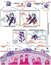
Figure 1:
Figure 1:. BAX activation pathway.
Cytosolic BAX (a, PDB: 1F16) is activated when a BH3-only protein such as BIM binds with its BH3 domain to the N-terminal BAX trigger site (b, PDB: 2K7W) initiating several conformational changes (c). Upon BH3 triggering, a conformational change of loop α1–α2 from a closed conformation, to an open conformation, contributes to exposure of the 6A7 epitope in α1 and BAX BH3-domain (α2) and subsequent dissociation of α9 from the C-terminal canonical pocket. Conformationally active BAX then translocates to the outer mitochondrial outer membrane where it can also bind to BH3-only proteins via its canonical site and separate its structure into a core (α1–α5) and latch (α6–α8) domains (d). BH3-in-groove symmetrical BAX homodimers in which the BH3-domain of one BAX protomer is bound by the canonical site of the other BAX protomer is believed to form the building block of the oligomeric BAX pore and (e) together with other BAX regions are then capable of forming higher oligomer pores which permeabilize the mitochondrial outer membrane to release cytochrome c and other apoptogens (f) which commit the cell to apoptosis.
Figure 2:
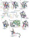
Figure 2:. Trigger site modulators of BAX.
a, Detailed diagram of the BAX/BIM-BH3 complex (PDB: 2K7W). BIM-BH3 forms hydrophobic contacts with α1 and α6 via h1–h4 (yellow). Ionic interactions between BIM and BAX are labeled red and blue with the critical BAX K21/BIM E158 interaction noted. Loop α1–α2 (orange), directly coupled to the BAX BH3-domain (pink), is maintained in an open conformation by tyrosine residues on BIM (green). b, Modelled structure of BTSA1 (light blue) bound to BAX trigger site, forming a critical hydrogen bond at K21 (blue) and using thiazole and phenyl group to open loop α1–α2 (orange). c, BAX activators (BAM7, BTSA1, and compound 8) and inhibitor (Eltrombopag) possess a similar core (blue). Substitution of a methyl for phenyl and thiazole group in BTSA1 (green) likely contributes to greater potency of BAX activation in vitro and in vivo. The biphenyl of compound 8 is proposed to make contacts with E17 and D142. d, Modelled structure of Eltrombopag (light blue) bound to BAX trigger such that loop α1–α2 remains in a closed conformation (green). Eltrombopag forms a critical interaction with R145 and a secondary interaction with R134. e, 3G11 inhibits BAX via a critical interaction with R134 (dark blue) and a secondary interaction with K21 (light blue) on the trigger site. HDX-MS shows 3G11 protects residues 36–45 on loop α1–α2 (lavender) from solvent exposure. (PDB: 1F16) f, 3C10 binds to residues 35–41 on cytosolic BAX and inhibits BAX activation. (PDB: 1F16) g, Co-crystal structure of BAX P168G (grey) and antibody 3C10 variable region (green). (PDB: 5W5X) Residues R35, R37 (blue), and M38 (orange) bind to the variable region. h–i, 3C10 activates membrane localized BAX mutant S184L by binding to residues 35–41(red). Potential conformations of BAX S184L include BAX with open loop α1–α2 (h, PDB: 2K7W) or core latch separated BAX (i, PDB: 4ZIE). Red dashed line corresponds to BAX residues to which 3C10 binds that are not resolved in PDB: 4ZIE.
Figure 3:
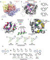
Figure 3:. Canonical site modulators of BAX.
a-b, Crystal structures of BAX bound to BIM-BH3 (a, light green, PDB: 4ZIE) and BID-BH3 (b, light blue, PDB: 4BD2). Canonical site helices are highlighted in lavender. Conserved hydrophobic BH3-domain residues h1–h4 are highlighted in yellow with secondary h0 residues highlighted in brown. Ionic interactions between BH3-domains and BAX are labeled red (anion) and blue (cation) with the critical BAX R109 interaction with conserved BH3-aspartate noted. c, Overlay of BID-BH3 (light blue) and BIM-BH3 (light green) highlighting similarities and differences in interaction with BAX canonical site. Notably, BIM-BH3 is capable of an additional ionic interaction with BAX D98/D102 via BIM R153. d, Proposed binding site of SMBA1, CYD-4-61, and GL0388 centered about S184 (green) on α9 featuring critical contacts with D102 (red) on α4 and R109 (blue) on α5. (PDB: 1F16) e, Structure of canonical site BAX activators SMBA1, CYD-4-61, and GL0388. Structural differences enhancing activity or reducing toxicity of CYD-4-61 and GL0388 compared to SMBA1 are highlighted in blue and green, respectively. f, Structure of proposed canonical site BAX activator compound 106. g, Small molecule inhibitors of BAX activation MSN-125, MSN-50, and DAN004 are predicted to bind to the BAX and BAK canonical site. The peptide like region and methoxyethyl methyl ether protected phenoxy group are highlighted in green and blue respectively. DAN004 features only a primary alkyl amine. h, BAX channel inhibitors Bci1 and Bci2 have an unknown binding site and feature a C12 lipid like tail (orange), a chiral core with two amide bonds (blue), and either C6 or C5 primary alkyl amines (black).
Figure 4:
Figure 4:. Non-canonical modulators of BAX.
a, BAX surface highlighting the N-terminal trigger site (yellow), C-terminal canonical site (lavender), and the non-canonical region (red). The non-canonical site can loosely be defined as including the hairpins of α3–α4 and α5–α6 as well as the N-terminal of loop α1–α2. b, NMR solution structure of BAX (grey) and BAX inhibiting peptide viral mitochondrial localized inhibitor of apoptosis (vMIA, pink) (PDB: 2LR1). Critical ionic interactions between BAX D84 and D86 and vMIA R139 and R146 are highlighted. c, Small molecule fragments BIF-44 and analogs bind to the vMIA binding site and sensitize BAX to activation by BH3-only activators. d, Binding site of the BCL-2 BH4 domain to inactive BAX (PDB: 1F16). Hydrophobic, hydrophilic, acidic, and basic residues are labeled as described in the figure. e, The BCL-2 BH4 domain (green) forms critical interactions with BAX via the hydrophobic residues I19 and L23 (purple). f, Allosteric BAX inhibitors (BAIs) bind to a site formed by α3, α5, α6 and loop α3–α4 making contacts with several hydrophobic residues highlighted in yellow. BAI1 makes critical contacts with K123 and D44 via its secondary alcohol and cationic piperazine, respectively. Obstructive mutagenesis of V83W/L120W (green) inhibited BAI1 binding mediated BAX inhibition. g, Structure-activity relationship studies of BAI1 highlighted the importance of steric bulk of the dibromo-carbazole (green) as well as the cationic piperazine (blue). h, BAX activators OICR77A and select analogs activate BAX at an unknown site but require the presence of BAX C126, which lies in the non-canonical region. The common phenyl substituted N-hydroxyindole core is highlighted in blue with various R and R’ groups displayed in black. The absence of the N-hydroxy group (red) in SR-1 (right) eliminates BAX activation activity.

Similar articles

Cited by

References

    1. Singh R, Letai A & Sarosiek K Regulation of apoptosis in health and disease: the balancing act of BCL-2 family proteins. Nat Rev Mol Cell Biol 20, 175–193, doi:10.1038/s41580-018-0089-8 (2019). - DOI - PMC - PubMed
    1. Delbridge AR, Grabow S, Strasser A & Vaux DL Thirty years of BCL-2: translating cell death discoveries into novel cancer therapies. Nat Rev Cancer 16, 99–109, doi:10.1038/nrc.2015.17 (2016). - DOI - PubMed
    1. Del Re DP, Amgalan D, Linkermann A, Liu Q & Kitsis RN Fundamental Mechanisms of Regulated Cell Death and Implications for Heart Disease. Physiol Rev 99, 1765–1817, doi:10.1152/physrev.00022.2018 (2019). - DOI - PMC - PubMed
    1. Kale J, Osterlund EJ & Andrews DW BCL-2 family proteins: changing partners in the dance towards death. Cell Death Differ 25, 65–80, doi:10.1038/cdd.2017.186 (2018). - DOI - PMC - PubMed
    1. Luna-Vargas MP & Chipuk JE The deadly landscape of pro-apoptotic BCL-2 proteins in the outer mitochondrial membrane. Febs j 283, 2676–2689, doi:10.1111/febs.13624 (2016). - DOI - PMC - PubMed

Publication types

MeSH terms

Substances