. 2021 Nov 16;9(11):1701.
doi: 10.3390/biomedicines9111701.

Canonical and Divergent N-Terminal HBx Isoform Proteins Unveiled: Characteristics and Roles during HBV Replication

Affiliations

Canonical and Divergent N-Terminal HBx Isoform Proteins Unveiled: Characteristics and Roles during HBV Replication

Sergio Hernández et al. Biomedicines. .

Abstract

Hepatitis B virus (HBV) X protein (HBx) is a viral regulatory and multifunctional protein. It is well-known that the canonical HBx reading frame bears two phylogenetically conserved internal in-frame translational initiation codons at Met2 and Met3, thus possibly generating divergent N-terminal smaller isoforms during translation. Here, we demonstrate that the three distinct HBx isoforms are generated from the ectopically expressed HBV HBx gene, named XF (full-length), XM (medium-length), and XS (short-length); they display different subcellular localizations when expressed individually in cultured hepatoma cells. Particularly, the smallest HBx isoform, XS, displayed a predominantly cytoplasmic localization. To study HBx proteins during viral replication, we performed site-directed mutagenesis to target the individual or combinatorial expression of the HBx isoforms within the HBV viral backbone (full viral genome). Our results indicate that of all HBx isoforms, only the smallest HBx isoform, XS, can restore WT levels of HBV replication, and bind to the viral mini chromosome, thereby establishing an active chromatin state, highlighting its crucial activities during HBV replication. Intriguingly, we found that sequences of HBV HBx genotype H are devoid of the conserved Met3 position, and therefore HBV genotype H infection is naturally silent for the expression of the HBx XS isoform. Finally, we found that the HBx XM (medium-length) isoform shares significant sequence similarity with the N-terminus domain of the COMMD8 protein, a member of the copper metabolism MURR1 domain-containing (COMMD) protein family. This novel finding might facilitate studies on the phylogenetic origin of the HBV X protein. The identification and functional characterization of its isoforms will shift the paradigm by changing the concept of HBx from being a unique, canonical, and multifunctional protein toward the occurrence of different HBx isoforms, carrying out different overlapping functions at different subcellular localizations during HBV genome replication. Significantly, our current work unveils new crucial HBV targets to study for potential antiviral research, and human virus pathogenesis.

Keywords: HBV; HBx; divergent N-terminal isoform; genome replication; hepatitis B virus; hepatitis B virus X protein; localization regulation; subcellular localization.


Conflict of interest statement

The authors declare no conflict of interest. The funders had no role in the design of the study; in the collection, analyses, or interpretation of data; in the writing of the manuscript, or in the decision to publish the results.

Figures
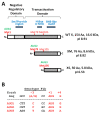
Figure 1
Figure 1
HBx domains organization, smaller isoforms, and their constructs for expression in Huh-7 hepatocarcinoma cells. (A) HBx functional domains organization [22,23,24,25,26]. N-terminal negative regulatory domain includes the AUG1 (green), Met1 (bold red), and Ser/Pro-rich region segment (blue). HBx C-terminal transactivation domain with AUG2 (green), Met79 for HBx Medium smaller isoform (XM), including H-box α-helix (blue). AUG3 (green), Met105 for HBx smaller isoform (XS), including the BH3-like motif (blue) are shown. Calculated molecular weight and basal isoelectric point (pI) for each HBx-derived isoform polypeptides are indicated to the right. (B) HBx in-frame AUG1, AUG2, and AUG3 initiation codons of HBV genotype F1b (Genebank KM233681.1) [27,28] compared with Kozak consensus sequences. Critical positions (−3, and +4) for optimal translation with respect to Kozak consensus are shown in red. (C) Scheme of HBx constructs for either canonical or individual HBx isoforms expression fused to the GFP marker. HBx constructs were made with the indicated regions of the HBx reading frame (white blocks), and with the substituted point changes (AUG -> GUG). For HBx XS expression (“mini-HBx”), the complete HBx reading frame was necessary for expression, introducing an in-frame early stop codon to prevent full HBx expression (UAA and red line) and a GUG change in the AUG2 to prevent the expression of XM. (D) Expression of the HBx isoforms upon transfection in Huh-7 cells, and Western blot to detect GFP expression. DNA constructs were transfected in Huh-7 cells, cell extracts were resolved by SDS-PAGE, and HBx isoform proteins were detected by Western blot against GFP protein marker. WT HBx (WT X, lane 1), HBx full-length (XF, lane 2), HBx medium-length (XM, lane 3), and HBx small-length (XS, lane 4) are indicated. Right panel is an overexposure of the signal showed to the left.
Figure 1
Figure 1
HBx domains organization, smaller isoforms, and their constructs for expression in Huh-7 hepatocarcinoma cells. (A) HBx functional domains organization [22,23,24,25,26]. N-terminal negative regulatory domain includes the AUG1 (green), Met1 (bold red), and Ser/Pro-rich region segment (blue). HBx C-terminal transactivation domain with AUG2 (green), Met79 for HBx Medium smaller isoform (XM), including H-box α-helix (blue). AUG3 (green), Met105 for HBx smaller isoform (XS), including the BH3-like motif (blue) are shown. Calculated molecular weight and basal isoelectric point (pI) for each HBx-derived isoform polypeptides are indicated to the right. (B) HBx in-frame AUG1, AUG2, and AUG3 initiation codons of HBV genotype F1b (Genebank KM233681.1) [27,28] compared with Kozak consensus sequences. Critical positions (−3, and +4) for optimal translation with respect to Kozak consensus are shown in red. (C) Scheme of HBx constructs for either canonical or individual HBx isoforms expression fused to the GFP marker. HBx constructs were made with the indicated regions of the HBx reading frame (white blocks), and with the substituted point changes (AUG -> GUG). For HBx XS expression (“mini-HBx”), the complete HBx reading frame was necessary for expression, introducing an in-frame early stop codon to prevent full HBx expression (UAA and red line) and a GUG change in the AUG2 to prevent the expression of XM. (D) Expression of the HBx isoforms upon transfection in Huh-7 cells, and Western blot to detect GFP expression. DNA constructs were transfected in Huh-7 cells, cell extracts were resolved by SDS-PAGE, and HBx isoform proteins were detected by Western blot against GFP protein marker. WT HBx (WT X, lane 1), HBx full-length (XF, lane 2), HBx medium-length (XM, lane 3), and HBx small-length (XS, lane 4) are indicated. Right panel is an overexposure of the signal showed to the left.
Figure 2
Figure 2
Subcellular localization of HBx canonical, and individual HBx isoforms in cultured hepatocarcinoma cells. (A) Fluorescence of the GFP-tagged HBx proteins in hepatoma cells transfected with high (H, left), medium (M, middle) or low (L, right) amounts of the plasmid DNA constructs [27,86,90]. Representative images of cells transfected with X WT (HBx WT), XF (HBx full-length), XM (HBx medium-length), XS (HBx short-length), each of them fused to the GFP protein are shown. Bar for scale of 50 µm. (B) 100 positive cells for the expression of GFP of WT or mutant HBx proteins were analyzed. Cells were all transfected with three different amounts of DNA, and after 24 h, coverslips were processed for fluorescence microscopy. Expression of HBx-GFP isoform proteins was associated with either the cytoplasm, nucleus, or nucleocytoplasmic (dual) compartments with respect to DAPI-positive nuclear staining.
Figure 3
Figure 3
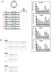
HBV DNA constructs for targeting the individual expression of the different HBx isoform proteins, and replication in HepG2 cells. (A) HBV DNA backbone containing HBx mutations of either individual HBx isoforms or all possible combinations of them [27,28]. To abolish the expression of HBx full-length (i.e., in the constructs XMS, 3X-, XM, XS) an early in-frame stop codon was introduced in the HBx reading frame. Additional point changes in the HBx reading frame are indicated by the GUG codon (green) replacing the corresponding AUG codon at either the second or third in-frame initiation codons. Protein expressions of each individual HBx isoforms for each HBV DNA construct are indicated in the columns to the right. (B) Point sequence changes on reading frames of either HBx or HBV P (polymerase) protein. Since the reading frames of HBx and P protein overlap but in a different phase (offset by one nucleotide), it is important that changes introduced into HBx reading frame do not affect HBV P protein. As shown, mutations in either AUG1 (top panel) or AUG2 (middle panel) of the HBV HBx do not affect the reading frame of the HBV P protein. HBx* and P* indicate the modified DNA and amino acid sequences. In the case of HBx AUG3 (bottom panel), it is overlapped by the basal core promoter (BCP) sequence, and the change in the sequence will introduce the indicated point mutation [118,119,120,121]. Analyses of intracellular HBV (C) Cyt DNA, and (D) cccDNA replicative intermediates after transfection of WT or mutant HBV genomes are shown, respectively. Analyses of secreted HBV (E) HBsAg, or (F) HBeAg viral antigen markers into supernatants are indicated, respectively. Results are shown as fold changes with respect to the WT HBV genome. The standard deviation was obtained from four independent experiments. *: p < 0.05, **: p < 0.01, ***: p < 0.001, Student’s t-test.
Figure 4
Figure 4
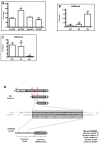
Roles and properties of the HBx XM and XS smaller isoforms. (A) The HBV XS isoform binds to the cccDNA and is sufficient to establish an active viral chromatin state. HepG2 cells were co-transfected with WT HBV DNA and one of the constructs, GFP-Vo (GFP empty vector), HBx XS-GFP, HBx XM-GFP or HBx XF-GFP, and the cells were analyzed by chromatin immunoprecipitation (ChIP) assays. HBx isoform binding to cccDNA was determined by ChIP analysis using GFP-conjugated Sepharose beads. Immunoprecipitated DNA was quantified by qPCR using specific primers for the core promoter. The results are expressed as % of input. The standard deviation was obtained from three PCR reactions, and the graphs are representative of three independent experiments. *: p < 0.05, **: p < 0.01, ***: p < 0.001, Student’s t-test. (B,C) Analysis of post-translational modifications of histones bound to the core viral promoter by ChIP. HepG2 cells were transfected with the viral full constructs, either WT HBV, HBV XS, or HBV 3X- genomes, and cells were analyzed 72 h later for ChIP assay. Covalent post-translational modifications on histone H3 were determined using specific antibodies H3K9me2 (B) and H3K4me3 (C). Immunoprecipitated DNA was quantified by qPCR using specific primers for the core promoter. The results are expressed as % of input and normalized against the ChIP H3 value. The standard deviation was obtained from three PCR reactions and the graphs are representative of three independent experiments. *: p < 0.05, **: p < 0.01, ***: p < 0.001, Student’s t-test. (D) Sequence similarity between the N-terminus of the COMMD8 protein and HBx XM isoform protein. On top, scheme showing domains and initiation codon positions M1, M2, and M3 (in red) of the three isoforms of the HBx protein, and the position of the similarity region within the isoforms. To the bottom, a scheme that depicts domain organization of COMMD8 protein, showing the conserved C-terminal COMM domain and the unique N-terminal extension (112 residues). Within its N-terminus, the similarity region is indicated. In the middle, the similarity region between HBx and COMMD8 is shown as well as amino acid residues involved in the alignment. The significant similarity region is boxed and highlighted in gray, which displays 25.53% identity, and 48.94% similarity. For these determinations, the website GenomeNet (genome.jp) was used, and the server utilized was MOTIF (sequence motif search), using the set of all databases.
Figure 5
Figure 5
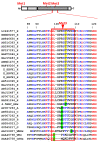
HBx XS small isoform is not expressed in HBV genotype H isolates. Multiple alignment of primary sequences of HBx proteins from all main human HBV genotypes, and chronic hepatitis B-type viruses from other organisms, such as woolly monkey hepatitis virus X protein (HMHx), woodchuck hepatitis virus X protein (WHx), and ground squirrel hepatitis virus X protein (GSHx) using multiple sequence comparison by log-expectation, the MUSCLE web server [132,133]. The alignment is magnified surrounding the Met3 position, between residues 85 to 122 of canonical HBx protein as indicated on top of the figure. HBx protein individual accession numbers are indicated in the left column, followed by the corresponding HBV genotype of each isolate or organism. The isolate 4.5HBV_HBx corresponds to our lab working HBV HBx genotype F1b isolate (GenBank: AIL83994.1) [27,28]. Individual residues across samples are colored as conserved (red), semi-conserved (blue) or non-conserved (black). Highlighted positions in bold and green indicate position of HBx Met3 in most of genotypes. Instead, highlighted position in bold and yellow indicate the corresponding conserved position of HBx Thr3, in HBV genotype H isolates, implicating the absence of Met3 in these isolates.

Similar articles

Cited by

References

    1. WHO Global Hepatitis Report, 2017. 2017. [(accessed on 15 June 2021)]. Available online: https://www.who.int/hepatitis/publications/global-hepatitis-report2017/en/
    1. Revill P., Tu T., Netter H., Yuen L., Locarnini S., Littlejohn M. The evolution and clinical impact of hepatitis B virus genome diversity. Nat. Rev. Gastroenterol. Hepatol. 2020;17:618–634. doi: 10.1038/s41575-020-0296-6. - DOI - PubMed
    1. Dias J., Sarica N., Neuveut C. Early Steps of Hepatitis B Life Cycle: From Capsid Nuclear Import to cccDNA Formation. Viruses. 2021;13:757. doi: 10.3390/v13050757. - DOI - PMC - PubMed
    1. Araujo N. Hepatitis B virus intergenotypic recombinants worldwide: An overview. Infect. Genet. Evol. 2015;36:500–510. doi: 10.1016/j.meegid.2015.08.024. - DOI - PubMed
    1. Pujol F., Jaspe R., Loureiro C., Chemin I. Hepatitis B virus American genotypes: Pathogenic variants? Clin. Res. Hepatol. Gastroenterol. 2020;44:825–835. doi: 10.1016/j.clinre.2020.04.018. - DOI - PubMed

LinkOut - more resources