Crotoxin Modulates Events Involved in Epithelial-Mesenchymal Transition in 3D Spheroid Model
- PMID: 34822613
- PMCID: PMC8618719
- DOI: 10.3390/toxins13110830
Crotoxin Modulates Events Involved in Epithelial-Mesenchymal Transition in 3D Spheroid Model
Abstract
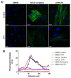
Epithelial-mesenchymal transition (EMT) occurs in the early stages of embryonic development and plays a significant role in the migration and the differentiation of cells into various types of tissues of an organism. However, tumor cells, with altered form and function, use the EMT process to migrate and invade other tissues in the body. Several experimental (in vivo and in vitro) and clinical trial studies have shown the antitumor activity of crotoxin (CTX), a heterodimeric phospholipase A2 present in the Crotalus durissus terrificus venom. In this study, we show that CTX modulates the microenvironment of tumor cells. We have also evaluated the effect of CTX on the EMT process in the spheroid model. The invasion of type I collagen gels by heterospheroids (mix of MRC-5 and A549 cells constitutively prepared with 12.5 nM CTX), expression of EMT markers, and secretion of MMPs were analyzed. Western blotting analysis shows that CTX inhibits the expression of the mesenchymal markers, N-cadherin, α-SMA, and αv. This study provides evidence of CTX as a key modulator of the EMT process, and its antitumor action can be explored further for novel drug designing against metastatic cancer.
Keywords: crotoxin; epithelial–mesenchymal transition; spheroid model; tumor stroma.
Conflict of interest statement
The authors declare no conflict of interest. The funders had no role in the design of the study; in the collection, analyses, or interpretation of data; in the writing of the manuscript, or in the decision to publish the results.
Figures


Similar articles
-
Crotoxin from Crotalus durissus terrificus venom: In vitro cytotoxic activity of a heterodimeric phospholipase A2 on human cancer-derived cell lines.Toxicon. 2018 Dec 15;156:13-22. doi: 10.1016/j.toxicon.2018.10.306. Epub 2018 Nov 2. Toxicon. 2018. PMID: 30395843
-
Neuromuscular paralysis by the basic phospholipase A2 subunit of crotoxin from Crotalus durissus terrificus snake venom needs its acid chaperone to concurrently inhibit acetylcholine release and produce muscle blockage.Toxicol Appl Pharmacol. 2017 Nov 1;334:8-17. doi: 10.1016/j.taap.2017.08.021. Epub 2017 Sep 1. Toxicol Appl Pharmacol. 2017. PMID: 28867438
-
Cytotoxic effect of crotoxin on cancer cells and its antitumoral effects correlated to tumor microenvironment: A review.Int J Biol Macromol. 2023 Jul 1;242(Pt 2):124892. doi: 10.1016/j.ijbiomac.2023.124892. Epub 2023 May 15. Int J Biol Macromol. 2023. PMID: 37196721 Review.
-
Crotoxin Modulates Macrophage Phenotypic Reprogramming.Toxins (Basel). 2023 Oct 17;15(10):616. doi: 10.3390/toxins15100616. Toxins (Basel). 2023. PMID: 37888647 Free PMC article.
-
Crotoxin: novel activities for a classic beta-neurotoxin.Toxicon. 2010 Jun 1;55(6):1045-60. doi: 10.1016/j.toxicon.2010.01.011. Epub 2010 Jan 28. Toxicon. 2010. PMID: 20109480 Review.
Cited by
-
Neurotoxins Acting at Synaptic Sites: A Brief Review on Mechanisms and Clinical Applications.Toxins (Basel). 2022 Dec 27;15(1):18. doi: 10.3390/toxins15010018. Toxins (Basel). 2022. PMID: 36668838 Free PMC article. Review.
-
Celebrating 120 Years of Butantan Institute Contributions for Toxinology.Toxins (Basel). 2022 Jan 21;14(2):76. doi: 10.3390/toxins14020076. Toxins (Basel). 2022. PMID: 35202104 Free PMC article.
References
-
- Kim K.K., Wei Y., Szekeres C., Kugler M.C., Wolters P.J., Hill M.L., Frank J.A., Brumwell A.N., Wheeler S.E., Kreidberg J.A., et al. Epithelial Cell Alpha3beta1 Integrin Links Beta-Catenin and Smad Signaling to Promote Myofibroblast Formation and Pulmonary Fibrosis. J. Clin. Investig. 2009;119:213–224. doi: 10.1172/JCI36940. - DOI - PMC - PubMed
-
- Ding S., Chen G., Zhang W., Xing C., Xu X., Xie H., Lu A., Chen K., Guo H., Ren Z., et al. MRC-5 Fibroblast-Conditioned Medium Influences Multiple Pathways Regulating Invasion, Migration, Proliferation, and Apoptosis in Hepatocellular Carcinoma. J. Transl. Med. 2015;13:237. doi: 10.1186/s12967-015-0588-8. - DOI - PMC - PubMed
Publication types
MeSH terms
Substances
LinkOut - more resources
Full Text Sources
Research Materials

