

High-resolution single-particle cryo-EM of samples vitrified in boiling nitro-gen
- PMID: 34804541
- PMCID: PMC8562666
- DOI: 10.1107/S2052252521008095
High-resolution single-particle cryo-EM of samples vitrified in boiling nitro-gen
Abstract
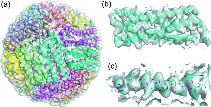
Based on work by Dubochet and others in the 1980s and 1990s, samples for single-particle cryo-electron microscopy (cryo-EM) have been vitrified using ethane, propane or ethane/propane mixtures. These liquid cryogens have a large difference between their melting and boiling temperatures and so can absorb substantial heat without formation of an insulating vapor layer adjacent to a cooling sample. However, ethane and propane are flammable, they must be liquified in liquid nitro-gen immediately before cryo-EM sample preparation, and cryocooled samples must be transferred to liquid nitro-gen for storage, complicating workflows and increasing the chance of sample damage during handling. Experiments over the last 15 years have shown that cooling rates required to vitrify pure water are only ∼250 000 K s-1, at the low end of earlier estimates, and that the dominant factor that has limited cooling rates of small samples in liquid nitro-gen is sample precooling in cold gas present above the liquid cryogen surface, not the Leidenfrost effect. Using an automated cryocooling instrument developed for cryocrystallography that combines high plunge speeds with efficient removal of cold gas, we show that single-particle cryo-EM samples on commercial grids can be routinely vitrified using only boiling nitro-gen and obtain apoferritin datasets and refined structures with 2.65 Å resolution. The use of liquid nitro-gen as the primary coolant may allow manual and automated workflows to be simplified and may reduce sample stresses that contribute to beam-induced motion.
Keywords: cryocooling; cryoelectron microscopy; vitrification.
© Engstrom et al. 2021.
Figures
Similar articles
-
A comparative study of single-particle cryo-EM with liquid-nitrogen and liquid-helium cooling.IUCrJ. 2019 Oct 22;6(Pt 6):1099-1105. doi: 10.1107/S2052252519011503. eCollection 2019 Nov 1. IUCrJ. 2019. PMID: 31709065 Free PMC article.
-
Cryo-EM structures from sub-nl volumes using pin-printing and jet vitrification.Nat Commun. 2020 May 22;11(1):2563. doi: 10.1038/s41467-020-16392-5. Nat Commun. 2020. PMID: 32444637 Free PMC article.
-
An Economical, Portable Manual Cryogenic Plunge Freezer for the Preparation of Vitrified Biological Samples for Cryogenic Electron Microscopy.Microsc Microanal. 2020 Jun;26(3):413-418. doi: 10.1017/S1431927620001385. Microsc Microanal. 2020. PMID: 32284082
-
Electron cryomicroscopy of membrane proteins: specimen preparation for two-dimensional crystals and single particles.Micron. 2011 Feb;42(2):107-16. doi: 10.1016/j.micron.2010.07.004. Epub 2010 Jul 16. Micron. 2011. PMID: 20678942 Free PMC article. Review.
-
Toward Compositional Contrast by Cryo-STEM.Acc Chem Res. 2021 Oct 5;54(19):3621-3631. doi: 10.1021/acs.accounts.1c00279. Epub 2021 Sep 7. Acc Chem Res. 2021. PMID: 34491730 Review.
Cited by
-
VitroJet: new features and case studies.Acta Crystallogr D Struct Biol. 2024 Apr 1;80(Pt 4):232-246. doi: 10.1107/S2059798324001852. Epub 2024 Mar 15. Acta Crystallogr D Struct Biol. 2024. PMID: 38488730 Free PMC article.
-
Effects of cryo-EM cooling on structural ensembles.Nat Commun. 2022 Mar 31;13(1):1709. doi: 10.1038/s41467-022-29332-2. Nat Commun. 2022. PMID: 35361752 Free PMC article.
-
Determining Protein Secondary Structures in Heterogeneous Medium-Resolution Cryo-EM Images Using CryoSSESeg.ACS Omega. 2024 Jun 8;9(24):26409-26416. doi: 10.1021/acsomega.4c02608. eCollection 2024 Jun 18. ACS Omega. 2024. PMID: 38911779 Free PMC article.
-
Millisecond mix-and-quench crystallography (MMQX) enables time-resolved studies of PEPCK with remote data collection.IUCrJ. 2021 Aug 4;8(Pt 5):784-792. doi: 10.1107/S2052252521007053. eCollection 2021 Sep 1. IUCrJ. 2021. PMID: 34584739 Free PMC article.
-
Folding of prestin's anion-binding site and the mechanism of outer hair cell electromotility.Elife. 2023 Dec 6;12:RP89635. doi: 10.7554/eLife.89635. Elife. 2023. PMID: 38054956 Free PMC article.
References
-
- Bald, W. B. (1984). J. Microsc. 134, 261–270.
-
- Bald, W. B. (1985). J. Microsc. 140, 17–40.
-
- Bald, W. B. (1986). J. Microsc. 143, 89–102. - PubMed
-
- Brüggeller, P. & Mayer, E. (1980). Nature, 288, 569–571.
Grants and funding
LinkOut - more resources
Full Text Sources
Other Literature Sources
