. 2021 Nov 15;17(11):e1009881.
doi: 10.1371/journal.pgen.1009881. eCollection 2021 Nov.

daf-16/FOXO blocks adult cell fate in Caenorhabditis elegans dauer larvae via lin-41/TRIM71

Affiliations

daf-16/FOXO blocks adult cell fate in Caenorhabditis elegans dauer larvae via lin-41/TRIM71

Matthew J Wirick et al. PLoS Genet. .

Abstract

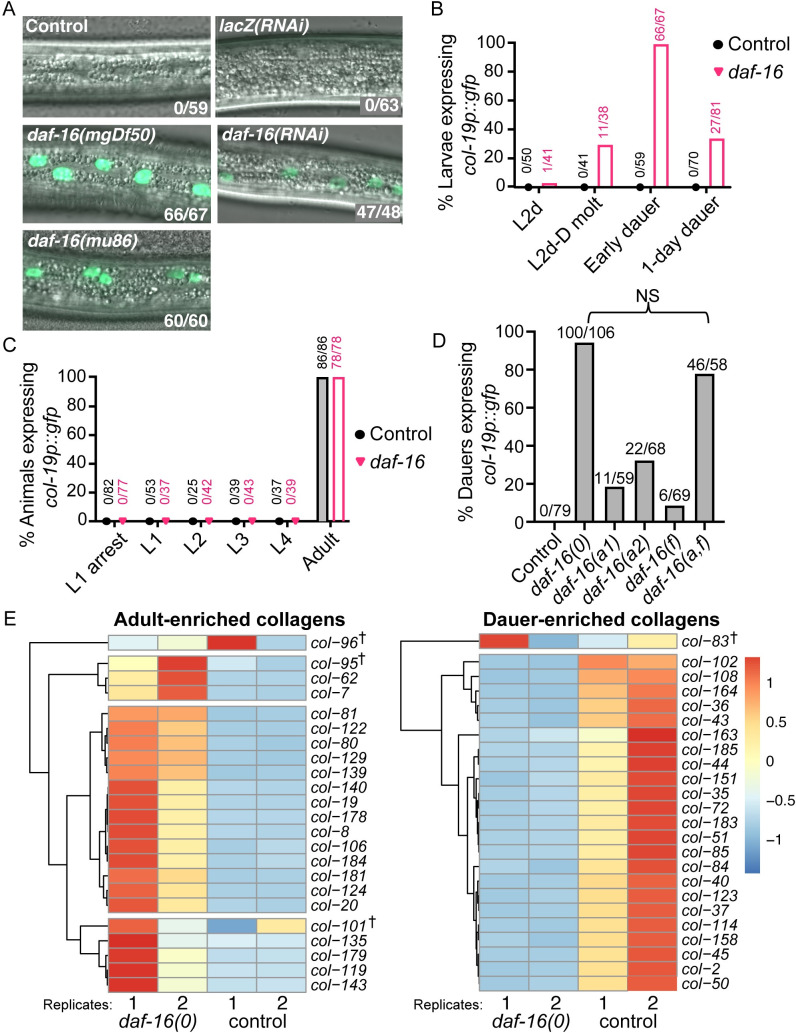
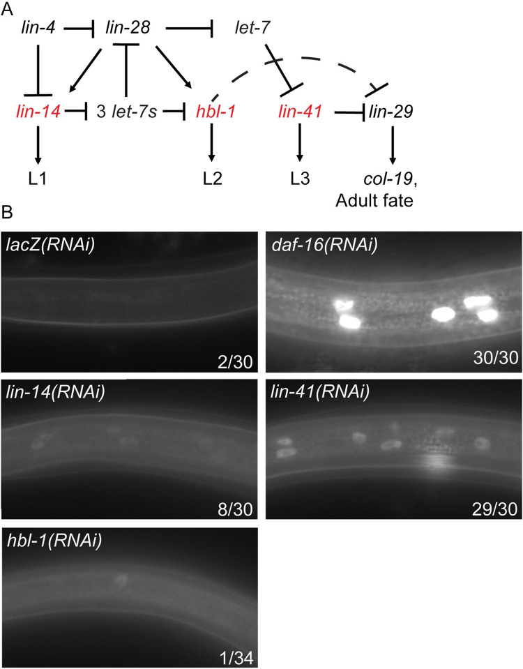
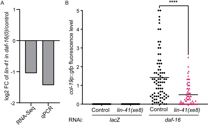
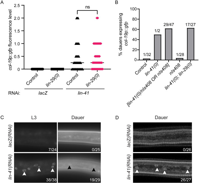
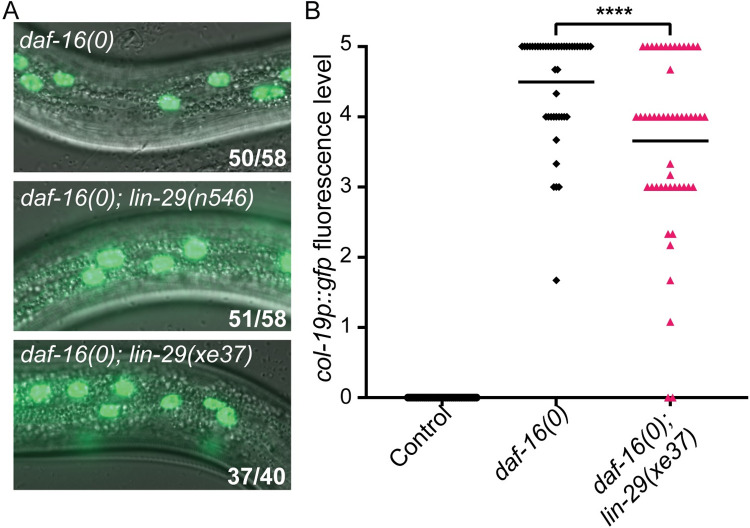
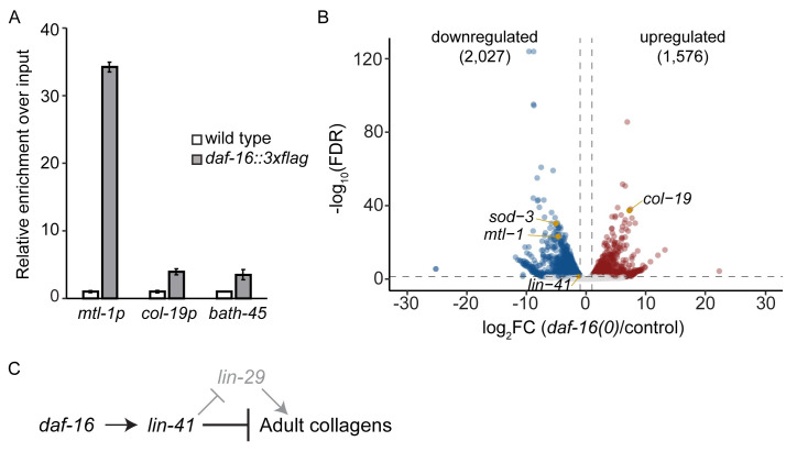
Many tissue-specific stem cells maintain the ability to produce multiple cell types during long periods of non-division, or quiescence. FOXO transcription factors promote quiescence and stem cell maintenance, but the mechanisms by which FOXO proteins promote multipotency during quiescence are still emerging. The single FOXO ortholog in C. elegans, daf-16, promotes entry into a quiescent and stress-resistant larval stage called dauer in response to adverse environmental cues. During dauer, stem and progenitor cells maintain or re-establish multipotency to allow normal development to resume after dauer. We find that during dauer, daf-16/FOXO prevents epidermal stem cells (seam cells) from prematurely adopting differentiated, adult characteristics. In particular, dauer larvae that lack daf-16 misexpress collagens that are normally adult-enriched. Using col-19p::gfp as an adult cell fate marker, we find that all major daf-16 isoforms contribute to opposing col-19p::gfp expression during dauer. By contrast, daf-16(0) larvae that undergo non-dauer development do not misexpress col-19p::gfp. Adult cell fate and the timing of col-19p::gfp expression are regulated by the heterochronic gene network, including lin-41 and lin-29. lin-41 encodes an RNA-binding protein orthologous to LIN41/TRIM71 in mammals, and lin-29 encodes a conserved zinc finger transcription factor. In non-dauer development, lin-41 opposes adult cell fate by inhibiting the translation of lin-29, which directly activates col-19 transcription and promotes adult cell fate. We find that during dauer, lin-41 blocks col-19p::gfp expression, but surprisingly, lin-29 is not required in this context. Additionally, daf-16 promotes the expression of lin-41 in dauer larvae. The col-19p::gfp misexpression phenotype observed in dauer larvae with reduced daf-16 requires the downregulation of lin-41, but does not require lin-29. Taken together, this work demonstrates a novel role for daf-16/FOXO as a heterochronic gene that promotes expression of lin-41/TRIM71 to contribute to multipotent cell fate in a quiescent stem cell model.


Conflict of interest statement

The authors have declared that no competing interests exist.

Figures

Fig 1
Fig 1. daf-16 is required to prevent precocious adoption of adult cell fate during dauer.
(A) Merged DIC and fluorescence images taken with a 63x objective displaying a portion of the lateral hypodermis in dauer larvae. Unless otherwise specified, the background for all strains in this and subsequent figures was daf-7(e1372); col-19p::gfp. (See S1 Table for full genotypes). Compromised daf-16 activity due to either of two null alleles (left) or RNAi (right) resulted in col-19p::gfp expression. Numbers indicate larvae expressing col-19p::gfp over the total number of dauer larvae scored. (B) Percent of daf-7 (control) and daf-16(0); daf-7 larvae that expressed col-19p::gfp before dauer formation and over time in dauer. GFP expression began during the molt, peaked soon after entry into dauer, and declined thereafter. (C) Percent of wild-type (control) and daf-16(0) animals that expressed col-19p::gfp at different stages. These worms were wild-type for daf-7 and therefore did not enter L2d or dauer. Larvae at L1 arrest were hatched in the absence of food and were monitored for GFP expression over seven days. (D) Percent of dauer larvae lacking some or all daf-16 isoforms that expressed any hypodermal col-19p::gfp (see S1 Table for full genotypes). Note that daf-16(a,f) displayed decreased expressivity compared to daf-16(0); the dauer larvae expressing col-19p::gfp were noticeably dimmer than in the null allele (S2 Fig). NS: p = 0.55, Fisher Exact Test. (E) Heat maps depicting relative expression of adult-enriched collagens (left) or dauer-enriched collagens (right) in daf-16(0); daf-7 dauer larvae or daf-7 (control) dauer larvae from mRNA-seq data. The row-normalized reads for each of two biological replicates per strain are depicted. These collagens were expressed at significantly different levels (FDR ≤ 0.05) in daf-16(0) vs control dauer larvae in all cases except those collagens indicated by †. Adult and dauer-enriched collagens were identified using modENCODE data (S3 Table). Adult-enriched collagens were upregulated in daf-16(0) dauer larvae whereas dauer-enriched collagens were downregulated.
Fig 2
Fig 2. lin-41 is required to prevent precocious adoption of adult cell fate during dauer.
(A) A simplified diagram of the network of heterochronic genes that regulates stage-specific seam cell fate during continuous development. “3 let-7s” indicates the let-7 family members, mir-48, mir-84, and mir-241. Genes in red are the most downstream positive regulators of larval cell fates [25]. (B) Precocious hypodermal expression of col-19p::gfp in dauer larvae in response to RNAi of heterochronic genes compared to the lacZ and daf-16 RNAi controls. Images were taken with a 63x objective. Numbers indicate the number of larvae expressing any hypodermal col-19p::gfp over the total number of dauer larvae scored. lin-14 RNAi induced only dim expression of col-19p::gfp, at low penetrance. hbl-1 RNAi did not induce hypodermal expression of col-19p::gfp. lin-41 RNAi caused highly penetrant expression of col-19p::gfp during dauer.
Fig 3
Fig 3. daf-16 promotes lin-41 expression during dauer to regulate col-19p::gfp.
(A) lin-41 was downregulated in daf-16(0); daf-7 dauer larvae compared to daf-7 (control) dauers, as measured by mRNA-seq (FDR < 0.05) and qPCR (p = 0.2, two-tailed Mann-Whitney test). At least two biological replicates per strain were used (see Methods). Raw qPCR data are shown in S5 Table. (B) Misexpression of lin-41 caused by lin-41(xe8[Δ3’UTR]) suppressed the col-19p::gfp phenotype in daf-16(RNAi) dauer larvae. Fluorescence levels were determined as described in S7 Fig. Each point represents an individual worm; horizontal lines indicate the mean fluorescence level. ****p<0.0001 (Two-tailed Mann-Whitney Test). n = 39–80; see S6 Table for complete underlying numerical data.
Fig 4
Fig 4. lin-41 regulates col-19p::gfp independently of lin-29 in dauer larvae.
(A) lin-29 is not required for col-19p::gfp expression in lin-41(RNAi) dauers. Fluorescence levels were determined as described in S7 Fig. ns = not significant (p = 0.4856, two-tailed Mann-Whitney Test). n = 33–41; see S7 Table for complete underlying numerical data. (B) lin-41(0); lin-29(0) dauer larvae expressed col-19p::gfp. nIs408 is a transgene used to balance lin-41(0) (see text). lin-41(0) larvae are dauer-defective [47]. Numbers indicate the number of dauers expressing col-19p::gfp over total dauers scored. (C, D) lin-29::gfp expression imaged on a compound microscope (C) or a confocal microscope (D). Images were taken with a 63x objective. Arrowheads indicate hypodermal nuclei with visible lin-29::gfp expression. Numbers indicate the number of larvae expressing any detectable lin-29::gfp over total number of larvae. lin-41(RNAi) produced substantial precocious expression of lin-29::gfp in continuously developing L3 staged larvae, easily discernable under the compound microscope. However, lin-41(RNAi) produced only very dim expression in dauer larvae. Dauer larvae produce substantial autofluorescence at the microscope settings required to visualize lin-29::gfp.
Fig 5
Fig 5. daf-16 acts at least partially independently of lin-29 to block adult cell fate during dauer.
(A) daf-16(0); lin-29(0) dauers expressed col-19p::gfp at a similar penetrance to daf-16(0) dauers. n546 is a nonsense mutation and xe37 is a deletion of all but 27 amino acids. Images were taken with a 63x objective. Numbers indicate the number of dauers expressing hypodermal col-19p::gfp over the total number of dauers. (B) Levels of col-19p::gfp are slightly reduced in daf-16(0); lin-29(xe37) dauer larvae compared to daf-16(0) dauers. Fluorescence levels were determined as described in S7 Fig. ****p<0.0001 (Two-tailed Mann-Whitney Test). n = 34–50; see S8 Table for complete underlying numerical data.
Fig 6
Fig 6. Gene expression changes in daf-16(0) dauer larvae.
(A) DAF-16 is not enriched at the col-19 promoter. ChIP-qPCR experiments were performed on N2 or daf-16(ar620[daf-16::zf1-wrmScarlet-3xFLAG]) dauer larvae. Binding of DAF-16-3xFLAG was first normalized to input, and then to the average of the respective wild-type value (mean +/- SD for two technical replicates) is shown. Binding to the col-19 promoter was not observed, whereas there was substantial binding to the promoter of the known DAF-16 target mtl-1 [52,53]. The coding region of the heterochromatinized gene bath-45 was used as negative control. A second biological replicate is shown in S11 Fig, and raw data are shown in S9 Table. (B) Volcano plot showing gene expression changes observed in daf-16(0) dauer larvae compared to control dauer larvae. Full mRNA-seq data can be found in NCBI under GEO accession number GSE179166. mtl-1 and sod-3 are known transcriptional targets of DAF-16 [52,53,74,75]. (C) Genetic model of the regulation of col-19p::gfp and adult cell fate during dauer, based on data presented in the text.

Similar articles

Cited by

References

    1. Orford KW, Scadden DT. Deconstructing stem cell self-renewal: genetic insights into cell-cycle regulation. Nature Reviews Genetics. 2008;9: 115–128. doi: 10.1038/nrg2269 - DOI - PubMed
    1. Cassada RC, Russell RL. The dauerlarva, a post-embryonic developmental variant of the nematode Caenorhabditis elegans. Developmental Biology. 1975;46: 326–342. doi: 10.1016/0012-1606(75)90109-8 - DOI - PubMed
    1. Liu Z, Ambros V. Alternative temporal control systems for hypodermal cell differentiation in Caenorhabditis elegans. Nature. 1991;350: 162–165. doi: 10.1038/350162a0 - DOI - PubMed
    1. Euling S, Ambros V. Reversal of cell fate determination in Caenorhabditis elegans vulval development. Development. 1996;122: 2507–2515. - PubMed
    1. Karp X, Greenwald I. Control of cell-fate plasticity and maintenance of multipotency by DAF-16/FoxO in quiescent Caenorhabditis elegans. PNAS. 2013;110: 2181–2186. doi: 10.1073/pnas.1222377110 - DOI - PMC - PubMed

Publication types

MeSH terms