. 2021 Dec 10;8(6):ENEURO.0406-21.2021.
doi: 10.1523/ENEURO.0406-21.2021. Print 2021 Nov-Dec.

Excitatory Amino Acid Transporter EAAT5 Improves Temporal Resolution in the Retina

Affiliations

Excitatory Amino Acid Transporter EAAT5 Improves Temporal Resolution in the Retina

Jana Gehlen et al. eNeuro. .

Abstract

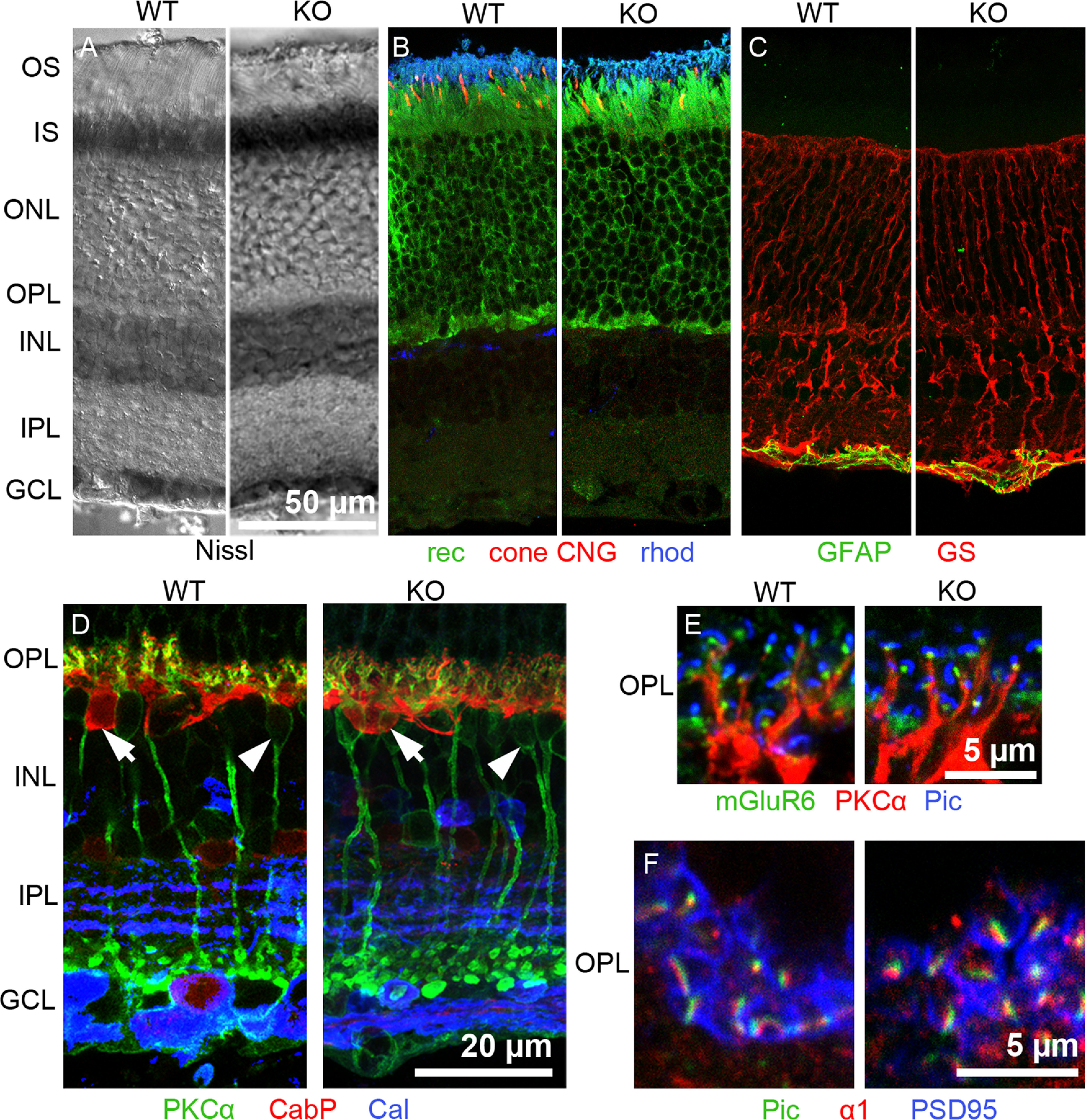
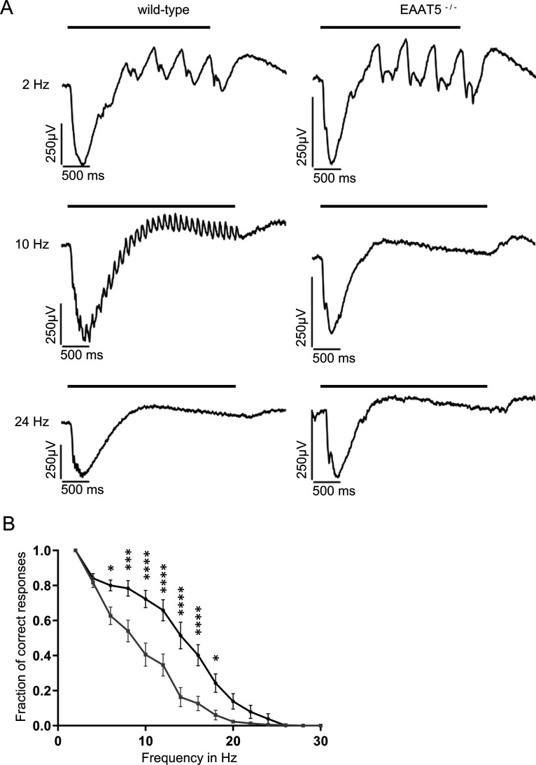
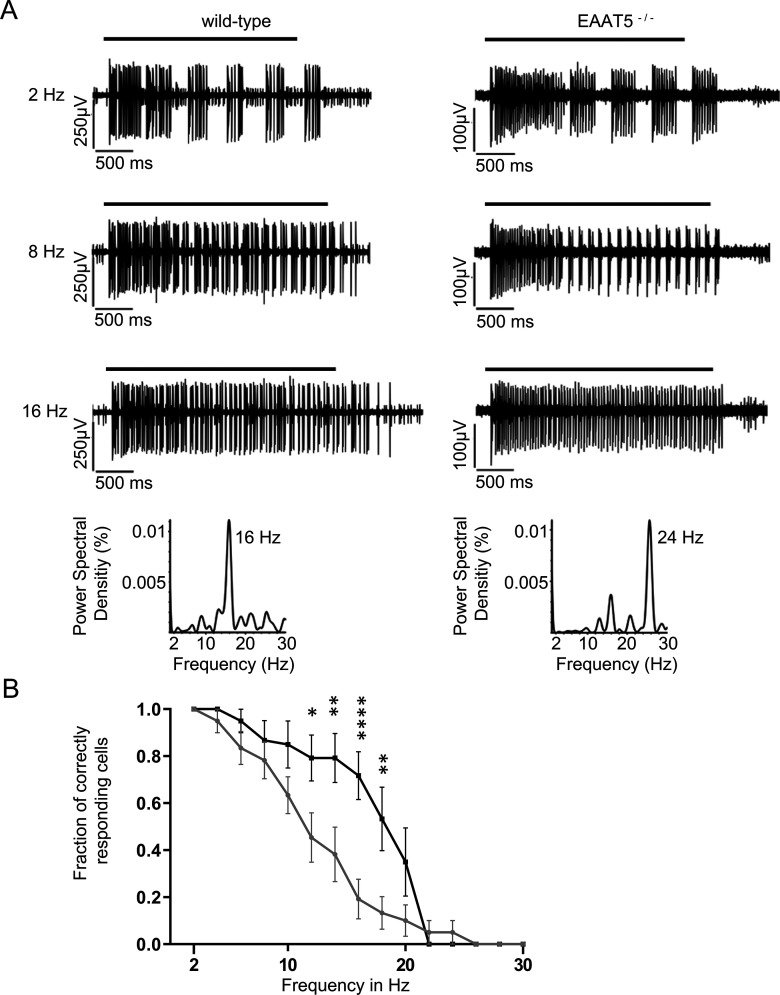
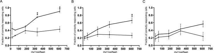
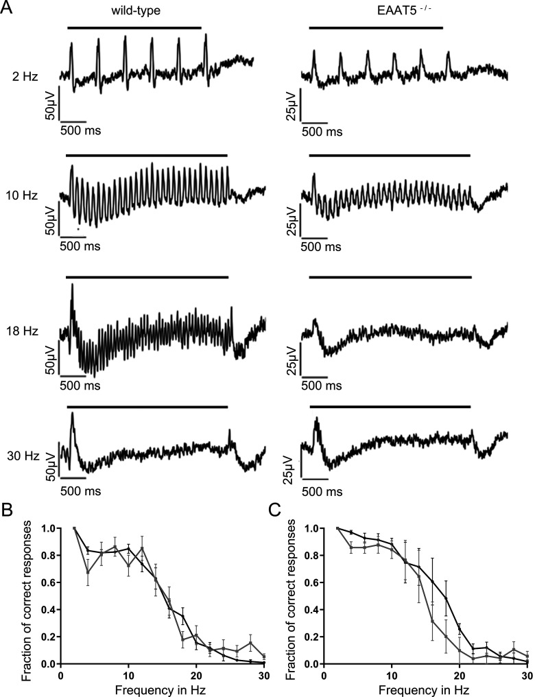
Excitatory amino acid transporters (EAATs) remove glutamate from the synaptic cleft. In the retina, EAAT1 and EAAT2 are considered the major glutamate transporters. However, it has not yet been possible to determine how EAAT5 shapes the retinal light responses because of the lack of a selective EAAT5 blocker or EAAT5 knock-out (KO) animal model. In this study, EAAT5 was found to be expressed in a punctate manner close to release sites of glutamatergic synapses in the mouse retina. Light responses from retinae of wild-type (WT) and of a newly generated model with a targeted deletion of EAAT5 (EAAT5-/-) were recorded in vitro using multielectrode arrays (MEAs). Flicker resolution was considerably lower in EAAT5-/- retinae than in WT retinae. The close proximity to the glutamate release site makes EAAT5 an ideal tool to improve temporal information processing in the retina by controlling information transfer at glutamatergic synapses.

Keywords: EAAT5; glutamate transporter; retina.


Figures

Figure 1.
Figure 1.
The EAAT5−/− retina appears histologically normal. A–F, left, WT retina (WT), right: EAAT5−/− retina (KO). A, Nissl staining reveals retinal thickness and retinal layering. B, Photoreceptors, recoverin (rec; green, photoreceptor somata and inner segments), cone cyclic nucleotide-gated (CNG) channel (red, cone outer segments), rhodopsin (rhod; blue, rod outer segments). C, Glial cells, glutamine synthetase (GS; red, Müller cells), glial fibrillary acidic protein (GFAP; green, astrocytes). D, Inner retinal cells, PKCα (green, rod bipolar cells, arrowheads), CabP (red, horizontal cells, arrows), calretinin (Cal; blue, several types of amacrine cells). E, OPL, mGluR6 (green, glutamate receptor on ON-bipolar cell dendrites), PKCα (red, rod bipolar cell dendrites), piccolo (Pic; blue, photoreceptor presynaptic ribbon). F, OPL, piccolo (Pic; green, photoreceptor presynaptic ribbon), pan α1 calcium channel subunit (red, calcium channel in rod photoreceptors), postsynaptic density protein 95 (PSD95; blue, plasma membrane marker for rod terminals). GCL: ganglion cell layer, INL: inner nuclear layer, IPL: inner plexiform layer, IS: inner segments, ONL: outer nuclear layer, OPL: outer plexiform layer, OS: outer segments. Scale bar in A also applies to B, C. WT and EAAT5−/−: n = 6 animals.
Figure 2.
Figure 2.
Localization of EAATs in mouse retinal sections. A, EAAT5 expression. In WT retina (left), arrows indicate brightly labeled puncta or short processes in the OPL and IPL, the arrowhead indicates bipolar cell soma in the INL, and asterisks indicate nonspecific staining of blood vessels by the secondary antibody (n = 10 animals). In EAAT5−/− (KO) retina (right), puncta are absent; only nonspecific staining in blood vessels is seen (n = 5 animals). B, EAAT1 (GLAST) was strongly expressed in Müller cells that span almost all retinal layers. No difference in expression pattern or level was observed in the EAAT5−/− retina (right; n = 3 animals for WT and for EAAT5−/−). C, EAAT2 (GLT1) was mainly expressed in photoreceptors and bipolar cells, with no differences in expression between WT (left) and EAAT5−/− (right) retinae (n = 3 animals for WT and for EAAT5−/−). D–J, Triple staining with antibodies against mGluR6, EAAT5, and piccolo (Pic) in a WT retina (n = 5 animals). D, In the OPL, EAAT5 (red) is closely associated with both the piccolo-positive ribbon (blue) of photoreceptors, and the mGluR6 label on ON-bipolar cell dendrites (green). Arrow, cone terminal; arrowheads, rod terminals. E–G, J, Higher magnification of rod spherules at different viewing angles, showing EAAT5-positive puncta (red) marked with arrows between the ribbon (blue) and mGluR6 (green) and marked with arrowheads between mGluR6 puncta. H, I, Single cone pedicles, showing close association of EAAT5-positive puncta with both ribbons and mGluR6. GCL: ganglion cell layer, INL: inner nuclear layer, IPL: inner plexiform layer, IS: inner segments, ONL: outer nuclear layer, OPL: outer plexiform layer, OS: outer segments. The scale bar in C also applies to A, B; the scale bar in G also applies to E, F, H–J.
Figure 3.
Figure 3.
Localization of EAAT5 on bipolar cell terminals in the mouse retina. A, Triple staining. EAAT5-positive puncta (red) were mostly found on vGluT1-positive (green) bipolar cell terminals in SL4 and SL5. ChAT-positive processes (blue) subdivide the IPL in a characteristic way (n = 3 animals). B, C, Triple staining. Close association of EAAT5-positive puncta (red, arrowheads) and ribbons [piccolo (Pic), green, arrows] on rod bipolar terminals (PKCα, blue; n = 6 animals). D–F, Acutely dissociated rod bipolar cells and photoreceptor terminals (three experiments, two retinae of one animal/experiment). D, Dissociated rod bipolar cell (PKCα, blue) with an axon terminal decorated with EAAT5 (red) puncta and ribbons (CTBP2, green). E, Axon terminal of the same cell. Insets are at higher magnification. Top right corner, Isolated rod terminal with horseshoe-shaped ribbon (CTBP2, green) and EAAT5 puncta (red). F, Isolated cone terminal [PEA (peanut agglutinin), blue] with closely associated ribbons (CTBP2 green) and EAAT5 puncta (red).
Figure 4.
Figure 4.
Temporal resolution of the EAAT5−/− retina is significantly impaired in the mesopic range. Flicker stimuli were applied for a series of frequencies from 2 to 30 Hz. A, The LFP of the EAAT5−/− retina (right) showed impaired resolution for the 10-Hz flicker stimulus compared with the WT retina (left). In neither genotype could the 24-Hz flicker stimulus be resolved. Bars: duration of flicker stimulus. B, When the fraction of correct responses was plotted against the stimulus frequency, a significant reduction in temporal resolution was apparent for EAAT5−/− (gray curve, mean ± SEM of 30 retinal pieces from 13 animals) compared with the WT (black curve, mean ± SEM of 33 retinal pieces from 13 animals). Two-way ANOVA with Bonferroni multiple comparisons. Flicker stimulus: 610 activated rhodopsin molecules (rhod*) per rod and flash (rhod*/rod/flash); total stimulus duration: 3 s; individual flash duration: 20 ms.
Figure 5.
Figure 5.
Temporal resolution of ON-ganglion cells is significantly impaired in the EAAT5−/− retina in the mesopic range. A, Typical response of one type of ON-ganglion cell (“ON-merge cell”) in the WT (left) and EAAT5−/− retina (right). Bars: duration of flicker stimulus. B, The fraction of correctly responding cells was significantly reduced in EAAT5−/− (gray curve, mean ± SEM of 32 cells of 10 animals) compared with WT (black curve, mean ± SEM of 40 cells of 10 animals) cells at frequencies of 10–20 Hz. Two-way ANOVA with Bonferroni multiple comparisons. Flicker stimulus: 610 activated rhodopsin molecules (rhod*) per rod and flash (rhod*/rod/flash; mesopic conditions); total stimulus duration: 3 s; individual flash duration: 20 ms.
Figure 6.
Figure 6.
The impact of EAAT5 on temporal resolution in ganglion cells increases with stimulus intensity under mesopic conditions. Black curves: WT; gray curves: EAAT5−/−. A, ON-merge cells (WT, 32 cells; EAAT5−/− 15 cells). B, ON-cells (except for ON-merge; WT, 92 cells; EAAT5−/− 96 cells). C, OFF-cells (WT, 39 cells; EAAT5−/−, 20 cells). Two-way ANOVA with Bonferroni multiple comparisons. Flicker stimulus: 12 Hz, total stimulus duration: 3 s; individual flash duration: 20 ms. Stimulus intensities: 17, 56, 183, 330, 610 activated rhodopsin molecules (rhod*) per rod and flash (rhod*/rod/flash).
Figure 7.
Figure 7.
Temporal resolution of the EAAT5−/− retina is not significantly impaired in the photopic range. Flicker stimuli were applied for a series of frequencies from 2 to 30 Hz. A, For the 2-Hz flicker stimulus, both genotypes showed a clear response. The LFP of EAAT5−/− retina (right) showed slightly impaired temporal resolution for 10- and 18-Hz flicker stimuli compared with the WT retina (left). Neither WT nor EAAT5−/− retinae could resolve the 30-Hz flicker stimulus. Bars: stimulus duration. B, The fraction of correct responses plotted against the stimulus frequency for stimulus intensity of 1000 activated rhodopsin molecules (rhod*/rod/flash) shows no significant reduction for EAAT5−/− (gray curve, mean ± SEM of eight retinal pieces from six animals) compared with the WT (black curve, mean ± SEM of 10 retinal pieces from six animals). Two-way ANOVA with Bonferroni multiple comparisons. C, The fraction of correct responses plotted against the stimulus frequency for stimulus intensity of 19,000 activated rhodopsin molecules (rhod*/rod/flash) shows a mild but not significant reduction for EAAT5−/− (gray curve, mean ± SEM of eight retinal pieces from six animals) compared with the WT (black curve, mean ± SEM of 10 retinal pieces from six animals). Two-way ANOVA with Bonferroni multiple comparisons. Total stimulus duration: 3 s; individual flash duration: 20 ms; background light: 10,000 rhod*/rod/s.

Similar articles

Cited by

References

    1. Baden T, Berens P, Franke K, Román Rosón M, Bethge M, Euler T (2016) The functional diversity of retinal ganglion cells in the mouse. Nature 529:345–350. 10.1038/nature16468 - DOI - PMC - PubMed
    1. Bligard GW, DeBrecht J, Smith RG, Lukasiewicz PD (2020) Light-evoked glutamate transporter EAAT5 activation coordinates with conventional feedback inhibition to control rod bipolar cell output. J Neurophysiol 123:1828–1837. 10.1152/jn.00527.2019 - DOI - PMC - PubMed
    1. Dalet A, Bonsacquet J, Gaboyard-Niay S, Calin-Jageman I, Chidavaenzi RL, Venteo S, Desmadryl G, Goldberg JM, Lysakowski A, Chabbert C (2012) Glutamate transporters EAAT4 and EAAT5 are expressed in vestibular hair cells and calyx endings. PLoS One 7:e46261. 10.1371/journal.pone.0046261 - DOI - PMC - PubMed
    1. Derouiche A, Rauen T (1995) Coincidence of L-glutamate/L-aspartate transporter (GLAST) and glutamine synthetase (GS) immunoreactions in retinal glia: evidence for coupling of GLAST and GS in transmitter clearance. J Neurosci Res 42:131–143. 10.1002/jnr.490420115 - DOI - PubMed
    1. Dowling J, Boycott B (1966) Organization of the primate retina: electron microscopy. Proc R Soc Lond B Biol Sci 166:80–111. 10.1098/rspb.1966.0086 - DOI - PubMed

Publication types

Substances