. 2021 Dec;28(1):2480-2494.
doi: 10.1080/10717544.2021.2000677.

Silmitasertib-induced macropinocytosis promoting DDP intracellular uptake to enhance cell apoptosis in oral squamous cell carcinoma

Affiliations

Silmitasertib-induced macropinocytosis promoting DDP intracellular uptake to enhance cell apoptosis in oral squamous cell carcinoma

Shaojuan Song et al. Drug Deliv. 2021 Dec.

Abstract

Cisplatin (DDP) is a first-line chemotherapeutic drug applied for the treatment of oral squamous cell carcinoma (OSCC). The anticancer activity of DDP is tightly linked to its intracellular uptake. It is unwise to increase the DDP intake by increasing the dose or shortening the dosing interval because of the severe systemic toxicity (nephrotoxicity, ototoxicity and neurotoxicity) in DDP application. The main uptake pathways of DDP include passive diffusion and active transporter transport. Therefore, finding additional uptake pathways that can improve the effective intracellular concentration of DDP is critical. Macropinocytosis, an endocytic mechanism for extracellular material absorption, contributes to the intracellular uptake of anticancer drugs. No research has been conducted to determine whether macropinocytosis can augment the intracellular uptake of DDP in OSCC cells or not. Based on that, we proved for the first time that silmitasertib (previously CX-4945) could trigger macropinocytosis, which may increase the intracellular uptake of DDP and enhance apoptosis via in vivo and in vitro experiments. We hope that our findings will inspire a new approach for the application of DDP in cancer treatment.

Keywords: Oral squamous cell carcinoma (OSCC); apoptosis; cisplatin (DDP) intracellular uptake; macropinocytosis; silmitasertib.


Conflict of interest statement

No potential conflict of interest was reported by the authors.

Figures

Figure 1.
Figure 1.
Silmitasertib-induced macropinocytosis promoting DDP intracellular uptake to enhance cell apoptosis. When silmitasertib acted on cells, the cell membrane recessed inward to form macropinosomes carrying DDP, which detached from the cell membrane and released DDP later. DDP induced strong apoptosis through the MAPK-caspase pathway in OSCC cells.
Figure 2.
Figure 2.
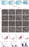
Silmitasertib can induce vacuolation in OSCC cells. (a) The process of macropinocytosis in cancer cells. (b) Cells were exposed respectively to different concentration of silmitasertib for 12 h and then their phase-contrast images were recorded by microscope. The number of vacuoles with different concentration of silmitasertib were measured in Cal-27 (c), HSC-3 (d), HSC-4 (e), and UM1 cells (f). The number of vacuoles in different cells with 10 μM (g), 20 μM (h), 40 μM (i) silmitasertib were measured. Data are shown as mean ± SD {*p<.05, **p<.01, ***p<.001} from three replicates. Scale bar: 50 μm.
Figure 3.
Figure 3.
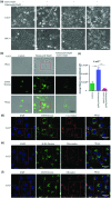
The vacuoles induced by silmitasertib were deriving from macropinocytosis. (a) Cells were exposed respectively to 20 μM silmitasertib or 10 nM BAF1(macropinocytosis inhibitor) or 20 μM silmitasertib + 10 nM BAF1 for 12 h and then their phase-contrast images were recorded by microscope. (b) Fluorescent images of macropinosomes in Cal-27 cells treated with 20 μM silmitasertib or 20 μM silmitasertib + 10 nM BAF1 were investigated by AF488 Dextran assay using microscopy. (c) Average fluorescence intensity in (b) were measured. (d) Fluorescent images of nucleus (DAPI), macropinosomes (AF488 Dextran) and lysosome (Lyso-Tracker) in Cal-27 cells treated with 10 μM silmitasertib for 12 h were investigated using microscopy. The red arrow indicated the macropinosomes co-localized with the lysosome, and the green arrow indicated the macropinosomes not co-localized with the lysosome. (e–f) Fluorescent images of nucleus (DAPI), macropinosomes (AF488 Dextran), mitochondria (Mito-Tracker) and endoplasmic reticulum (ER-Tracker) were investigated like (d). The red arrow indicated the macropinosomes not co-localized with the mitochondria (e) or endoplasmic reticulum (f). Data are shown as mean ± SD {*p<.05, **p<.01, ***p<.001} from three replicates. Scale bar: 30 μm or 50 μm.
Figure 4.
Figure 4.
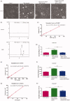
Silmitasertib could increase small molecule drugs intake in OSCC cells. (a) Cal-27 cells treated with 1 mg/mL DDP, 1 mg/mL DDP + 100 μM silmitasertib or 1 mg/mL DDP + 100 μM silmitasertib + 10 nM BAF1 for 4 h were observed by microscopy. (b) Peak figure of DDP was investigated by HPLC. (c, e, g) The standard curve of the drugs (DDP, 5-FU, isoG) was made with concentration as the abscissa and the peak area as the ordinate. (d, f, h) Intracellular drug (DDP, 5-FU, isoG) concentration in three groups were measured. Data are shown as mean ± SD {*p<.05, **p<.01, ***p<.001} from three replicates. Scale bar: 50 μm.
Figure 5.
Figure 5.
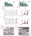
Combination of silmitasertib with DDP could promote OSCC cell apoptosis. (a, b) Cell viability of Cal-27 and UM1 cells treated with 2 μg/mL DDP, 10/20/40 μM silmitasertib or 2 μg/mL DDP + 10/20/40 μM silmitasertib for 24 and 48 h were measured by CCK8 assay. (c, e) Apoptosis in Cal-27 and UM1 cells treated with 2 μg/mL DDP, 10 μM silmitasertib or 2 μg/mL DDP + 10 μM silmitasertib for 24 and 48 h were conducted using Annexin V-FITC and propidium iodide (PI) double staining. (d, f) Rates of cell apoptosis in (c, e) were measured. (g, h) Cal-27 and UM1 cells were treated with 2 μg/mL DDP, 10/20/40 μM silmitasertib or 2 μg/mL DDP + 10/20/40 μM silmitasertib for 12 h, and the protein level of cleaved Caspase 3, cleaved Caspase 8, ERK, p-ERK, p38, JNKs, p-JNKs were assessed by WB. Data are shown as mean ± SD {* p<.05, ** p<.01, *** p<.001} from three replicates.
Figure 6.
Figure 6.
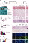
Combination of silmitasertib with DDP could enhance OSCC tumor suppression in vivo. (a) The drugs were administered once every five days for four weeks, after which the mice were sacrificed and the tumor tissues were collected. (b) The image of Cal-27 xenografts at the end of the experiment. (c) Average tumor growth curves in control and treatment groups receiving the indicated treatment. (d) Average animal body weight curves in control and treatment groups. (e) Hematoxylin and eosin (H&E) staining images were observed in main organs (heart, kidney, liver, lung and spleen) at the end of the experiment. (f) The histological, cell proliferation, and cell apoptosis of Cal-27 xenografts tissues in different groups were performed by H&E staining, immunohistochemical method (Ki67) and the TUNEL assay, respectively. (g, h) Rates of Ki67 positive cells and apoptosis cells in Cal-27 xenograft tissues of different groups were measured. Data are shown as mean ± SEM {*p<.05, **p<.01, ***p<.001}. Scale bar: 100 μm, 200 μm.

Similar articles

Cited by

  • Cellular Regulation of Macropinocytosis.
    Wu Y, Hu X, Wei Z, Lin Q. Wu Y, et al. Int J Mol Sci. 2024 Jun 26;25(13):6963. doi: 10.3390/ijms25136963. Int J Mol Sci. 2024. PMID: 39000072 Free PMC article. Review.
  • The Role and Therapeutic Potential of Macropinocytosis in Cancer.
    Qiu Z, Liu W, Zhu Q, Ke K, Zhu Q, Jin W, Yu S, Yang Z, Li L, Sun X, Ren S, Liu Y, Zhu Z, Zeng J, Huang X, Huang Y, Wei L, Ma M, Lu J, Chen X, Mou Y, Xie T, Sui X. Qiu Z, et al. Front Pharmacol. 2022 Aug 15;13:919819. doi: 10.3389/fphar.2022.919819. eCollection 2022. Front Pharmacol. 2022. PMID: 36046825 Free PMC article. Review.

References

    1. Afzal M, Kren BT, Naveed AK, et al. (2020). Protein kinase CK2 impact on intracellular calcium homeostasis in prostate cancer. Mol Cell Biochem 470:131–43. - PMC - PubMed
    1. Belaid A, Filippakis H. (2021). Quantitative assessment of macropinocytosis in mTORC1-hyperactive cells using flow cytometry. J Vis Exp 2:174. - PubMed
    1. Buckley CM, Pots H, Gueho A, et al. (2020). Coordinated ras and rac activity shapes macropinocytic cups and enables phagocytosis of geometrically diverse bacteria. Curr Biol 30:2912–26.e2915. - PMC - PubMed
    1. Chen F, Pei S, Wang X, et al. (2020). Emerging JWA-targeted Pt (IV) prodrugs conjugated with CX-4945 to overcome chemo-immune-resistance. Biochem Biophys Res Commun 521:753–61. - PubMed
    1. Cullis J, Siolas D, Avanzi A, et al. (2017). Macropinocytosis of nab-paclitaxel drives macrophage activation in pancreatic cancer. Cancer Immunol Res 5:182–90. - PMC - PubMed

MeSH terms

Grants and funding

This work was supported by the National Natural Science Foundations of China [No. 81922020, 81970950], the funding from the Sichuan Provincial Department of Science and Technology [2021-YJ-0021], the Postdoctoral Research and Development Funding of Sichuan University [2020SCU12016], the Research Funding for Talents Developing, West China Hospital of Stomatology Sichuan University [No. RCDWJS2020-4, RCDWJS2020-14], and the CAMS Innovation Fund for Medical Sciences [CIFMS,2019-I2M-5-004].

LinkOut - more resources