. 2021 Dec;12(1):2883-2901.
doi: 10.1080/21505594.2021.2000689.

Site-specific SUMOylation of viral polymerase processivity factor: a way of localizingtoND10 subnuclear domains for restricted and self-controlled reproduction of herpesvirus

Affiliations

Site-specific SUMOylation of viral polymerase processivity factor: a way of localizingtoND10 subnuclear domains for restricted and self-controlled reproduction of herpesvirus

Shuyan Lai et al. Virulence. 2021 Dec.

Abstract

Lytic replication of human cytomegalovirus (HCMV), a member of β-herpesvirus, is a highly complicated and organized process that requires its DNA polymerase processivity factor, UL44, the first-reported HCMV replication protein subjected to SUMO post-translational modification (PTM). SUMOylation plays a pleiotropic role in protein functions of host cells and infecting viruses. Particularly, formation of herpesviral replication compartments (RCs) upon infection is induced in proximity to ND10 subnuclear domains, the host cell's intrinsic antiviral immune devices and hot SUMOylation spots, relying just on SUMOylation of their protein components to become mature and functional in restriction of the viral replication. In this study, to unveil the exact role of SUMO PTM on UL44 involved in HCMV replication, we screened and identified PIAS3, an annotated E3 SUMO ligase, as a novel UL44-interacting protein engaged in cellular SUMOylation pathway. Co-existence of PIAS3 could enhance the UBC9-based SUMO modification of UL44 specifically at its conserved 410lysine residue lying within the single canonical ψKxE SUMO Conjugation Motif (SCM). Intriguingly, we found this SCM-specific SUMOylation contributes to UL44 co-localization and interaction with subnuclear ND10 domains during infection, which in turn exerts an inhibitory effect on HCMV replication and growth. Together, these results highlight the importance of SUMOylation in regulating viral protein subnuclear localization, representing a novel way of utilizing ND10-based restriction to achieve the self-controlled slower replication and reproduction of herpesviruses.

Keywords: E3 SUMO ligase; Human cytomegalovirus (HCMV); PIAS3; SUMO conjugation motif (SCM); SUMOylation; UL44; subnuclear localization, ND10; viral polymerase processivity factor.


Conflict of interest statement

The authors have declared no competing financial interests exist.

Figures

Figure 1.
Figure 1.
UL44, HCMV DNA polymerase processivity factor, interacts with human E3 SUMO ligase PIAS3. (a) Co-IP analysis of the association between UL44 and PIAS3 in transfected cells. pCMV-Myc-UL44 and pRK-Flag-PIAS3 plasmids were co-introduced into 293 T cells. At 48 h post transfection, the supernatant of cell lysate was taken to IP (immuno-precipitate) with anti-Myc or anti-Flag mAbs. The immunoprecipitants, after heat denaturation, were analyzed by Western blot with anti-Myc or anti-Flag mAbs. Meanwhile, negative controls were set up by cell co-transfection with pCMV-Myc-UL44 and a plasmid expressing the Flag tag, or with pRK-Flag-PIAS3 and a plasmid expressing the Myc tag. (b) His and GST pull-down analyses of the direct interaction between UL44 and PIAS3. Expressed in E. coli BL21 (DE3), His-tagged UL44 and GST-tagged PIAS3 were individually purified by affinity chromatography using Ni2+-NTA or GST column. Then, the reciprocal GST and His pull-down assays were performed to verify the direct UL44-PIAS3 interaction in vitro. (c) Co-IP analysis of UL44 association with endogenous PIAS3 under HCMV infection. U251 cells were infected with wt-HCMV at an MOI of 1, before preparation of the total cell lysates at 48 hpi. The input proteins and IP samples were analyzed by Western blot, using as the indicated anti-UL84, anti-UL44 or anti-PIAS3 antibody, respectively. For all panels, results were representative of three independent experiments
Figure 2.
Figure 2.
Co-localization of UL44 with PIAS3 in human transfected cells. U251 cells were transfected individually with (a) the single pCMV-Myc-UL44 construct, (b) the single pRK11-Flag-PIAS3 construct, or (c) both of pCMV-Myc-UL44 and pRK11-Flag-PIAS3 together. At 48 h post transfection, cells were fixed and permeabilized, followed by incubation with anti-Flag/anti-Myc primary Abs and with Dylight 488/549-conjugated fluorescent secondary Abs. The subcellular localization of UL44 (green) and PIAS3 (red) were visualized, with the cell nuclei stained by DAPI (blue). For a same field of view, “Merge” denoted the overlapping fluorescence from both the nuclei and expressed proteins. The displayed images were representative ones from three independent experiments
Figure 3.
Figure 3.
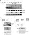
PIAS3 exerts an enhancing effect on UL44 SUMOylation. (a) The in vivo SUMOylation of UL44 in transfection assays, in the presence of exogenous PIAS3 using different SUMO peptides. pCMV-Myc-UL44 and pRK-Flag-SUMO-1/2/3, along with or without pcDNA-His-PIAS3, were co-introduced into HEK293T cells. At 48 h post transfection, Western blot with anti-Myc antibody was performed to detect UL44 and UL44-SUMO in total cell lysates. Free SUMO-1/2/3 peptides were probed with anti-Flag antibody. GAPDH was set as the internal control. (b) The in vivo SUMOylation of UL44 in transfection assays, in the presence of exogenous UBC9 and PIAS3 (WT or C334S mutant). pCMV-Myc-UL44, pRK-Flag-SUMO-1 and pcDNA-HA-UBC9, along with or without pcDNA-His-PIAS3 (WT or C334S mutant) were co-introduced into HEK293T cells. At 48 h post-transfection, Western blot with anti-Myc or anti-Flag antibody was performed to analyze UL44 and UL44-SUMO in total cell lysates. SUMO-1, UBC9 and PIAS3/PIAS3-C334S were probed with anti-Flag, anti-HA and anti-His antibodies, respectively. GAPDH served as the internal control. (c) The in vivo SUMOylation of UL44 during wt-HCMV infection, upon interference of endogenous PIAS3. U251 cells, after transfection with siControl (a negative control siRNA molecule) or siPIAS3 (a chemically synthesized siRNA that specially targets the CDS region of PIAS3 mRNA) for 24 h, were infected with wt-HCMV at an MOI of 1. At 48 hpi, the supernatant of total cell lysates, either immunoprecipitated with anti-UL44 antibody or not, was analyzed by Western blot using anti-UL44 or anti-SUMO1 antibody as indicated to detect UL44 and UL44-SUMO proteins. (d) The in vitro SUMOylation assays on UL44 protein. His-UL44, GST-PIAS3 and GST-PIAS3-C334S were individually expressed in E. coli BL21 (DE3), before further protein purification by affinity chromatography and SEC (size exclusion chromatography). Enzymatic reactions were set up with the SUMOylation-related components as indicated, and the production of SUMOylated UL44 was determined by Western blot with anti-His or anti-SUMO1 antibody. For all panels, each set of assays was repeated three times and a representative one was shown
Figure 4.
Figure 4.
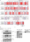
The SUMOylation-promoting effect of PIAS3 on UL44 is site-specific at stringently conserved 410lysine residue within the single canonical SCM (SUMO Conjugation Motif; ψ410KxE). (a) The multiple sequence alignment among UL44 and other CMV homologs. Subdomain-1 and Subdomain-2 denote the two topologically conserved subdomains connected by a loop [7]. The two sequences indicated by black arrows and boxes, lying as a conserved region at the C-terminus, represent the single canonical SCM (SUMO Conjugation Motif; ψKxE) site and the NLS (nuclear localization signal), respectively. (b) Influence of the K410R mutation on SUMOylation of UL44 in transfection assays, in the presence of exogenous PIAS3. pCMV-Myc-UL44 (WT or K410R) and pRK-Flag-SUMO-1, along with or without pcDNA-His-PIAS3, were co-introduced into HEK293T cells. At 48 h post transfection, UL44 and UL44-SUMO proteins in total cell lysates were analyzed by Western blot with anti-Myc or anti-Flag antibody. PIAS3 and SUMO-1 were probed with anti-His and anti-Flag antibody, respectively. (c) The E3 SUMO ligase activity of endogenous PIAS3 toward UL44 during HCMV (WT or K410R mutant) infection. U251 cells, after transfection with siControl or siPIAS3 for 24 h, were infected with HCMV (WT or K410R mutant) at an MOI of 1. At 48 hpi, the supernatant of total cell lysates, either immunoprecipitated with anti-UL44 antibody or not, was analyzed by Western blot using antibodies as indicated (Left: anti-UL44; Right: anti-SUMO-1) to detect UL44 and UL44-SUMO proteins. The endogenous PIAS3 was probed with anti-PIAS3 antibody. In both (b) and (c), GAPDH served as the internal control and results were representative of three repeated experiments
Figure 5.
Figure 5.
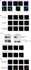
SUMOylation at 410lysine of SCM site guides UL44 co-localization and interaction with ND10 structures during HCMV infection. (a) Effect of K410 SUMOylation on UL44 nuclear localization in transfected cells. (i) pCMV-Myc-UL44 alone, (ii) pCMV-Myc-UL44-K410R alone, (iii) pCMV-Myc-UL44 together with pcDNA-HA-UBC9-C93S, or (iv) pCMV-Myc-UL44wt-ΔNLS alone was individually introduced into U251 cells. At 48 h post transfection, cells were fixed and permeabilized, followed by incubation with anti-UBC9/anti-Myc primary Abs and with Dylight 488/549-conjugated fluorescent secondary Abs. Through a confocal microscope, the subcellular localization of UL44 (WT or derivative forms, green) or UBC9-C93S (red) was visualized, with the cell nuclei stained by DAPI (blue). (b) Effect of K410 SUMOylation on UL44 subnuclear localization in HCMV infected cells. (v, vi) U251-C (control cells, carrying the empty pcDNA3.1(+) vector) or (vii) U251-PIAS3 cells (stably overexpressing PIAS3, carrying the pcDNA3.1-PIAS3 plasmid) were infected with wt-HCMV or v-K410R at an MOI of 1. At 48 hpi, fluorescent localization assays of UL44 (green) and PIAS3 (red) were performed with those infected cells as described above, using anti-UL44 or anti-PIAS3 as primary antibody. In a same field of view, “Merge” denoted the overlapping fluorescence from both the nuclei (blue) and expressed proteins. (c) The SCM-SUMOylation mediated UL44 protein co-localization and association with PML, the scaffold protein of ND10 subnuclear domains. (viii) Co-IP assays of UL44 with the cellular PML. U251 cells were infected with wt-HCMV or v-K410R at an MOI of 1, and 48 h later subjected to Co-IP assays using the Abs as indicated. (ix, x) Effect of endogenous PML interference on UL44 subnuclear localization in HCMV infected cells. U251-siControl (control cells, stably transduced using a control siRNA called siControl) or U251-siPML cells (stably transduced using siPML, a chemically synthesized siRNA that specially targets the CDS region of PML mRNA) were infected with wt-HCMV at an MOI of 1, followed by fluorescent localization assays of UL44 (green) and PIAS3 (red) at 48 hpi, using anti-UL44 and anti-PIAS3 as primary Abs, respectively. (d) The nuclear locations of UL44 and UL70 in HCMV infected cells. U251-siControl cells were infected with (xi) wt-HCMV or (xii) v-K410R at an MOI of 1, followed by fluorescent localization analyses of UL44 (green) and UL70 (red) at 48 hpi, using anti-UL44 and anti-UL70 as primary Abs, respectively. For each panel, the images were representative ones form three independent experiments
Figure 6.
Figure 6.
SCM SUMOylation-mediated UL44 localization to ND10 structure restricts the replication and reproduction of HCMV. U251-siControl cells and U251-siPML cells were individually infected with the indicated type of HCMV strain (wt-HCMV, v-K410R or v-K410rev) at an MOI of 0.5. At the indicated time points, (a) the viral titers and (b) HCMV DNA levels were determined by counting of the viral plaque numbers and by Real-Time PCR quantitation, respectively. The copies of viral genome were normalized to those of the cellular RNase P. In both (a) and (b), three repetitions were conducted for each set of assays in triplicate. Data represented the Average ± SD (standard deviation) from three repeated experiments. The Averages, between 2 groups or among more (≥3) groups, were appraised by usage of unpaired Student’s t-test or one-way ANOVA (analysis of variance) with Tukey’s post hoc test. NS denoted no significant difference. Differences were considered statistically significant when * denoted p < 0.05, ** denoted p < 0.01, and *** denoted p < 0.001

Similar articles

Cited by

References

    1. Mocarski ES, Shenk T, Griffiths PD, et al. Cytomegaloviruses. In: Knipe DM, Howley PM, editors. Fields Virology. 6th ed. Vol. 2. Lippincott Williams and Williams,Philadelphia, PA, USA; 2013, p. 1960–2014.
    1. Masse MJ, Karlin S, Schachtel GA, et al. Human cytomegalovirus origin of DNA replication (oriLyt) resides within a highly complex repetitive region. Proc Natl Acad Sci U S A. 1992;89(12):5246–5250. - PMC - PubMed
    1. Pari GS, Anders DG.. Eleven loci encoding trans-acting factors are required for transient complementation of human cytomegalovirus oriLyt-dependent DNA replication. J Virol. 1993;67(12):6979–6988. - PMC - PubMed
    1. Loregian A, Appleton BA, Hogle JM, et al. Specific residues in the connector loop of the human cytomegalovirus DNA polymerase accessory protein UL44 are crucial for interaction with the UL54 catalytic subunit. J Virol. 2004;78(17):9084–9092. - PMC - PubMed
    1. Weiland KL, Oien NL, Homa F, et al. Functional analysis of human cytomegalovirus polymerase accessory protein. Virus Res. 1994;34(3):191–206. - PubMed

Publication types

MeSH terms

Substances

Grants and funding

This work was supported in part by grants from Guangdong Innovative and Entrepreneurial Research Team Program [no. 2014ZT05S136] and Project for Construction of Guangzhou Key Laboratory of Virology [no. 201705030003]. We are also grateful for financial support from NSFC [31100128, 31500137, 82070406], the Department of Science and Technology of Guangdong [2020A1515011158], the Department of Education of Guangdong [2020KTSCX106], as well as Guangdong Basic and Applied Basic Research Foundation [2019A1515011742].

LinkOut - more resources