. 2022 Jul;18(7):1599-1612.
doi: 10.1080/15548627.2021.1994296. Epub 2021 Nov 6.

Bunyavirus SFTSV exploits autophagic flux for viral assembly and egress

Affiliations

Bunyavirus SFTSV exploits autophagic flux for viral assembly and egress

Jia-Min Yan et al. Autophagy. 2022 Jul.

Abstract

Severe fever with thrombocytopenia syndrome virus (SFTSV) is an emerging negatively stranded enveloped RNA bunyavirus that causes SFTS with a high case fatality rate of up to 30%. Macroautophagy/autophagy is an evolutionarily conserved process involved in the maintenance of host homeostasis, which exhibits anti-viral or pro-viral responses in reaction to different viral challenges. However, the interaction between the bunyavirus SFTSV and the autophagic process is still largely unclear. By establishing various autophagy-deficient cell lines, we found that SFTSV triggered RB1CC1/FIP200-BECN1-ATG5-dependent classical autophagy flux. SFTSV nucleoprotein induced BECN1-dependent autophagy by disrupting the BECN1-BCL2 association. Importantly, SFTSV utilized autophagy for the viral life cycle, which not only assembled in autophagosomes derived from the ERGIC and Golgi complex, but also utilized autophagic vesicles for exocytosis. Taken together, our results suggest a novel virus-autophagy interaction model in which bunyavirus SFTSV induces classical autophagy flux for viral assembly and egress processes, suggesting that autophagy inhibition may be a novel therapy for treating or releasing SFTS.

Keywords: Autophagy; bunyavirus; sftsv; viral assembly; viral egress.


Conflict of interest statement

No potential conflict of interest was reported by the author(s).

Figures
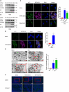
Figure 1.
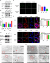
Figure 1.
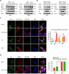
Autophagy is induced under SFTSV infection. (A and B) Vero and HeLa cells were infected with SFTSV at an MOI of 5 at different time points. Cell lysates were evaluated via western blot (WB). (C and D) Vero and HeLa cells were infected with SFTSV at an MOI of 5. Nuclear DNA, endogenous LC3, and SFTSV NP were stained as blue, green, and violet, respectively. Rapamycin served as the positive control of autophagy. For rapamycin (100 nM), cells were treated for 6 h without SFTSV infection before harvest. The number of LC3 puncta in each cell was counted, and at least 20 cells were included for each group. Scale bar: 20 μm. (E) Mock-infected, CQ-treated, or SFTSV-infected Vero cells at 24 h were processed and analyzed for the formation of autophagosomes or autolysosomes via electron transmission microscopy. For CQ (100 µM), cells were treated for 6 h without SFTSV infection before harvest. Black arrows indicate autophagic vacuoles. Scale bar: 2 μm. (F) WT C57BL/6 J mice were intraperitoneal injected with SFTSV at 106 TCID50/animal for 24 h and 48 h. The spleen sections underwent immunofluorescence assays (IFA) with LC3 (green) and DAPI (blue). Scale bar: 50 μm. Error bars, mean ± SD of three experiments. Student’s t test; *p < 0.05; **p < 0.01; ***p < 0.005; ****p < 0.001.
Figure 2.
Figure 2.
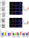
SFTSV-induced autophagy is a complete process. (A) Vero or HeLa cells were mock infected or infected with SFTSV at MOI of 5 for 24 h and 36 h and then treated with CQ for 6 h. Cell lysates were evaluated via WB. (B) Vero or HeLa cells were mock infected or infected with SFTSV for 24 h and then treated with Baf-A1 for 6 h. Cell lysates were evaluated via WB. (C) Vero cells were transfected with mCherry-GFP-LC3 for 12 h and then were mock infected or infected with SFTSV, or treated with CQ (100 µM), or starved in EBSS medium for 6 h. The number of mcherry-GFP-LC3 dots in each cell was counted, and at least 20 cells were included for each group. Single plane type of images was present. Scale bar: 20 μm. (D) Vero cells were infected with SFTSV for 24 h for analyzing the colocalization of endogenous LC3 and LAMP1. Single plane type of images was present. Scale bar: 20 μm.
Figure 3.
Figure 3.
Autophagy is essential for the replication of SFTSV. (A-E) BECN1, atg5, atg7, RB1CC1, ATG16L1 knockout MEF or HeLa cells and WT cells were infected with SFTSV at an MOI of 1 for 24 h and 36 h. Cell lysates were evaluated via WB. (F-G) BECN1, atg5, atg7, RB1CC1, ATG16L1 knockout MEF or HeLa cells and WT cells were infected with SFTSV at an MOI of 1 for 24 h. Nuclear DNA, endogenous LC3, and SFTSV NP were stained as blue, green, and violet respectively. Single plane type of images was present. Scale bar: 20 μm. (H) BECN1, atg5, atg7, RB1CC1, ATG16L1 knockout MEF or HeLa cells and WT cells were infected with SFTSV at an MOI of 1 for 2 h. Then cells were washed once by PBS for three times and internalized SFTSV were detected via RT-qPCR. (I) BECN1, atg5, atg7, RB1CC1, ATG16L1 knockout MEF or HeLa cells and WT cells were infected with SFTSV at an MOI of 1 for 24 h. Endpoint 10-fold dilutions of an SFTSV stock were titrated. Values presented in the graph are calculated and expressed as the log10 of TCID50 units per ml of supernatant. Error bars, mean ± SD of three experiments. Student’s t test; *p < 0.05; **p < 0.01; ***p < 0.005; ****p < 0.001.
Figure 4.
Figure 4.
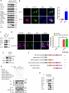
SFTSV NP triggers BECN1-dependent autophagy. (A) Vero cells were transfected with pVAX1, pVAX1-NSs-HA, or pVAX1-NP-HA for 24 h. The cell lysates were evaluated via WB.(B) Representative images of Vero cells co-transfected with EGFP-LC3 and pVAX1 or pVAX1-NP-HA for 24 h respectively. Single plane type of images was present. Scale bar: 20 μm. (C) BECN1 knockout cells or WT HeLa cells were transfected with pVAX1-NP-HA for 24 h. The cell lysates were evaluated via WB. (D) Representative images of Vero cells infected with SFTSV at an MOI of 5 for 24 h and stained for SFTSV NP and endogenous BECN1. Nuclei were stained with DAPI. Single plane type of images was present. Scale bar: 20 μm. (E) HEK293T cells were transfected with pVAX1-NP-HA for 24 h. The cell lysates were immunoprecipitated with anti-BECN1 antibody and then immunoblotted with the indicated antibody. (F) The schematic diagram of BECN1 truncation mutants and the BECN1-NP interacting domain. (G) HEK293T cells were co-transfected with pVAX1-NP-HA and Flag-BECN1, Flag-BECN1 BD, Flag-BECN1 BD+CCD, Flag-BECN1 BD+CCD+ECD, Flag-BECN1 CCD+ECD, or Flag-BECN1 ECD. The cell lysates were immunoprecipitated with anti-HA antibody and then immunoblotted with the indicated antibody. (H) Vero cells were transfected with pVAX1 or pVAX1-NP-HA for 24 h. The cell lysates were immunoprecipitated with anti-BECN1 antibody and then immunoblotted with the indicated antibody.
Figure 5.
Figure 5.
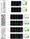
The autophagosome serves as SFTSV assembly platform. (A) HeLa cells were infected with SFTSV at an MOI of 1 for 5 days and SFTSV in supernatant were harvested and purified for WB detection. (B) PEG8000 purified SFTSV particles were analyzed via electron transmission microscopy. Scale bar: 200 nm. (C) Representative images of Vero cells infected with SFTSV at an MOI of 5 for 24 h and stained for endogenous LC3, SFTSV NP, and Gn. Nuclei were stained with DAPI. Single plane type of images was present. Scale bar: 20 μm. (D) 10-nm gold particles were used to label endogenous LC3. The red arrows refer to LC3, and the black arrows refer to SFTSV virions. Single plane type of images was present. Scale bar: 500 nm. (E) Vero cells were infected with SFTSV at an MOI of 1 for 24 h. The colocalization of Gn and LAMP1 to the ER, ERGIC and Golgi was analyzed. Single plane type of images was present. Scale bar: 20 μm.
Figure 6.
Figure 6.
Autophagic vacuoles is exploited by SFTSV for exocytosis. (A and B) STX17 orVAMP7 knockout HeLa cells and WT cells were infected with SFTSV at an MOI of 1 for 24 h and 36 h. Cell lysates were evaluated via WB. (C) STX17 or VAMP7 knockout HeLa cells and WT cells were infected with SFTSV at an MOI of 1 for 24 h and 36 h. Viral titer was measured by TCID50. (D) Vero cells were mock infected or infected with SFTSV at MOI of 5 for 24 h. Pro-form and mature-form of CTSB and CTSD were evaluated via WB. (E) Vero cells were mock infected or infected with SFTSV at MOI of 5 for 24 h or treated with Baf-A1 for 6 h. Then autolysosome acidification was determined by LysoSensor Green DND-189 staining. Single plane type of images was present. The mean density was calculated via ImageJ. Error bars, mean ± SD of three experiments. Student’s t test; *p < 0.05; **p < 0.01; ***p < 0.005; ****p < 0.001. (F) Representative images of Vero cells infected with SFTSV at an MOI of 5 for 24 h and then underwent immunofluorescence assay to mark endogenous LAMP1, SFTSV NP, and Gn. Nuclei were stained with DAPI. Single plane type of images was present. Scale bar: 20 μm. (G) 10-nm gold particles were used to label endogenous LAMP1 in SFTSV infected Vero cells. The red arrows refer to LAMP1, and the black arrows refer to SFTSV virions. Scale bar: 500 nm. (H) 10-nm gold particles were used to label endogenous LC3 and 4-nm gold particles were used to label endogenous LAMP1 in SFTSV infected HeLa cells. The red arrows refer to LC3, the green arrows refer to LAMP1 and the black arrows refer to SFTSV virions. Scale bar: 500 nm.
Figure 7.
Figure 7.
Schematic presentation of the SFTSV replication cycle. SFTSV Gp is synthesized by membrane bound ribosomes at the ER and then translocated to ERGIC and Golgi complex. Gp is expected to recruit SFTSV RNP complex, comprising SFTSV NP, RdRp, and genomic RNA, for SFTSV assembly and maturation. Traditionally, virus is considered to be formed by budding into Golgi complex lumen. Then matured virus particles are released from cell via exocytosis. Here, we provided a novel model of the exocytosis of bunyavirus SFTSV. ERGIC and Golgi complex were recruited as autophagic vesicles for SFTSV assembly and exocytosis via autolysosome. Alternatively, Golgi complex-originated secretory vesicle containing virions might fuse with autophagosome and egressed via autolysosome.

Similar articles

Cited by

References

    1. Yu XJ, Liang MF, Zhang SY, et al. Fever with thrombocytopenia associated with a novel bunyavirus in China. N Engl J Med. 2011;364:1523–1532. - PMC - PubMed
    1. Li H, Lu QB, Xing B, et al. Epidemiological and clinical features of laboratory-diagnosed severe fever with thrombocytopenia syndrome in China, 2011-17: a prospective observational study. Lancet Infect Dis. 2018;18:1127–1137. - PubMed
    1. Kuhn JH, Adkins S, Alioto D, et al. taxonomic update for phylum Negarnaviricota (Riboviria: orthornavirae), including the large orders Bunyavirales and Mononegavirales. Arch Virol. 2020;165:3023–3072. - PMC - PubMed
    1. Liu S, Chai C, Wang C, et al. Systematic review of severe fever with thrombocytopenia syndrome: virology, epidemiology, and clinical characteristics. Rev Med Virol. 2014;24:90–102. - PMC - PubMed
    1. Novoa RR, Calderita G, Cabezas P, et al. Key Golgi factors for structural and functional maturation of bunyamwera virus. J Virol. 2005;79:10852–10863. - PMC - PubMed

Publication types

Grants and funding

This study was supported by the National Natural Science Foundation of China[81971939 and 31570167] and the Fundamental Research Funds for the Central Universities [2042021kf0046]. The funders had no role in the study design, data collection and analysis, decision to publish, or the preparation of the manuscript.

LinkOut - more resources