Review
. 2021 Oct 6:12:100086.
doi: 10.1016/j.mbplus.2021.100086. eCollection 2021 Dec.

Molecular conformations and dynamics in the extracellular matrix of mammalian structural tissues: Solid-state NMR spectroscopy approaches

Affiliations
Review

Molecular conformations and dynamics in the extracellular matrix of mammalian structural tissues: Solid-state NMR spectroscopy approaches

Adrian Murgoci et al. Matrix Biol Plus. .

Abstract

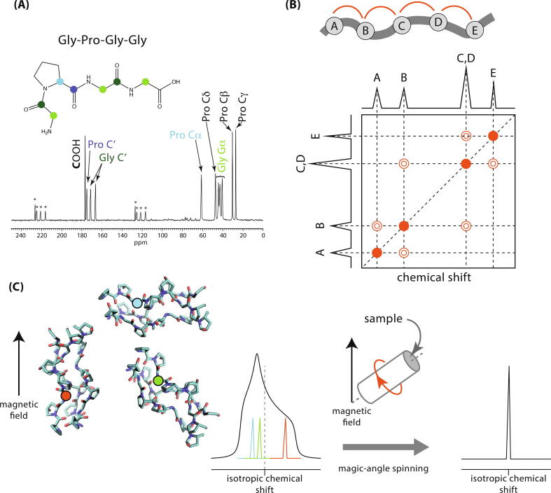
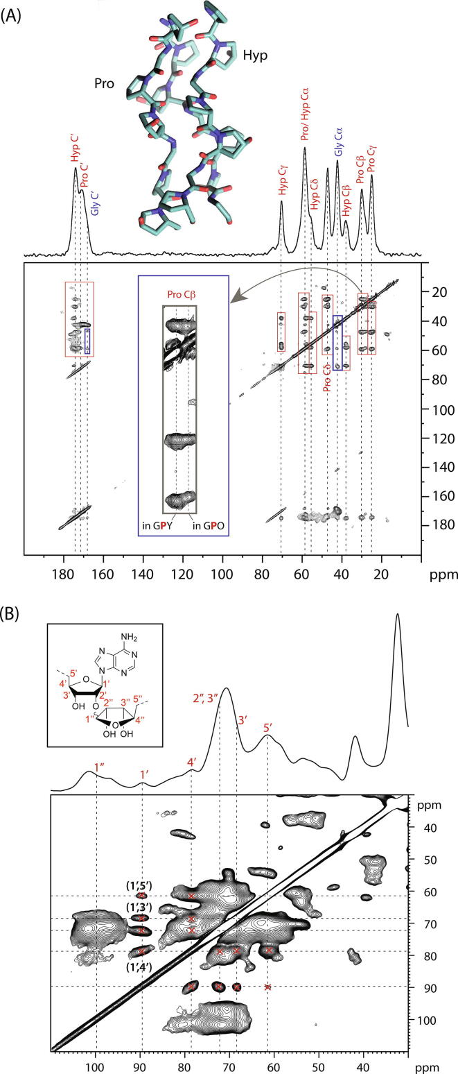
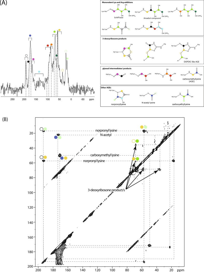
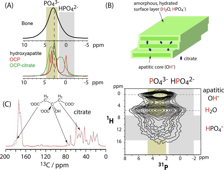
Solid-state NMR spectroscopy has played an important role in multidisciplinary studies of the extracellular matrix. Here we review how solid-state NMR has been used to probe collagen molecular conformations, dynamics, post-translational modifications and non-enzymatic chemical changes, and in calcified tissues, the molecular structure of bone mineral and its interface with collagen. We conclude that NMR spectroscopy can deliver vital information that in combination with data from structural imaging techniques, can result in significant new insight into how the extracellular matrix plays its multiple roles.

Keywords: Biomineralization; Bone mineral; Collagen; Extracellular matrix structure; Multidimensional solid-state NMR spectroscopy; Proline conformation.


Conflict of interest statement

The authors declare that they have no known competing financial interests or personal relationships that could have appeared to influence the work reported in this paper.

Figures

Fig. 1
Fig. 1
(A) A 1D 13C (MAS) NMR spectrum of a lyophilised sample of Gly-Pro-Gly-Gly peptide. The backbone carbons are indicated with coloured circles and their respective NMR signals assigned on the spectrum. Signals marked * are spinning sidebands, necessary artefacts of magic-angle spinning (see (C)). (B) Schematic of a 2D homonuclear correlation spectrum, e.g. 13C - 13C for the molecule at the top with nuclear (atomic) sites A, B, C, D, E. In this hypothetical spectrum, cross-peaks, indicated as contours (open circles), occur between signals from nuclei that are physically close in space, i.e. A-B, B-C, etc. The diagonal signals in the 2D spectrum (filled circles) are typical for this type of 2D spectrum and do not indicate that e.g. site As are close together with other site As. (C) Solid-state NMR spectroscopy typically requires magic-angle spinning (MAS) to remove the effects of anisotropic nuclear spin interactions. The strength of the effect of these interactions on the NMR signal frequency depends on the orientation of the molecule containing the nucleus with respect to the magnetic field applied in the NMR experiment. Thus in a sample where, e.g. there are collagen molecules with multiple orientations, there will be multiple, overlapping signals, resulting in a broad line (exemplified here with the lineshape resulting from shielding anisotropy - interaction of e.g. 13C nuclei with surrounding electrons in the collagen molecules) which limits resolution. Spinning the whole sample at the so-called magic-angle (54.74) with respect to the magnetic field, removes the molecular orientation dependence of the chemical shift, resulting in a sharp line at the isotropic chemical shift for each nuclear site. If the spinning rate is slower than width of the NMR signal, so-called spinning sidebands also appear in the spectrum, radiating out from the isotropic signal at the spinning frequency apart.
Fig. 2
Fig. 2
(A) A 2D 13C-13C correlation NMR spectrum (proton-driven spin-diffusion) of in vitro ECM from foetal sheep osteoblasts in which U-13C-Pro, Gly were used in the cell culture media, resulting in ECM proteins in which Gly, Pro and Hyp are extensively 13C-labelled. Correlation signals from these (primarily collagen) residues thus appear in this 2D correlation spectrum. Intraresidue signals are indicated in red rectangles and inter-residue correlations in blue rectangles. The inset (grey rectangle) is an expansion of the Pro Cβ signals, showing they can be resolved into those from Pro in GPO and more general GPY triplets. The 1D 13C MAS NMR spectrum of the same sample is shown at the top. (B) 2D 13C-13C correlation NMR spectrum (proton-driven spin-diffusion) of in vitro calcifying (bovine) vascular smooth muscle cell ECM showing assignments of some of the correlation signals from poly(ADP ribose) (inset: structure of one monomer unit of poly(ADP ribose) showing the 13C site labelling scheme). The 1D 13C projection of the spectrum is shown above the 2D spectrum. The poly(ADP ribose) signals are not resolved from other glycan species in this 1D spectrum, although they are well-resolved in the 2D spectrum, showing the power of 2D NMR correlation spectroscopy.
Fig. 3
Fig. 3
(A) Left: 1D double-quantum-filtered solid-state 13C NMR spectrum of bovine collagen type I fibrils glycated with U-13C-R5P. The spectra contain only signals from 13C bonded to another 13C and so contain only signals from molecular species derived from the respective 13C-labelled sugar. Possible assignments for each signal/ range of signals are indicated with coloured circles; the similarity of chemical structure between possible glycation products means that there is both overlap of signals from different glycation products and several possible assignments for most signals. Dotted lines are to allow easy comparison of chemical shifts of signals for the different glycation sugars. Right: possible glycation products with 13C site chemical shifts expected indicated with coloured circles that correspond to the colour scheme used to designate signals in the spectra on the left. (B) 2D 13C-13C proton-driven spin-diffusion correlation spectrum for U-13C-R5P-glycated collagen type I, showing the assignment of norpronyl-lysine, (carboxymethyl) lysine, N-acetyl and products of Lys-Arg glycation with 3-deoxyribosone-like molecules as the major collagen - R5P glycation products. Products of 3-deoxyribosone-like molecules are expected to include products, such as DOPDIC, with a -CH(N)....CH2-CH(OH)- fragment . The 13CH2 component in such fragments has a distinctive 37 ppm signal .
Fig. 4
Fig. 4
(A) 1D 31P MAS NMR spectrum of bone (top) and representative synthetic calcium phosphate phases to indicate the chemical shift ranges expected for orthophosphate (inorganic PO43-, olive box) and hydrogen phosphate (HPO42-, grey box). (B) The current model of bone mineral chemical structure (top) comes largely from solid-state NMR spectroscopy such as the 2D 1H - 31P correlation spectrum of intact sheep cortical bone (bottom), where the correlation signals are assigned to apatitic hydroxyl groups and non-apatitic water and hydrogen phosphate ions. The expected chemical shift ranges for orthophosphate (inorganic PO43-, olive box) and hydrogen phosphate (HPO42-, grey box) are indicated as for (A). From TEM, bone mineral consists of stacks of 2–4 nanoscopic mineral platelets , . The same work shows that the mineral platelets schematically depicted here likely derive from sideways-aggregated needle-like structures rather than being homogeneous platelets. Citrate anions are hypothesised to bridge between the mineral platelets . (C) A 13C{31P} REDOR spectrum of sheep bone. Signals in the REDOR spectrum (red) with lower intensity than those in the reference specrtum (black) are from carbons close in space to phosphorus. The strongest reductions in signal intensity are from signals assigned to the various carbons of citrate, showing that citrate is physically close to bone mineral phosphate.

Similar articles

Cited by

References

    1. Bernd Reif, Sharon E. Ashbrook, Lyndon Emsley, and Mei Hong. Solid-state NMR spectroscopy. Nature Reviews Methods Primers, 1(1), 2021. - PMC - PubMed
    1. Wishart D.S., Sykes B.D., Richards F.M. The Chemical shift index. A fast and simple method for the assignment of protein secondary structure through NMR spectroscopy. Biochemistry. 1992;31(1992):1647–1651. - PubMed
    1. K J Fritzsching, Y Yang, K Schmidt-Rohr, and Mei Hong. Practical use of chemical shift databases for protein solid-state NMR: 2D chemical shift maps and amino-acid assignment with secondary-structure information. Journal of Biomolecular NMR, 56(2):155–167, 2013. - PMC - PubMed
    1. Andrea Cavalli, Xavier Salvatella, Christopher M Dobson, and Michele Vendruscolo. Protein structure determination from NMR chemical shifts. Proceedings of the National Academy of Sciences of the United States of America, 104(23):9615–9620, 2007. - PMC - PubMed
    1. Böckmann Anja. Structural and dynamic studies of proteins by high-resolution solid-state NMR. C. R. Chim. Mar 2006;9(3–4):381–392.

LinkOut - more resources