

Traumatic Brain Injury: Mechanisms of Glial Response
- PMID: 34744783
- PMCID: PMC8569708
- DOI: 10.3389/fphys.2021.740939
Traumatic Brain Injury: Mechanisms of Glial Response
Abstract
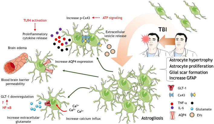
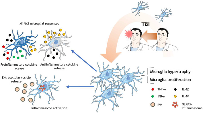
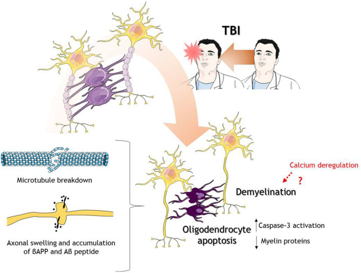
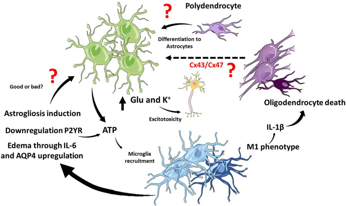
Traumatic brain injury (TBI) is a heterogeneous disorder that involves brain damage due to external forces. TBI is the main factor of death and morbidity in young males with a high incidence worldwide. TBI causes central nervous system (CNS) damage under a variety of mechanisms, including synaptic dysfunction, protein aggregation, mitochondrial dysfunction, oxidative stress, and neuroinflammation. Glial cells comprise most cells in CNS, which are mediators in the brain's response to TBI. In the CNS are present astrocytes, microglia, oligodendrocytes, and polydendrocytes (NG2 cells). Astrocytes play critical roles in brain's ion and water homeostasis, energy metabolism, blood-brain barrier, and immune response. In response to TBI, astrocytes change their morphology and protein expression. Microglia are the primary immune cells in the CNS with phagocytic activity. After TBI, microglia also change their morphology and release both pro and anti-inflammatory mediators. Oligodendrocytes are the myelin producers of the CNS, promoting axonal support. TBI causes oligodendrocyte apoptosis, demyelination, and axonal transport disruption. There are also various interactions between these glial cells and neurons in response to TBI that contribute to the pathophysiology of TBI. In this review, we summarize several glial hallmarks relevant for understanding the brain injury and neuronal damage under TBI conditions.
Keywords: astrocytes; glia; microglia; oligodendrocytes; traumatic brain injury.
Copyright © 2021 Mira, Lira and Cerpa.
Conflict of interest statement
The authors declare that the research was conducted in the absence of any commercial or financial relationships that could be construed as a potential conflict of interest.
Figures
Similar articles
-
Techniques and Methods of Animal Brain Surgery: Perfusion, Brain Removal, and Histological Techniques.In: Kobeissy FH, editor. Brain Neurotrauma: Molecular, Neuropsychological, and Rehabilitation Aspects. Boca Raton (FL): CRC Press/Taylor & Francis; 2015. Chapter 15. In: Kobeissy FH, editor. Brain Neurotrauma: Molecular, Neuropsychological, and Rehabilitation Aspects. Boca Raton (FL): CRC Press/Taylor & Francis; 2015. Chapter 15. PMID: 26269921 Free Books & Documents. Review.
-
Glial Cells: Role of the Immune Response in Ischemic Stroke.Front Immunol. 2020 Feb 26;11:294. doi: 10.3389/fimmu.2020.00294. eCollection 2020. Front Immunol. 2020. PMID: 32174916 Free PMC article. Review.
-
Lack of NG2 exacerbates neurological outcome and modulates glial responses after traumatic brain injury.Glia. 2016 Apr;64(4):507-23. doi: 10.1002/glia.22944. Epub 2015 Dec 6. Glia. 2016. PMID: 26638112
-
Neuronal injury in chronic CNS inflammation.Best Pract Res Clin Anaesthesiol. 2010 Dec;24(4):551-62. doi: 10.1016/j.bpa.2010.11.001. Epub 2010 Nov 29. Best Pract Res Clin Anaesthesiol. 2010. PMID: 21619866 Review.
-
Reactive gliosis in traumatic brain injury: a comprehensive review.Front Cell Neurosci. 2024 Feb 28;18:1335849. doi: 10.3389/fncel.2024.1335849. eCollection 2024. Front Cell Neurosci. 2024. PMID: 38481632 Free PMC article. Review.
Cited by
-
Loss of Mitochondrial Tusc2/Fus1 Triggers a Brain Pro-Inflammatory Microenvironment and Early Spatial Memory Impairment.Int J Mol Sci. 2024 Jul 5;25(13):7406. doi: 10.3390/ijms25137406. Int J Mol Sci. 2024. PMID: 39000512 Free PMC article.
-
Inflammatory response in traumatic brain and spinal cord injury: The role of XCL1-XCR1 axis and T cells.CNS Neurosci Ther. 2024 Jun;30(6):e14781. doi: 10.1111/cns.14781. CNS Neurosci Ther. 2024. PMID: 38887195 Free PMC article. Review.
-
Cell-specific spatial profiling of targeted protein expression to characterize the impact of intracortical microelectrode implantation on neuronal health.J Mater Chem B. 2024 Dec 4;12(47):12307-12319. doi: 10.1039/d4tb01628a. J Mater Chem B. 2024. PMID: 39479901 Free PMC article.
-
Ginkgolide A attenuated apoptosis via inhibition of oxidative stress in mice with traumatic brain injury.Heliyon. 2024 Jan 14;10(2):e24759. doi: 10.1016/j.heliyon.2024.e24759. eCollection 2024 Jan 30. Heliyon. 2024. PMID: 38304806 Free PMC article.
-
Regional variances depict a unique glial-specific inflammatory response following closed-head injury.Front Cell Neurosci. 2023 Feb 15;17:1076851. doi: 10.3389/fncel.2023.1076851. eCollection 2023. Front Cell Neurosci. 2023. PMID: 36909284 Free PMC article.
References
Publication types
LinkOut - more resources
Full Text Sources